Review
. 2024 Dec;14(12):100925.
doi: 10.1016/j.jpha.2023.12.015. Epub 2023 Dec 21.

3D-printed constructs deliver bioactive cargos to expedite cartilage regeneration

Affiliations
Review

3D-printed constructs deliver bioactive cargos to expedite cartilage regeneration

Rong Jiao et al. J Pharm Anal. 2024 Dec.

Abstract

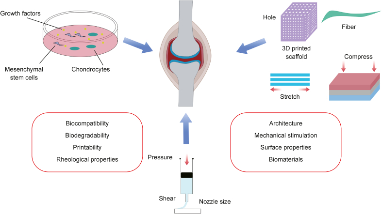
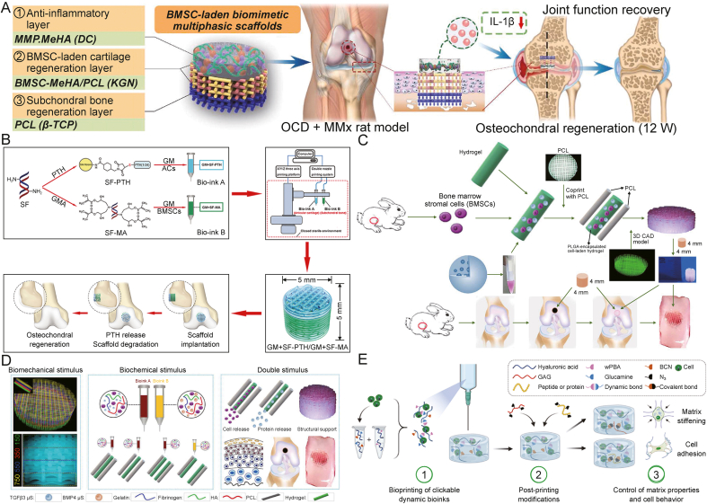
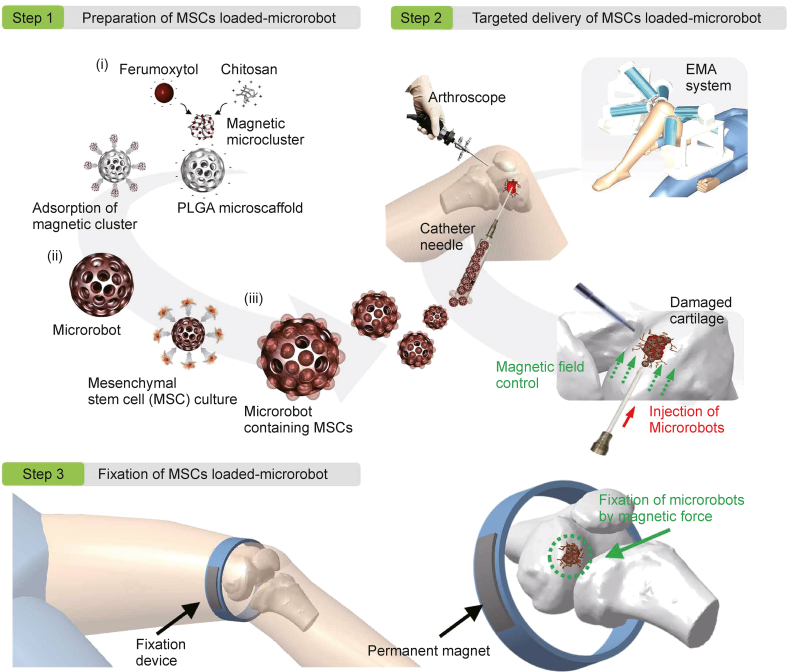
Cartilage is solid connective tissue that recovers slowly from injury, and pain and dysfunction from cartilage damage affect many people. The treatment of cartilage injury is clinically challenging and there is no optimal solution, which is a hot research topic at present. With the rapid development of 3D printing technology in recent years, 3D bioprinting can better mimic the complex microstructure of cartilage tissue and thus enabling the anatomy and functional regeneration of damaged cartilage. This article reviews the methods of 3D printing used to mimic cartilage structures, the selection of cells and biological factors, and the development of bioinks and advances in scaffold structures, with an emphasis on how 3D printing structure provides bioactive cargos in each stage to enhance the effect. Finally, clinical applications and future development of simulated cartilage printing are introduced, which are expected to provide new insights into this field and guide other researchers who are engaged in cartilage repair.

Keywords: 3D bioprinting; Articular cartilage; Cartilage regeneration; Tissue engineering.


Conflict of interest statement

The authors declare that there are no conflicts of interest.

Figures

Image 1
Graphical abstract
Fig. 1
Fig. 1
Structure and microenvironment of the cartilage.
Fig. 2
Fig. 2
Schematic diagram of various attributes to be considered for the design and development of 3D bioprinting widely used with cartilage regeneration.
Fig. 3
Fig. 3
Strategies for enhancing cell acquisition, proliferation and differentiation. (A) Schematic diagram of the dual-stage crosslinking mechanism and workflow of transforming growth factor (TGF-β1) tethered construct generation for chondrogenic tissue culture. Reprinted with permission from Ref. [54]. (B) Schematic illustration showing the rationale of the study and covalent immobilization of TGF on polycaprolactone melt electrowriting (PCL MEW) microfibers. Reprinted with permission from Ref. [55]. (C) Schematic illustration of the bilayered scaffold loaded with TGF-β and bone morphogenetic protein (BMP)-2 for osteochondral repair. Reprinted with permission from Ref. [58]. (D) Concept for a core–shell system with two specified coaxially-extruded zones with central factor depots in one scaffold. Reprinted with permission from Ref. [59]. (E) Schematic illustration of 3D printing of microenvironment-specific biomimetic hydrogel scaffolds for the repairing of osteochondral (OC) defects; (F) 3D-printed bilayer microenvironment-specific biomimetic scaffolds (Bi-Hydrogel-Exos) plays a role via sustained release of bioactive exosomes from the double-network hydrogel system. Reprinted with permission from Ref. [34]. PCL: polycaprolactone; MEW: melt elec trowriting; APPJ: atmospheric-pressure plasma; SF: silk fibroin; hChon: human articular chondrocytes; hOB: human pre-osteoblasts; BMP-2: bone morphogenetic protein type 2; GelMA: methacrylated gelatin; OHA: oxidative hyaluronic acid; HA-DA: hyaluronic acid-dopamine-conjugated; DCM: derived from cartilage: DBM: derived from bone; hADSCs: human adipose-derived stem cells; PEG: poly(ethylene) glycol; Exos: exosome.
Fig. 4
Fig. 4
New biological ink improves cartilage repair and regeneration. (A) Schematic of the 3D bioprinting approach for the engineering of articular cartilage. Reprinted with permission from Ref. [87]. (B) Schematic diagram of platelet-rich plasmagelatin methacrylated (PRP-GelMA) hydrogel promoting osteochondral regeneration in rabbits by polarization of M2 macrophages. Reprinted with permission from Ref. [95]. (C) Schematic illustration of supramolecular interactions in polycarbonate polyurethane methysulfobetaine (PCU-MeSB) polyurethane network. Reprinted with permission from Ref. [98]. (D) Schematic diagrams of the 3D silk fibroin tyramine-substituted gelatin (SF-GT) hydrogel scaffold synthesis. Reprinted with permission from Ref. [101]. (E) Schematic representation of the fixed fused deposition modeling (FDM) scaffolds and remote ultrasound stimulation. Reprinted with permission from Ref. [107]. hMSC: human mesenchymal stem cell; PEG: polyethylene glycol; UV: ultraviolet; PRP: platelet-rich plasma.
Fig. 5
Fig. 5
Schematic design diagram of widely used DNA hydrogel. Reprinted with permission from Ref. [108]. PCR: polymerase chain reaction; X, Y, T: shapes of DNA ; NdeI and EcoRI: two different restriction sites.
Fig. 6
Fig. 6
Multifunctional layer-by-layer continuous 3D bioprinting can be applied to cartilage regeneration. (A) Schematic diagram of repairing cartilage defect with bone marrow-derived mesenchymal stem cell (BMSC) loaded bionic multiphase scaffold. Reprinted with permission from Ref. [117]. (B) Schematic diagram of the preparation of the gelatin methacryloyl (GM)+silk fibroin with parathyroid hormone (SF-PTH) /GM+SF-gelatin methacryloyl (MA) biphasic scaffold and its application. Reprinted with permission from Ref. [119]. (C) Schematic presentation of the 3D bioprinting dual-factor releasing for anisotropic cartilage regeneration; (D) Schematic presentation of the gradient-structured constructs ready to implant. Reprinted with permission from Ref. [123]. (E) General concept of clickable dynamic bioinks. Reprinted with permission from Ref. [127]. MMP: matrix metalloproteinase; OCD: osteochondral defects; MeHA: methacrylated hyaluronic acid; PCL: polycaprolactone ; MMx: medial meniscectomy; BMP: bone morphogenetic protein; HA: hyaluronic acid; GAG: glycosaminoglycans; BCN: bicyclononyne; wPBA: Wulff-type phenylboronic acid.
Fig. 7
Fig. 7
Schematic diagram of articular cartilage regeneration program mediated by magnetic micro-robot. Reprinted with permission from Ref. [134].

Similar articles

Cited by

References

    1. Coughlin T.R., Kennedy O.D. The role of subchondral bone damage in post-traumatic osteoarthritis. Ann. N. Y. Acad. Sci. 2016;1383:58–66. - PubMed
    1. Trivedi J., Betensky D., Desai S., et al. Post-traumatic osteoarthritis assessment in emerging and advanced pre-clinical meniscus repair strategies: A review. Front. Bioeng. Biotechnol. 2021;9 - PMC - PubMed
    1. Jiang W., Liu H., Wan R., et al. Mechanisms linking mitochondrial mechanotransduction and chondrocyte biology in the pathogenesis of osteoarthritis. Ageing Res. Rev. 2021;67 - PubMed
    1. Primorac D., Molnar V., Rod E., et al. Knee osteoarthritis: A review of pathogenesis and state-of-the-art non-operative therapeutic considerations. Genes. 2020;11 - PMC - PubMed
    1. Lieberthal J., Sambamurthy N., Scanzello C.R. Inflammation in joint injury and post-traumatic osteoarthritis. Osteoarthritis Cartilage. 2015;23:1825–1834. - PMC - PubMed

LinkOut - more resources