Review
. 2024 Dec 6;36(1):102416.
doi: 10.1016/j.omtn.2024.102416. eCollection 2025 Mar 11.

Splicing to orchestrate cell fate

Affiliations
Review

Splicing to orchestrate cell fate

Xurui Zhang et al. Mol Ther Nucleic Acids. .

Abstract

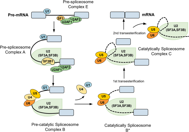
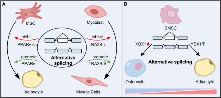
Alternative splicing (AS) plays a critical role in gene expression by generating protein diversity from single genes. This review provides an overview of the role of AS in regulating cell fate, focusing on its involvement in processes such as cell proliferation, differentiation, apoptosis, and tumorigenesis. We explore how AS influences the cell cycle, particularly its impact on key stages like G1, S, and G2/M. The review also examines AS in cell differentiation, highlighting its effects on mesenchymal stem cells and neurogenesis, and how it regulates differentiation into adipocytes, osteoblasts, and chondrocytes. Additionally, we discuss the role of AS in programmed cell death, including apoptosis and pyroptosis, and its contribution to cancer progression. Importantly, targeting aberrant splicing mechanisms presents promising therapeutic opportunities for restoring normal cellular function. By synthesizing recent findings, this review provides insights into how AS governs cellular fate and offers directions for future research into splicing regulatory networks.

Keywords: MT: Bioinformatics; alternative splicing; apoptosis; cell cycle; cell differentiation; neurogenesis; tumorigenesis.


Conflict of interest statement

The authors declare no competing interests.

Figures

None
Graphical abstract
Figure 1
Figure 1
The schematic view of the spliceosome assembly The assembly of the spliceosome begins with U1 small nuclear ribonucleoprotein (snRNP) recognizing and binding to the 5′ splice site (SS) through base pairing, while splicing factor 1 (SF1) binds to the branchpoint sequence (BPS), U2 auxiliary factor 2 (U2AF2) binds to the polypyrimidine tract, and U2 auxiliary factor 1 (U2AF1) binds to the 3′ SS, forming complex E. Subsequently, U2 snRNP, aided by U2AF, replaces SF1 by base pairing with the BPS, forming complex A. Next, the U5/U4/U6 tri-snRNP is recruited, leading to the rearrangement of complex A. U4 and U6 snRNPs are linked through cRNA pairing, and U5 snRNP is loosely bound through protein interactions, resulting in the formation of complex B. Following a series of conformational changes, U1 snRNP exits, U6 snRNP binds to the 5′ SS, and U4 snRNP departs, allowing U6 snRNP to pair with U2 snRNP via small nuclear RNA interactions. This rearrangement forms the pre-catalytic spliceosome complex B, which undergoes two transesterification reactions. The first transesterification reaction produces complex C, where further rearrangements promote the second transesterification, yielding a post-spliceosomal complex. Consequently, the exons are joined to form mature mRNA, the introns are degraded, and the snRNPs are recycled.
Figure 2
Figure 2
Alternative splicing regulates the process of the cell cycle The cell-cycle progression of somatic stem cells is tightly regulated by cyclins, cyclin-dependent kinases (CDKs), and their inhibitors. In the active state, somatic stem cells move through the G1, S, G2, and M phases. The transition into and out of the cell cycle is critically dependent on the phosphorylation status of retinoblastoma (RB) proteins. In pro-growth conditions, cyclin D activates CDK4 and CDK6, which phosphorylate RB, repressing its activity and enabling the transcription of E2F target genes essential for cell proliferation. CDK2/cyclin E further phosphorylates RB to drive the cell into the S phase. During the late S phase, CDK2/cyclin A phosphorylates CDC6, facilitating exit from the S phase. Additionally, CDK2/cyclin E phosphorylates the splicing factor CDC5L, which regulates the splicing of numerous genes involved in the cell cycle. Somatic stem cells enter a quiescent state early in the G1 phase due to the downregulation of cyclin D and cyclin E and the upregulation of CDK inhibitors such as p21, p27, and p57. These inhibitors prevent the activity of CDK4 and CDK6. Quiescent stem cells can shift to an “alert” state, characterized by mammalian target of rapamycin signaling, which reduces the time required to re-enter the cell cycle.
Figure 3
Figure 3
Alternative splicing directs the differentiation of stem cells (A) Alternative splicing (AS) plays a critical role in regulating key transcription factors during stem cell differentiation, impacting cellular fate decisions. The diagram illustrates how AS modulates the expression of transcription factors such as PPARγ in mesenchymal stem cells (MSCs), guiding lineage specification. PPARγ, subject to AS, produces multiple isoforms, including both promotive and inhibitory variants that influence adipogenesis. Similarly, the splicing factor TRA2B undergoes AS during myoblast differentiation, with its isoforms TRA2B-L and TRA2B-S exerting opposing effects on myocyte maturation. This figure highlights the complex regulatory role of AS in orchestrating the balance between differentiation pathways across various cell types. (B) The spatiotemporal-specific expression of Y-box binding protein 1 (YBX1) during stem cell differentiation. YBX1 demonstrates temporal heterogeneity, with its expression rising during osteogenesis and decreasing during adipogenesis in bone marrow stromal cells (BMSCs). Over time, the decline of YBX1 in aging BMSCs leads to aberrant splicing of genes involved in bone formation and aging, promoting adipogenic differentiation at the expense of osteogenesis. This temporal regulation of YBX1 highlights its crucial role in maintaining proper differentiation pathways and balancing stem cell fate decisions across developmental stages.
Figure 4
Figure 4
AS regulates neurogenesis Neuronal progenitor cells primarily express PTBP1, but when induced to differentiate, PTBP1 is repressed and PTBP2 is induced. This switch alters the splicing of exons sensitive to PTBP1, while exons sensitive to both PTBP1 and PTBP2 remain repressed during early neuron differentiation. As neurons mature and form synapses, PTBP2 is downregulated, leading to changes in splicing regulatory programs affecting exons in genes, which are associated with adult brain functions. Additionally, RBM4 regulates PTBP1 expression or splicing activity by modulating exon selection during the differentiation of both non-neuronal and neuronal cells. RBM4 suppresses PTBP1 and PTBP2 levels in non-neuronal differentiation by promoting exon 11/10 skipping, leading to mRNA degradation via nonsense-mediated decay. During neuronal differentiation, miR-124 downregulates PTBP1, and RBM4-induced exon 9 skipping generates a PTBP1 isoform with reduced splicing activity, mitigating the effect of PTBP1. This intricate regulation ensures proper neuron differentiation and maturation, and disruptions in these processes can lead to the premature expression of adult brain mRNA isoforms and subsequent cell death.
Figure 5
Figure 5
Multiple roles of AS in tumorigenesis AS significantly contributes to tumorigenesis by generating isoforms that support cancer hallmarks like proliferation, apoptosis, invasion, metastasis, angiogenesis, and metabolism. For example, AS of ITGA6 produces ITGA6A, promoting colorectal cancer cell proliferation, while splicing of PKM favors PKM2, enhancing glycolysis in cancer cells. PTBP1 influences the splicing of Bcl-x, generating pro-apoptotic Bcl-xs or anti-apoptotic Bcl-xl isoforms. In angiogenesis, AS of VEGF-A produces various isoforms with pro- or anti-angiogenic properties.

References

    1. Bonnal S.C., López-Oreja I., Valcárcel J. Roles and mechanisms of alternative splicing in cancer - implications for care. Nat. Rev. Clin. Oncol. 2020;17:457–474. doi: 10.1038/s41571-020-0350-x. - DOI - PubMed
    1. Chow L.T., Gelinas R.E., Broker T.R., Roberts R.J. An amazing sequence arrangement at the 5' ends of adenovirus 2 messenger RNA. Cell. 1977;12:1–8. - PubMed
    1. Berget S.M., Moore C., Sharp P.A. Spliced segments at the 5' terminus of adenovirus 2 late mRNA. Proc. Natl. Acad. Sci. USA. 1977;74:3171–3175. - PMC - PubMed
    1. Kalsotra A., Xiao X., Ward A.J., Castle J.C., Johnson J.M., Burge C.B., Cooper T.A. A postnatal switch of CELF and MBNL proteins reprograms alternative splicing in the developing heart. Proc. Natl. Acad. Sci. USA. 2008;105:20333–20338. doi: 10.1073/pnas.0809045105. - DOI - PMC - PubMed
    1. Kent O.A., Ritchie D.B., Macmillan A.M. Characterization of a U2AF-independent commitment complex (E') in the mammalian spliceosome assembly pathway. Mol. Cell Biol. 2005;25:233–240. - PMC - PubMed

LinkOut - more resources