. 2024 Dec 5;33(1):101389.
doi: 10.1016/j.omtm.2024.101389. eCollection 2025 Mar 13.

Increased functional potency of multi-edited CAR-T cells manufactured by a non-viral transfection system

Affiliations

Increased functional potency of multi-edited CAR-T cells manufactured by a non-viral transfection system

Aine O'Sullivan et al. Mol Ther Methods Clin Dev. .

Abstract

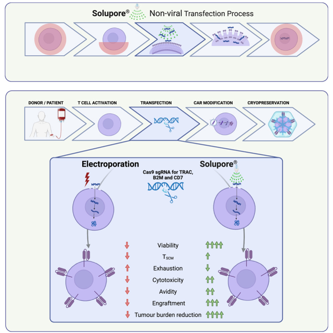
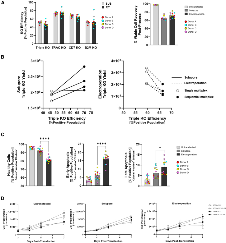
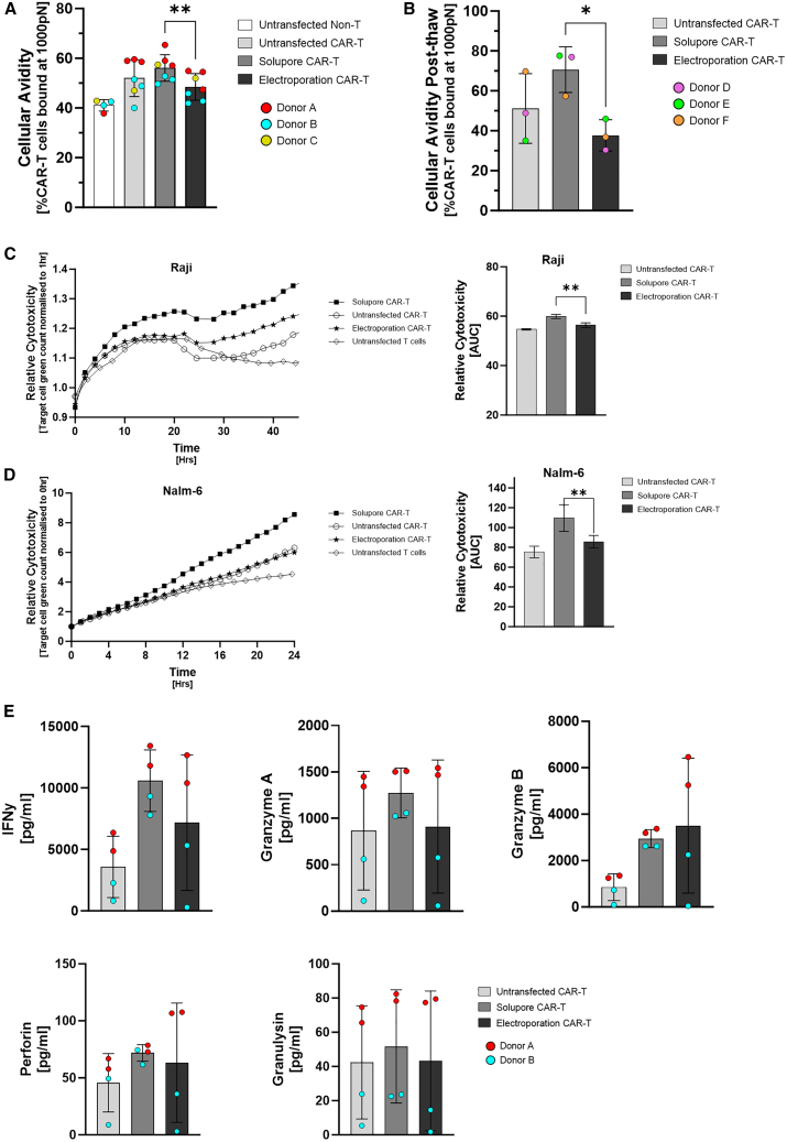
Chimeric antigen receptor (CAR)-T cell therapy represents a breakthrough for the treatment of hematological malignancies. However, to treat solid tumors and certain hematologic cancers, next-generation CAR-T cells require further genetic modifications to overcome some of the current limitations. Improving manufacturing processes to preserve cell health and function of edited T cells is equally critical. Here, we report that Solupore, a Good Manufacturing Practice-aligned, non-viral physicochemical transfection system, can be used to manufacture multi-edited CAR-T cells using CRISPR-Cas9 ribonucleoproteins while maintaining robust cell functionality. When compared to electroporation, an industry standard, T cells that were triple edited using Solupore had reduced levels of apoptosis and maintained similar proportions of stem cell memory T cells with higher oxidative phosphorylation levels. Following lentiviral transduction with a CD19 CAR, and subsequent cryopreservation, triple-edited CAR-T cells manufactured using Solupore demonstrated improved immunological synapse strength to target CD19+ Raji cells and enhanced cellular cytotoxicity compared with electroporated CAR-T cells. In an in vivo mouse model (NSG), Solupore triple-edited CAR-T cells enhanced tumor growth inhibition by more than 30-fold compared to electroporated cells.

Keywords: CAR-T; CAR-T manufacturing; Solupore; adoptive cell therapy; cell avidity; cell cytotoxicity; genetic engineering; metabolism; non-viral transfection; stem cell memory.


Conflict of interest statement

All authors are current or former Avectas employees. Avectas has filed patents covering the Solupore technology described in this paper. M.W.L. is a member of the scientific advisory board of Avectas and supervised experiments performed in the Royal Free Hospital, London and UCL. Avectas has received funding for elements of this work under the Disruptive Technologies Innovation Fund (DTIF), owned by the Department of Enterprise, Trade, and Employment under Call 3, DTIF reference number DT2020224.

Figures

None
Graphical abstract
Figure 1
Figure 1
Solupore demonstrates efficient delivery of complex cargo, while maintaining cell expansion and viability (A) Solupore-based CRISPR-Cas9 RNP KO of TRAC, CD7, and B2M in single transfection of multiplexed guide RNAs (gRNAs) using either the research tool (RT) or the clinical-grade single-use system (SUS), measured 72 h post-transfection. (B) Sequential transfection (a second transfection with multiplexed gRNAs) of the same edited population of cells, examining yield and triple KO (TKO) efficiency following 7 days of proliferation of two independent healthy donors, each in duplicate. Solupore single TKO transfection efficiency = 46.23 ± 1.66, sequential efficiency = 70.10 ± 4.42. Electroporation single TKO transfection efficiency = 59.61 ± 6.42, sequential efficiency = 66.30 ± 1.06. (C) Healthy and apoptotic cells were measured by flow cytometry using a dual-color live/dead stain as well as a caspase-3 activity dye (Nucview-405). Samples were measured 30 min immediately post-transfection using a non-transfected sample as a control for healthy cells from each donor (three donors in duplicate, n = 6). Unpaired t test; healthy cells p < 0.0001, early apoptosis p < 0.0001, and late apoptosis p = 0.0389. (D) Untransfected and Solupore- and electroporation-edited cells from n = 3 donors were cultured at 1 × 106 cells per well on day 0 in a 24-well G-Rex in either TexMACS or CTSOpTmizer and stimulatory cytokines and expanded for 7 days. Cell counts were performed on days 3, 4, 5, and 7 post-transfection to compare cell proliferation between growth conditions. Data represented as mean +/- SD.
Figure 2
Figure 2
Analysis of TSCM phenotype over time (A and B) CD4+ (A) and (B) CD8+ T cells were expanded for 0–4 days post-transfection in a 24-well G-Rex plate and stimulation with TransACT and IL-2 (N = 3 donors in duplicate). TSCM phenotype (CD45RA+ CD45RO CD62L+ CCR7+ CD95+) was measured by flow cytometry, and a comparison of results for Solupore and electroporation TKO transfected T cells and untransfected control cells is shown. (C and D) CAR-T cells were generated via lentiviral transduction, and the TSCM phenotype of CD4+ and CD8+ CAR-T cells was analyzed 24 h post-transduction (pre-cryopreservation; C) and (D) post-thaw. n = 3 donors, paired t test, p = 0.0396 (C). Data represented as mean +/- SD.
Figure 3
Figure 3
Metabolic health and function of Solupore-transfected cells (A) Untransfected, Solupore-transfected, or electroporation-transfected cell metabolic potential at 24 h post-thaw (cells were cryopreserved 3 days post-transfection) (n = 3 individual donors, two-way ANOVA statistical analysis, p < 0.05). (B) Representative OCR profile at 24 h post-thaw. (C) Cytokine profiling 4 days post-transfection. Data were normalized to cell counts on day 4 of the experiment to correlate with relative cytokine production per cell (paired t test, p = 0.0085 [IL-2], p = 0.0498 [IFN-γ]). (D) Surface marker phenotype data for CD8+ T cells (CTLA-4, LAG-3, PD-1, TIGIT, TIM-3) was 7 days post-transfection after three rounds of stimulation with PMA (10 ng/mL) and ionomycin (250 ng/mL) (stimulation on days 0, 3, and 5 post- transfection). Unpaired t test, untransfected versus Solupore p = 0.0267, untransfected versus electroporation p = 0.0047 (CTLA-4), Solupore versus electroporation p = 0.0461 (LAG-3), untransfected versus Solupore p = 0.0005, untransfected versus electroporation p = 0.005, Solupore versus electroporation p < 0.0001 (PD-1). Data represented as mean +/- SD.
Figure 4
Figure 4
Comparison of effector function of CAR-T product generated from Solupore and electroporation triple-edited T cells (A and B) Cellular avidity was measured using the z-Movi and is represented as percentage of cells bound to target cells after a maximal force of 1,000 pN has been applied (n = 3 donors; donors A–C were used for [A], while donors D–F were used for [B]). Paired t test, p = 0.001 (A), 0.0486 (B). (C and D) Raji and Nalm-6 cells (CD19+) co-culture with triple-edited and cryopreserved T cells (CD19 CAR+). Cytotoxicity was measured using the Incucyte S3 Live-Cell Analysis System for 48 h. Representative figure for 1:1 ratio (Raji) and 4:1 ratio (Nalm-6); n = 3 (mean of three donors). AUC analysis was performed to compare the differences between sample cytotoxicity (unpaired t test, p = 0.0089 [C], 0.0015 [D]). (E) Cytokine profiling from supernatants of a 1:1 co-culture between Raji cells and untransfected, Solupore, and electroporation CAR-T cells. Supernatants were harvested 24 h post-co-culture. Data represented as mean +/- SD.
Figure 5
Figure 5
Tumor growth analysis in NSG mice inoculated with CD19+ Raji-LUC cells measured over 25 days (A and B) IVIS imaging and tumor burden measurement using luminescent target Raji cells expressing CD19 (Raji-LUC) in an NSG mouse model over 26 days (n = 8), comparing Solupore TKO CAR-T cells with untransfected CAR-T cells, untransfected cells (negative for CAR), electroporation CAR-T cells, and PBS/vehicle control. (C–E) Dose-dependent response of Solupore TKO CAR-T cells, untransfected CAR-T cells, and electroporated TKO CAR-T cells. (D) Day 18 Welch’s t test, p = 0.0005, day 25 unpaired t test, p < 0.0001. 2 × 106 day 25 p = 0.0088; 4 × 106 day 13 Mann-Whitney, p = 0.003; day 18 Welch’s t test, p = 0.0005; day 25 unpaired t test, p < 0.0001. Data represented as mean +/- SD.
Figure 6
Figure 6
In vivo study analysis of cell product engraftment and activation Flow cytometry analysis of blood draws from mice on study days 7, 13, 18, and 26 (A, B, and F–H) or homogenized spleens at the study endpoint (day 26, C–E). (A) The percentage of engrafted CAR-T cells in mice based on human CD45-expressing (B and C) KO of TRAC across untransfected (UT), Solupore (SOL), and electroporation (EP) groups by flow cytometry. unpaired T-test p = 0.0008 (A) and unpaired T-test p <0.0001 (C). (D and E) TKO quantification from homogenized spleen cells. (F) Early activation marker quantification by expression of the surface markers CD25 and CD69. (G and H) Cytotoxic populations determined by expression of CD8+ CD107a+ T cells (G) Mann-Whiteny test p = 0.0281 and exhaustion assessed by expression of PD-1 (H) unpaired T-test p <0.0001 Day 18. p = 0.0051 Day 26. Data represented as mean +/- SD.

References

    1. Davila M.L., Riviere I., Wang X., Bartido S., Park J., Curran K., Chung S.S., Stefanski J., Borquez-Ojeda O., Olszewska M., et al. Efficacy and toxicity management of 19-28z CAR T cell therapy in B cell acute lymphoblastic leukemia. Sci. Transl. Med. 2014;6:224ra25. - PMC - PubMed
    1. Maude S.L., Frey N., Shaw P.A., Aplenc R., Barrett D.M., Bunin N.J., Chew A., Gonzalez V.E., Zheng Z., Lacey S.F., et al. Chimeric antigen receptor T cells for sustained remissions in leukemia. N. Engl. J. Med. 2014;371:1507–1517. - PMC - PubMed
    1. Lee D.W., Kochenderfer J.N., Stetler-Stevenson M., Cui Y.K., Delbrook C., Feldman S.A., Fry T.J., Orentas R., Sabatino M., Shah N.N., et al. T cells expressing CD19 chimeric antigen receptors for acute lymphoblastic leukaemia in children and young adults: a phase 1 dose-escalation trial. Lancet. 2015;385:517–528. - PMC - PubMed
    1. Turtle C.J., Hanafi L.A., Berger C., Gooley T.A., Cherian S., Hudecek M., Sommermeyer D., Melville K., Pender B., Budiarto T.M., et al. CD19 CAR-T cells of defined CD4+:CD8+ composition in adult B cell ALL patients. J. Clin. Invest. 2016;126:2123–2138. - PMC - PubMed
    1. Maude S.L., Laetsch T.W., Buechner J., Rives S., Boyer M., Bittencourt H., Bader P., Verneris M.R., Stefanski H.E., Myers G.D., et al. Tisagenlecleucel in Children and Young Adults with B-Cell Lymphoblastic Leukemia. N. Engl. J. Med. 2018;378:439–448. - PMC - PubMed

LinkOut - more resources