

Control values of intraocular pressure in different species: a review of literature
- PMID: 39812101
- PMCID: PMC11748451
- DOI: 10.3325/cmj.2024.65.518
Control values of intraocular pressure in different species: a review of literature
Abstract
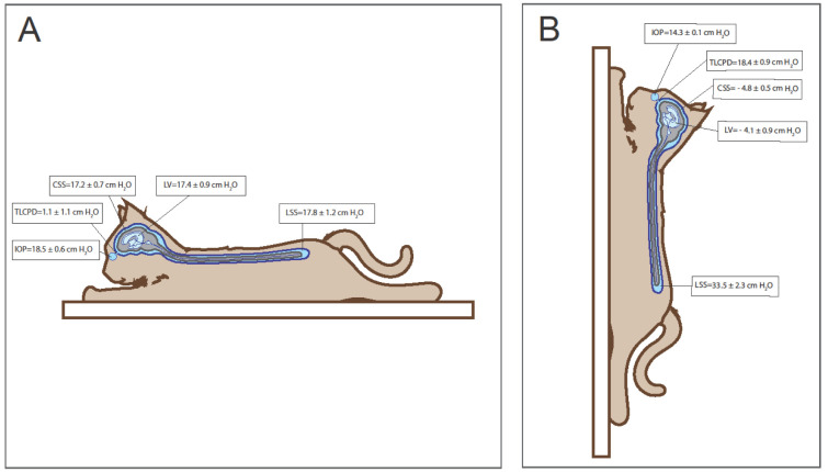
It is generally accepted that intraocular pressure (IOP) depends on the rate of aqueous humor production, system outflow resistance, and episcleral venous pressure. Therefore, control IOP values are expected to be within the strict and predictable limits in specific animal species, and there should be no vast differences between species. However, in the literature the control IOP values significantly vary (from potentially "hypotensive" to "hypertensive") within the same species, and especially between species depending on the measurement technique, head position in relation to the rest of the body, circadian rhythm, age, and topical and systemic drugs (anesthetics) applied. These variations make it difficult to compare different therapeutic approaches for intraocular hypertension, investigate the correlation between IOP and intracranial pressure, and determine target IOP values in glaucoma research. We recommend that different IOP physiology and pathophysiology studies take into account all the mentioned factors when describing IOP measurement methodology.
Figures

References
-
- Janssen SF, Gorgels TG, Ten Brink JB, Jansonius NM, Bergen AA. Gene expression-based comparison of the human secretory neuroepithelia of the brain choroid plexus and the ocular ciliary body: potential implications for glaucoma. Fluids Barriers CNS. 2014;11:2. doi: 10.1186/2045-8118-11-2. - DOI - PMC - PubMed
Publication types
MeSH terms
LinkOut - more resources
Full Text Sources