. 2025 Feb 5;14(1):e240248.
doi: 10.1530/ETJ-24-0248. Print 2025 Feb 1.

Sciatic nerve analysis in thyroid hormone transporters Mct8 and Oatp1c1 knockout mice

Sciatic nerve analysis in thyroid hormone transporters Mct8 and Oatp1c1 knockout mice

Steffen Mayerl et al. Eur Thyroid J. .

Abstract

Objective: Mutations in the thyroid hormone (TH) transporter monocarboxylate transporter 8 (MCT8) cause Allan-Herndon-Dudley syndrome (AHDS), a severe form of psychomotor retardation with muscle hypoplasia and spastic paraplegia as key symptoms. These abnormalities have been attributed to impaired TH transport across brain barriers and into neural cells, thereby affecting brain development and function. Likewise, Mct8/Oatp1c1 (organic anion-transporting polypeptide 1c1) double knockout (M/Odko) mice, a well-established murine AHDS model, display a strongly reduced TH passage into the brain as well as locomotor abnormalities. To which extent the peripheral nervous system is affected by combined MCT8/OATP1C1 deficiency has not been addressed.

Methods: Using the sciatic nerve as a model, we studied the spatiotemporal expression of TH transporters as well as the sciatic thyroidal state, sciatic nerve myelination and function in M/Odko mice by immunofluorescence, qPCR, Western blotting and electrophysiology.

Results: We detected MCT8 protein expression in sciatic nerve axons, whereas OATP1C1 expression was observed in a subset of endothelial cells early in postnatal development. The absence of MCT8 and OATP1C1 did not alter the thyroidal state of isolated nerves at P12. Moreover, electrophysiological studies did not disclose any significant alteration in sciatic nerve signal propagation parameters in adult M/Odko mice. Although Schwann cell numbers were similar, Western blot analysis showed a mild form of hypermyelination in adult M/Odko mice.

Conclusions: Altogether, our data point to a largely unaffected sciatic nerve structure and function in the absence of MCT8 and OATP1C1.

Keywords: LAT1; LAT2; MCT10; Slc16a2; Slco1c1; T3; T4; sciatic nerve.


Conflict of interest statement

The authors declare that there is no conflict of interest that could be perceived as prejudicing the impartiality of the work.

Figures
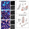
Figure 1
Figure 1
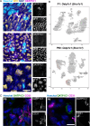
MCT8 expression in the sciatic nerve. (A) Immunohistochemical analysis on Wt sciatic nerve cryo-sections obtained from P8, P12 and P180 mice following fixation in methanol. Axonal marker NFH is depicted in yellow, myelin marker MBP in blue, Hoechst33258 to label cell nuclei in cyan, and MCT8 in magenta. Single-channel grayscale pictures are shown for NFH and MCT8. n = 3. Scale bar: 10 μm. (B) Visualization of Mct8 (Slc16a2) transcript expression in single-cell RNA sequencing data of the P1 and P60 sciatic nerve. pmSC, pro-myelinating Schwann cells; mSC, myelinating Schwann cells; nm(R)SC, non-myelinating (Remak) Schwann cells; iSC, immature Schwann cells; prol. SC, proliferating Schwann cells; IC, immune cells; Prol. Fb, proliferating fibroblast-like cells; Fb.Rel*, fibroblast-related cluster (tentative cluster); EnC, endoneurial cells; PnC, perineurial cells; EpC, epineurial cells; EC, endothelial cells; Per, pericytes; and VSMC, vascular smooth muscle cells.
Figure 2
Figure 2
OATP1C1 expression in a subset of sciatic nerve endothelial cells. (A) Wt sciatic nerve sections were fixed in methanol and co-stained for OATP1C1 (in magenta), MBP (in blue), NFH (in yellow) and Hoechst33258 (in cyan). Single-channel grayscale pictures are shown for NFH and OATP1C1. n = 3. Scale bar: 10 μm. (B) Visualization of Oatp1c1 (Slc01c1) transcript expression in single-cell RNA sequencing data of the P1 and P60 sciatic nerve. pmSC, pro-myelinating Schwann cells; mSC, myelinating Schwann cells; nm(R)SC, non-myelinating (Remak) Schwann cells; iSC, immature Schwann cells; prol. SC, proliferating Schwann cells; IC, immune cells; Prol. Fb, proliferating fibroblast-like cells; Fb.Rel*, fibroblast-related cluster (tentative cluster); EnC, endoneurial cells; PnC, perineurial cells; EpC, epineurial cells; EC, endothelial cells; Per, pericytes; and VSMC, vascular smooth muscle cells. (C) Sciatic nerve sections obtained from P8 Wt animals were stained for OATP1C1 (in green) and CD31 (in magenta). Representative pictures for double-positive blood vessels (left) and structures positive for only one of these proteins (right) are displayed. Hoechst33258 counter-stained cell nuclei appear blue. Single-channel grayscale pictures are presented for CD31 and OATP1C1. n = 3. Scale bar: 10 μm.
Figure 3
Figure 3
Sciatic nerve thyroidal state. Sciatic nerves from P12 mice were subjected to qPCR analysis, and the expression of TH signaling components (A) and TH-responsive marker genes (B) was evaluated. Expression levels were normalized to Gapdh, cyclophilin A and cyclophilin D expression as housekeeping genes. Wt values were set as 1.0. n = 4–5.
Figure 4
Figure 4
Electrophysiological properties of the sciatic nerve. Sciatic nerves of 1-year-old female mice were stimulated using repetitive single square-wave pulses. Signals were recorded at different distances to the stimulus, and maximum responses were averaged. CMAP amplitudes at the proximal (A) and distal (B) positions as well as maximum nerve conduction velocity (C) are displayed. n = 8 (Wt, Mct8ko); 5 (Oatp1c1ko); 7 (M/Odko).
Figure 5
Figure 5
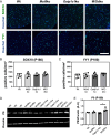
Schwann cell analysis in developing animals. Cross sections of PFA-fixed sciatic nerves obtained from P12 animals were immunostained for the Schwann cell marker SOX10 and the myelinating Schwann cell marker YY1 (both in green) (A). Hoechst33258-labeled cell nuclei appear in blue. Numbers of SOX10+ cells (B) and YY1+ cells (C) were quantified and are shown as density per mm2. n = 3. Scale bar: 100 μm.
Figure 6
Figure 6
Schwann cell and myelination analysis in adult animals. Cross sections of PFA-fixed sciatic nerves obtained from P180 female mice were immunostained for the Schwann cell marker SOX10 and for YY1 (both in green) (A). Hoechst33258-labeled cell nuclei are shown in blue. Cells positive for SOX10 (B) and YY1 (C) were enumerated and are presented as density per mm2. n = 5. Scale bar: 100 μm. (D) Sciatic nerves of 6-month-old females were homogenized and subjected to Western blot analysis. The mature myelin marker P0 was assessed. Protein levels were quantified by measuring the integrated density of the respective bands and normalizing it to Vinculin as a housekeeping protein. Wt values were set as 1.0. n = 3. *P < 0.05.

Similar articles

References

    1. Bernal J. Thyroid hormones and brain development. Vitam Horm 2005. 71 95–122. (10.1016/s0083-6729(05)71004-9) - DOI - PubMed
    1. Groeneweg S, van Geest FS, Peeters RP, et al. . Thyroid hormone transporters. Endocr Rev 2020. 41 146–201. (10.1210/endrev/bnz008) - DOI - PubMed
    1. Friesema EC, Grueters A, Biebermann H, et al. . Association between mutations in a thyroid hormone transporter and severe X-linked psychomotor retardation. Lancet 2004. 364 1435–1437. (10.1016/s0140-6736(04)17226-7) - DOI - PubMed
    1. Dumitrescu AM, Liao XH, Best TB, et al. . A novel syndrome combining thyroid and neurological abnormalities is associated with mutations in a monocarboxylate transporter gene. Am J Hum Genet 2004. 74 168–175. (10.1086/380999) - DOI - PMC - PubMed
    1. Schwartz CE, May MM, Carpenter NJ, et al. . Allan-Herndon-Dudley syndrome and the monocarboxylate transporter 8 (MCT8) gene. Am J Hum Genet 2005. 77 41–53. (10.1086/431313) - DOI - PMC - PubMed

MeSH terms

Supplementary concepts