Review
. 2025 Jan 31;39(2):e70326.
doi: 10.1096/fj.202402816RR.

SKP1-CUL1-F-box: Key molecular targets affecting disease progression

Affiliations
Review

SKP1-CUL1-F-box: Key molecular targets affecting disease progression

Xiangrong Zeng et al. FASEB J. .

Abstract

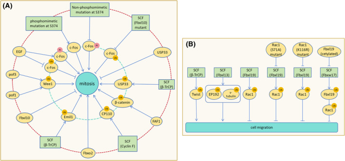
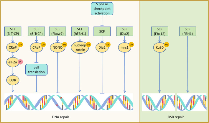
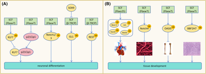
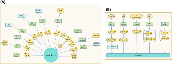
The correct synthesis and degradation of proteins are vital for numerous biological processes in the human body, with protein degradation primarily facilitated by the ubiquitin-proteasome system. The SKP1-CUL1-F-box (SCF) E3 ubiquitin ligase, a member of the Cullin-RING E3 ubiquitin ligase (CRL) family, plays a crucial role in mediating protein ubiquitination and subsequent 26S proteasome degradation during normal cellular metabolism. Notably, SCF is intricately linked to the pathogenesis of various diseases, including malignant tumors. This paper provides a comprehensive overview of the functional characteristics of SCF complexes, encompassing their assembly, disassembly, and regulatory factors. Furthermore, we discuss the diverse effects of SCF on crucial cellular processes such as cell cycle progression, DNA replication, oxidative stress response, cell proliferation, apoptosis, cell differentiation, maintenance of stem cell characteristics, tissue development, circadian rhythm regulation, and immune response modulation. Additionally, we summarize the associations between SCF and the onset, progression, and prognosis of malignant tumors. By synthesizing current knowledge, this review aims to offer a novel perspective for a holistic and systematic understanding of SCF complexes and their multifaceted functions in cellular physiology and disease pathogenesis.

Keywords: CUL1; F‐box; SKP1; disease progression; molecular target.


Figures

FIGURE 1
FIGURE 1
F‐box proteins are categorized into three subfamilies. Fbxl (F‐box with leucine‐rich repeats), Fbxw (F‐box with WD‐40 amino acid repeat sequence), and Fbxo (F‐box with uncharacterized domains).
FIGURE 2
FIGURE 2
Functional characteristics of the SCF complex. (A) Both SCF and ARIH1 serve as E3 ubiquitin ligases. A ‘E3‐E3’ super‐domain is formed, involving the CR3 and 4HB fragments of the CUL1 component of SCF and the RING of TBX1 with the Ariaden fragment of ARIH1. Ubiquitin is transferred from the E2 ubiquitin enzyme UBE2L3, which binds to ARIH1, to the catalytic cysteine of ARIH1, and subsequently to the substrate of SCF. (B) During the degradation of substrates mediated by SCF through the 26S proteasome, CUL1 binds to the 20S subcomplex, and the ubiquitinated CUL1 binds to the 19S subcomplex. (C) SKP1 can form SKP1‐SKP2 and SKP1‐SKP2‐cks1 complexes to regulate the polyubiquitination of substrates.
FIGURE 3
FIGURE 3
The assembly and disassembly of SCF and its influencing factors. (A) CAND1 binds to and allosterically modulates inactive SCF complexes, causing the separation of CUL1 from RBX1 and SKP1, thereby facilitating the recovery of CUL1. It can also replace the substrate receptor module F‐box, promoting the synthesis of new SCF complexes. (B) Rig‐G inhibits the assembly of SCF (β‐TrCP) by down‐regulating the levels of CUL1 and β‐TrCP. (C) Roc1 promotes the binding of Nedd8 to CUL1, leading to the degradation of CUL1 by the 26S proteasome, thereby inhibiting the assembly of SCF. (D) SmRBx binds to CUL1 to facilitate the assembly of SCF complexes and then dissociates from CUL1.
FIGURE 4
FIGURE 4
SCF regulation of cell cycle progression. SCF (β‐TrCP) ubiquitinates and degrades Mis18β, inhibiting centromere function to limit cell entry into the G1 phase. SCF (β‐TrCP) also promotes the ubiquitin‐dependent degradation of cyclin D1, promoting G1 phase progression. CDK inhibits SCF (Fbxo11) recognition of CDT2 to prevent cells from exiting the G0/G1 phase. SCF (Fbx6) ubiquitinates and degrades Chk1 to terminate the S‐phase checkpoint and promote cell entry into the S phase. AKT phosphorylation of cyclin F accelerates G0/G1 arrested cells into the S phase. Upon activation by ATM and ATR, Chk1/2 phosphorylates Cdc25A to stimulate SCF (β‐TrCP) to down‐regulate Cdc25A, delaying cells in the S phase. Phosphorylation site mutant USP37 resists the ubiquitination and degradation of SCF (β‐TrCP), hindering the transition of cells from the G2 phase to the M phase. SCF (cyclin F) ubiquitinates and degrades E2F1, impeding cell G2/M phase progression. SCF (β‐TrCP) mediates the ubiquitination and degradation of Cdc25B, which promotes cell G2/M phase progression by binding to the non‐phosphorylated motif of Cdc25B.
FIGURE 5
FIGURE 5
SCF regulation of cell proliferation and migration. (A) After SCF (β‐TrCP) ubiquitinates and degrades Emil1, APC activity increases to promote cell proliferation. Fbxl10 inhibits cell proliferation by suppressing ribosomal RNA expression. Pof1 and Pof3 co‐downregulate Wee1 to increase the rate of cell mitosis entry. Phosphorylation of c‐Fos by EGF antagonizes c‐Fos ubiquitination and promotes cell proliferation. Pseudophosphorylation mutation of c‐Fos S374 stabilizes c‐Fos, promoting cell proliferation, while dephosphorylation mutation destabilizes c‐Fos, hindering cell proliferation. SCF (Fbxl10) mutation loses the ability to inhibit cell proliferation. By mediating USP33 degradation, SCF (β‐TrCP) relieves USP33 inhibition on cell proliferation and promotes proliferation. FAF1 promotes SCF (β‐TrCP) to down‐regulate β‐catenin, preventing β‐catenin from inhibiting cell proliferation. SCF (cyclin F) mediates CP110 degradation to ensure DNA replication correctness and integrity during mitosis. Translocation expression of Fbxo2 promotes osteosarcoma cell proliferation. (B) The degradation of Twist by SCF (β‐TrCP) inhibits the motility and migration of cancer cells. Overexpression of Fbxl13 leads to the ubiquitination and degradation of CEP192 and γ‐tubulin, disrupting the nucleation of centrosome microtubule arrays and promoting cell migration. Ubiquitination and degradation of Rac1 by Fbxl19 reduces the formation of cell migration fronts and inhibits cell migration. S71A‐Rac1 and K166R‐Rac1 mutants resist Fbxl19 degradation and promote cell migration. Fbxw17 promotes cell migration by degrading Fbxl19 and reducing Rac ubiquitination, while acetylation of Fbxl19 resists Fbxw17‐mediated degradation.
FIGURE 6
FIGURE 6
SCF affects DNA repair. SCF (β‐TrCP) promotes the phosphorylation of eIF2α after degrading CReP, leading to the expression of DNA repair proteins such as DDR and promoting DNA repair. Degradation of CReP by SCF (β‐TrCP) can also decrease cell expression during DNA repair, aiding DNA repair. The substrate NONO of SCF (Fbxw7) assists in the DNA repair process. SCF (hFBH1) degrades DNA binding proteins like nucleoprotein, facilitating the binding of DNA to repair proteins. Activation of the S‐phase checkpoint reduces Dia2 degradation, assisting in DNA repair at damaged sites. SCF (Dia2) degrades mrc1, aiding in the repair of S‐phase DNA damage. SCF (Fbx12) degrades Ku80, relieving Ku80's inhibition on DNA double‐strand break (DSB) repair, and promoting DSB repair. SCF (FBH1) repairs DSBs produced by Rec12.
FIGURE 7
FIGURE 7
SCF involves in maintaining stem cell properties, affecting cell differentiation, and tissue development. (A) SCF (Fbxw7) overexpression degrades KLF7, inhibiting p21Cip1 gene expression and impairing neuronal differentiation by reducing the interaction between KLF7 and p21Cip1. SCF (Fbxw7)‐mediated degradation of Notch1/3 is necessary for neuronal differentiation. SOX9 prevents SCF (β‐TRCP)‐mediated degradation of GL1, maintaining the stem cell properties of cancer cells. Normal degradation of REST by SCF (β‐TRCP) promotes normal neuronal differentiation. (B) SCF (Fbxo25) degrades Nkx2‐5, Isl1, Hand1, and Mef2C to assist in heart development. SCF (Fbxw7)‐mediated degradation of Notch4 is required for vascular network development. Degradation of OASIS and BBF2H7 by SCF (Fbxw7) inhibits bone and cartilage formation, while depletion of Fbxw7 promotes bone and cartilage development facilitated by OASIS and BBF2H7.
FIGURE 8
FIGURE 8
Effects of SCF complex on oxidative stress, apoptosis, and autophagy. (A) Fbxw7 degrades Nrf1, promoting neuronal apoptosis during endoplasmic reticulum stress. The S350A‐Nrf1 mutant resists Fbxw7 degradation and apoptosis. SCF (Fbxl7) mediates the polyubiquitination and degradation of survivin, impairing mitochondrial function and promoting apoptosis. Deletion of JKF fails to degrade p53, resulting in cell cycle blockade and apoptosis induction. Inactivation of Fbxw7 halts the cell cycle and induces apoptosis. USP47 deletion inhibits SCF (β‐TrCP) ubiquitination and degradation of Cdc25A, leading to apoptosis. PTEN prevents Fbxl2 from degrading IP3R3, leading to apoptosis. Fbxo31 plays an anti‐apoptotic role in ESCC. SCF (Fbxo31) degrades MKK6, preventing activation of p38 and apoptosis. Calyxin Y promotes apoptosis of hepatocellular carcinoma cells by enhancing SCF (β‐TrCP) degradation of eEF2K. Fbxo22 deletion fails to degrade BAG3, resulting in high levels of BAG3 and preventing apoptosis. The S337A‐BAG3 mutant, resistant to degradation, inhibits apoptosis. (B) Phosphorylated p85 resists SCF (Fbx12)‐mediated ubiquitination and degradation, hindering PI(3)K binding to IRS1 and inhibiting PI(3)K signaling cascades, ultimately promoting autophagy. P53 promotes SCF (Fbxl20) degradation of Vps34 and inhibits PtdIns3P‐induced autophagy. N‐myristoylation promotes SCF (Fbxo27) ubiquitination of glycoproteins, facilitating lysosomal autophagy. TBL1 promotes SCF‐mediated degradation of MYC and BECLIN‐1, promoting autophagy in damaged cells to stabilize them. SCF (Fbxl4) ubiquitination degrades NIX and BNIP3, inhibiting mitophagy.
FIGURE 9
FIGURE 9
SCF involves in the regulation of immune response. SCF (Fbxo3) ubiquitination of AIRE promotes AIRE binding to b (P‐TEFb) and increases AIRE expression, thereby facilitating the expression of specific antigens in the thymus. LPS promotes SCF (β‐TrCP) to degrade PKD1, inhibiting the TLR inflammatory signaling pathway. SCF (Fbxl19) targets ISGylation of p65 to reduce inflammation and inflammation‐induced damage in mice. Following Fbxl19 deletion, p65 phosphorylation increases, enhancing the inflammatory response. Fbxw11 deletion promotes the phosphorylation of PKR and eIF2α, leading to the shutdown of protein synthesis in RVFV‐infected cells and inhibition of viral proliferation. Fbxo2 ubiquitination of GAS promotes cell heterophagic degradation of bacterial pathogens. SCF (Fbxo21) mediates polyubiquitination of ASK1, promoting the production of interferon IFNα/β.

Similar articles

Cited by

References

    1. Horn‐Ghetko D, Krist DT, Prabu JR, et al. Ubiquitin ligation to F‐box protein targets by SCF‐RBR E3‐E3 super‐assembly. Nature. 2021;590(7847):671‐676. - PMC - PubMed
    1. Gazdoiu S, Yamoah K, Wu K, et al. Proximity‐induced activation of human Cdc34 through heterologous dimerization. Proc Natl Acad Sci USA. 2005;102(42):15053‐15058. - PMC - PubMed
    1. Sadowski M, Suryadinata R, Lai X, Heierhorst J, Sarcevic B. Molecular basis for lysine specificity in the yeast ubiquitin‐conjugating enzyme Cdc34. Mol Cell Biol. 2010;30(10):2316‐2329. - PMC - PubMed
    1. Bloom J, Peschiaroli A, DeMartino G, Pagano M. Modification of Cul1 regulates its association with proteasomal subunits. Cell Div. 2006;1:5. - PMC - PubMed
    1. Chandra Dantu S, Nathubhai Kachariya N, Kumar A. Molecular dynamics simulations elucidate the mode of protein recognition by Skp1 and the F‐box domain in the SCF complex. Proteins. 2016;84(1):159‐171. - PubMed

LinkOut - more resources