. 2025 Mar;66(2):189-206.
doi: 10.1007/s10329-024-01177-x. Epub 2025 Jan 15.

A three-dimensional kinematic analysis of bipedal walking in a white-handed gibbon (Hylobates lar) on a horizontal pole and flat surface

Affiliations

A three-dimensional kinematic analysis of bipedal walking in a white-handed gibbon (Hylobates lar) on a horizontal pole and flat surface

Takafumi Fujiwara et al. Primates. 2025 Mar.

Abstract

Gibbons, a type of lesser ape, are brachiators but also walk bipedally and without forelimb assistance, not only on the ground but also on tree branches. The arboreal bipedal walking strategy of the gibbons has been studied in previous studies in relation to two-dimensional (2D) kinematic analysis. However, because tree branches and the ground differ greatly in width, leading to a constrained foot contact point on the tree branches, gibbons must adjust their 3D joint motions of trunk and hindlimb on the tree branches. Furthermore, these motor adjustments could help minimize the center of mass (CoM) mediolateral displacement. This study investigated the kinematic adjustment mechanism necessary to enable a gibbon to walk bipedally on an arboreal-like substrate using 3D measurements. Trials were recorded with eight video cameras that were placed around the substrate. The CoM position on the body, the Cardan angles of the hindlimb joints and trunk, and spatiotemporal parameters were calculated. Asymmetry of thorax, pelvis, trunk, and left and right hindlimb joint motion was observed in the pole and flat conditions. In the pole condition, the narrower step width and the smaller range of motion of the mediolateral CoM displacement were observed with increased hip adduction and knee eversion angles. These kinematic adjustments might place the knee and foot directly under the body during the single support phase, producing a reduced step width and the amount of the mediolateral CoM displacement of a gibbon.

Keywords: Bipedal walking; Gibbon; Joint angle; Pole and flat condition; Step width.


Conflict of interest statement

Declarations. Conflict of interest: The authors declare no conflicts of interest.

Figures

Fig. 1
Fig. 1
Landmarks set in a global coordinate system in the sagittal (a) and frontal (b) view. The landmarks are denoted only on the right side here, but were marked on both sides in experiments. Red arrows represent the local coordinate systems that align with the cardinal anatomical axes: mediolateral (x), anteroposterior (y), and superoinferior (z). a OP: occipital protuberance, OR: orbital ridge, T1: first thoracic spinous process, T13: thirteenth thoracic spinous process, NI: nipple, IT: ischial tuberosity, IC: the most cranial points of iliac crest, HGT: humeral greater tubercle, UO: ulnar olecranon process, SP: ulnar styloid process, HP: the tip of third hand phalange, FGT: femoral greater trochanter, LE: femoral lateral epicondyle, LC: tibial lateral condyle, LM: lateral malleolus, CR: the tip of calcaneal ridge, and FP: third foot phalange. b ME: femoral medial epicondyle and MM: medial malleolus
Fig. 2
Fig. 2
Experimental setup of the pole (a) and flat (b) condition. Bipedal walking on the flat and pole substrates were recorded using eight cameras placed around the substrates. The substrates were placed so that their long axis coincided with the direction of travel
Fig. 3
Fig. 3
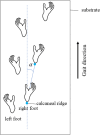
Schematic image of the foot contact positions in the horizontal plane in the flat condition showing right stride cycle. To control the direction of walking in the flat condition, the coordinates of each landmark in all trials were transformed α degrees between the line segment that connected the right calcaneal ridge at the initial contact and at the next initial contact (solid blue line), and a line parallel to the substrate (broken blue line) in the horizontal plane
Fig. 4
Fig. 4
Trunk flexion/extension (a), lateral flexion (b), axial rotation (c), thorax flexion/extension (d), lateral flexion (e), axial rotation (f), pelvis tilt (g), obliquity (h) and axial rotation (i) angles in the left stride cycle between the pole (red) and flat (black) substrate conditions. The thick line represents the mean value, and the shaded area represents the standard deviation ( ±). Each stride cycle begins with a right foot contact and ends on the next right foot contact. The vertical dotted, broken, and solid lines represent the terminal stance of the left foot, its initial contact, and terminal stance of the right foot, respectively
Fig. 5
Fig. 5
Trunk flexion/extension (a), lateral flexion (b), axial rotation (c), thorax flexion/extension (d), lateral flexion (e), axial rotation (f), pelvis tilt (g), obliquity (h) and axial rotation (i) angles in the right stride cycle between the pole (red) and flat (black) substrate conditions. The thick line represents the mean value, and the shaded area represents the standard deviation ( ±). Each stride cycle begins with a right foot contact and ends on the next right foot contact. The vertical dotted, broken, and solid lines represent the terminal stance of the left foot, its initial contact, and terminal stance of the right foot, respectively
Fig. 6
Fig. 6
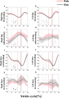
The left and right hip flexion/extension (a, b), adduction/abduction (c, d), knee flexion/extension (e, f) and inversion/eversion (g, h) angles in the stride cycle between the pole (red) and flat (black) substrate conditions. The thick line represents the mean value, and the shaded area represents the standard deviation ( ±). Each stride cycle begins with a right foot contact and ends on the next right foot contact. The vertical dotted, broken, and solid lines represent the terminal stance of the left foot, its initial contact, and terminal stance of the right foot, respectively
Fig. 7
Fig. 7
CoM height during stance phase in the pole (red) and flat (black) conditions (a). Thick lines represent mean values and shaded areas represent standard deviations ( ±). The vertical dotted and broken lines represent the terminal stance of the left foot and the initial contact of the left foot, respectively. Stick picture of the whole body in the sagittal plane at the initial contact (0% of the stance phase; IC), midstance (50% of the stance phase; Mst), and terminal stance (100% of the stance phase; Tst) (b). The trunk segment is represented as a line segment that connected the first thoracic spinous process and the midpoint of both ischial tuberosities. For the limbs, the solid line shows the right side, and the broken line shows the left side. The ‘plus’ signs represent the CoM position. The black horizontal line represents the substrate. Stick picture was plotted as the anteroposterior position of the right calcaneal ridge in both conditions matched in each phase
Fig. 8
Fig. 8
CoM mediolateral displacement on stride cycle during the pole (red) and flat (black) conditions (a). Thick lines represent mean values and shaded areas represent standard deviations ( ±). The vertical dotted, broken, and solid lines represent the terminal stance of the left foot, the initial contact of the left foot, and the terminal stance of the right foot, respectively. Relationship between the CoM and both feet in the horizontal plane in the flat and pole conditions (b). The filled diamonds indicate the position of the CoM where orange (flat condition) and blue (pole condition) indicate the CoM positions at the initial contact of the left foot. The line segments indicate the feet (connecting the calcaneal ridge and the third foot phalange). The filled triangles indicate the third foot phalange. The gibbon’s initial contact was set as the intersection of the vertical and horizontal lines at zero
Fig. 9
Fig. 9
The mediolateral distance between the CoM and the feet (left: broken; right: solid) during stride cycle in the pole (red) and flat (black) conditions. Thick lines represent mean values and shaded areas represent standard deviations ( ±). The vertical dotted, broken, and solid lines represent the terminal stance of the left foot, the initial contact of the left foot, and the terminal stance of the right foot, respectively
Fig. 10
Fig. 10
Thorax (dotted) and pelvis (solid) mediolateral displacements during stride cycle in the pole (red) and flat (black) conditions. Thick lines represent mean values and shaded areas represent standard deviations ( ±). The vertical dotted, broken, and solid lines represent the terminal stance of the left foot, the initial contact of the left foot, and the terminal stance of the right foot, respectively
Fig. 11
Fig. 11
Schematic relationships between the CoM and hindlimb in the frontal plane at midstance (top row) and between the contact point of the left foot and the axial rotation of the trunk in the horizontal plane at the terminal stance (bottom row). The solid lines in the thigh and shank represent the rough placement of the bones, and the dashed lines represent the gravity lines (top row). Note that increased hip adduction and knee eversion in arboreal walking brings the knee and foot closer to the gravity line. The solid horizontal line in the trunk represents the rough line segment that connects both iliac crests (bottom row). Note that the increased right rotation angle of pelvis and trunk in arboreal walking allows the feet to be placed in a straight line

Similar articles

References

    1. Aerts P, Damme RV, Elsacker LV, Duchene V (2000) Spatio-temporal gait characteristics of the hind-limb cycles during voluntary bipedal and quadrupedal walking in bonobos (Pan paniscus). Am J Phys Anthropol 111:503–517. 10.1002/(SICI)1096-8644(200004)111:4%3c503::AID-AJPA6%3e3.0.CO;2-J - PubMed
    1. Baldwin LA, Teleki G (1976) Patterns of gibbon behavior on Hall’s Island, Bermuda: a preliminary ethogram for Hylobates lar. In: Rumbaugh D (ed) Gibbon and siamang, vol 4. Karger, Basel, pp 21–105
    1. Carlson KJ, Demes B (2010) Gait dynamics of Cebus apella during quadrupedalism on different substrates. Am J Phys Anthropol 142:273–286. 10.1002/ajpa.21224 - PubMed
    1. Cartmill M (1974) Rethinking primate origins. Science 184:436–443 - PubMed
    1. Cartmill M (1985) Climbing. In: Bramble DM, Liem KF, Wake DB (eds) Functional vertebrate morphology. Belknap Press, Cambridge, pp 73–88

LinkOut - more resources