. 2025 Jan 21;122(3):e2418766122.
doi: 10.1073/pnas.2418766122. Epub 2025 Jan 15.

Soil microbiome bacteria protect plants against filamentous fungal infections via intercellular contacts

Affiliations

Soil microbiome bacteria protect plants against filamentous fungal infections via intercellular contacts

Long Lin et al. Proc Natl Acad Sci U S A. .

Abstract

Bacterial-fungal interaction (BFI) has significant implications for the health of host plants. While the diffusible antibiotic metabolite-mediated competition in BFI has been extensively characterized, the impact of intercellular contact remains largely elusive. Here, we demonstrate that the intercellular contact is a prevalent mode of interaction between beneficial soil bacteria and pathogenic filamentous fungi. By generating antibiotics-deficient mutants in two common soil bacteria, Lysobacter enzymogenes and Pseudomonas fluorescens, we show that antibiotics-independent BFI effectively inhibits pathogenic fungi. Furthermore, transcriptional and genetic evidence revealed that this antibiotics-independent BFI relies on intercellular contact mediated by the type VI secretion system (T6SS), which may facilitate the translocation of bacterial toxic effectors into fungal cells. Finally, by using a "conidia enrichment" platform, we found that T6SS-mediated fungal inhibition resulting from intercellular contact naturally occurs within the soil microbiome, particularly represented by Pseudomonas fulva. Overall, these results demonstrate that bacteria from the soil microbiome can protect host plants from fungal infection through antibiotics-independent intercellular contacts, thus revealing a naturally occurring and ecologically important mode of BFI in agricultural contexts.

Keywords: T6SS; bacterial–fungal interaction; contact-dependent antifungal activity; filamentous fungi.


Conflict of interest statement

Competing interests statement:The authors declare no competing interest.

Figures
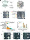
Fig. 1.
Fig. 1.
Discovery of contact-dependent inhibition of filamentous fungal growth by L. enzymogenes OH11 independent of antifungal antibiotics. (A) L. enzymogenes OH11 secretes HSAF to inhibit F. graminearum PH-1. The lafB gene is critical for HSAF biosynthesis, with red dots indicating HSAF secretion by OH11. (B) The HSAF-defective mutant ΔlafB exhibited a nearly complete loss of antagonistic activity against PH-1. Bacterial strains were cultured in 10% TSB media to induce HSAF production, which was subsequently tested for antagonistic activity on 10% TSA plates. (C) L. enzymogenes inhibited conidial germination of F. graminearum PH-1 through a HSAF-independent and contact-dependent mechanism. Both the WT OH11 and ΔlafB hindered the growth of PH-1. Filter separation impeded cell-to-cell contact, resulting in diminished antifungal activity of ΔlafB, while WT OH11 displayed a small zone of inhibition outside the filter. Again, L. enzymogenes strains were cultured in 10% TSB media to induce HSAF production for antagonistic testing on 10% TSA. The blue dotted lines indicate the 0.22-μM PVDF filter, while the red arrows point to the inhibition zones. (D) WT OH11 inhibited PH-1 growth prior to cell contact, whereas ΔlafB did not. Red circles denote PH-1, while yellow circles represent L. enzymogenes. PH-1 and L. enzymogenes were inoculated at varying distance marked by circles. Bacteria cells suspended in water were inoculated onto PDA media to evaluate antifungal activity. (E) The number of differentially expressed genes (DEGs) categorized into various GO terms is illustrated. Orange columns represent up-regulated genes, while blue columns denote down-regulated genes. (F) Expression levels of selected genes were validated by qRT-PCR. Three replicates of each sample were analyzed with a t test. Asterisks indicate significant differences (P < 0.01). (G) L. enzymogenes strains inhibited growth of multiple filamentous fungi in a contact-dependent manner. Both WT OH11 and ΔlafB inhibited growth of F. oxysporum, A. alternata, and C. gloeosporioides, while filter separation obstructed cell-to-cell contact, thereby hindering antifungal activity of ΔlafB. Red arrows indicate zones of inhibition, and blue dotted lines depict the 0.22-μM PVDF filters.
Fig. 2.
Fig. 2.
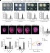
T6SS is essential for contact-dependent inhibition of fungi by L. enzymogenes. (A) T6SS is critical for the contact-dependent inhibition of various filamentous fungi by L. enzymogenes without HSAF. ΔlafBΔtssM exhibited significantly reduced inhibitory activity against all tested fungi compared with ΔlafB, whereas the tssM complementation strain ΔlafBΔtssM-c restored this defect. Bacteria cells suspended in water were inoculated onto PDA media to assess antifungal activity. The down panels were fungal colony sizes quantified from the upper panels. Three replicates of each sample were analyzed using a one-way ANOVA, and different letters indicated the significant differences (P < 0.01). (B) L. enzymogenes prevents soybean infection by F. graminearum through T6SS. Hyphal plugs of F. graminearum were combined with culture of L. enzymogenes–related strains. After coculturing for 1 h, the hyphal plugs were inoculated onto soybean leaves for 2 d, and images were captured under UV light. (C) Quantification of lesion sizes in panel B. Three replicates of each sample were analyzed with a one-way ANOVA test. Different letters indicate the significant differences (P < 0.01). (D) The relative biomass of F. graminearum significantly increased during interactions with ΔlafBΔtssM in soil. Conidia of F. graminearum were mixed with L. enzymogenes strains and subsequently inoculated into sterile soil, which was cultured for 7 d. The relative biomass of F. graminearum and L. enzymogenes was measured by qPCR using total soil DNA. Three replicates of each sample were analyzed with a one-way ANOVA test. Different letters indicate the significant differences (P < 0.01). (E) L. enzymogenes inhibited fungal biomass in natural soil via T6SS. Strains ΔlafB and ΔlafBΔtssM were individually inoculated into the natural soil planted with banana for 7 d. The relative biomass of total fungi and bacteria was measured by qPCR using total soil DNA, with three replicates of each sample analyzed with a one-way ANOVA, and different letters indicating the significant differences (P < 0.01). (F) Principal coordinates analysis (PCoA) of microbiome sequencing indicated that the fungal communities derived from the ΔlafB-treated soil were clustered separately from those derived from the LB-treated soil and the ΔlafBΔtssM-treated soil.
Fig. 3.
Fig. 3.
L. enzymogenes inhibits the elongation of germinating hyphae of F. graminearum through T6SS. (A) Elongation of F. graminearum PH-1 germinating hyphae was inhibited by ΔlafB, whereas the inhibitory capacity of ΔlafBΔtssM was significantly diminished. The hyphae were stained by CFW to enhance visibility. (B) Quantification of hyphae length from panel A. Three replicates of each sample were analyzed with a one-way ANOVA test. Different letters indicate the significant differences (P < 0.01). (C) SEM analysis demonstrated that ΔlafB resulted in partial hyphal degradation of strain PH-1, while this defect was less pronounced in the ΔlafBΔtssM double mutant. Red arrows indicate the attachment of L. enzymogenes cells to F. graminearum hypha. Yellow arrows indicate F. graminearum hypha was partially degraded. (D) Transmission electron microscopy analysis demonstrated that ΔlafB caused deformations in the cell wall and plasma membrane of strain PH-1, while these deformations were partially restored during interactions with ΔlafBΔtssM. Black arrows indicate cell wall, and white arrows indicate cell membrane. (E) The number of DEGs categorized into various GO terms. Orange spots indicate up-regulated genes, while blue spots denote down-regulated genes. (F) Expression levels of selected genes were assessed by qRT-PCR. Three replicates of each sample were analyzed with a t test. Asterisks indicate the significant differences (P < 0.01).
Fig. 4.
Fig. 4.
The L. enzymogenes T6SS effector Le1893 demonstrates antifungal activity. (A) L. enzymogenes inhibited yeast growth in a manner that was both HSAF-independent and dependent on cell-to-cell contacts. The HSAF-defective mutant ΔlafB(mCherry) inhibited the growth of yeast strain W303(GFP) after coculturing for 24 h. Blocking of intercellular contact with a filter resulted in a loss of antifungal activity. The blue dotted line represented the 0.22-μM PVDF filter. Bacterial cells suspended in water were inoculated onto YPD media to assess their antifungal activity. (B) ΔlafBΔtssM exhibited reduced antifungal activity against yeast. Normalized bacterial strains and yeast strain W303(GFP) were mixed in various volumetric ratios and cultured for 24 h prior to analysis. (C) The living number of CFU of W303 (bacteria: yeast = 5:1) in panel B was quantified. Three replicates of each sample were analyzed using a one-way ANOVA test. Different letters indicate the significant differences (P < 0.01). (D) The structure of Le1893 was predicted by AlphaFold2. Color codes: PAAR domain (green), RHS domain (yellow), C-terminal toxin domain (red). (E) Mutation of the predicted self-cleavage sites in Le1893 resulted in the loss of antifungal activity. W303-derived yeast strains were diluted to a series of ODs and cultured for 48 h on either noninducing (glucose) or inducing (galactose) media. (F) Transmission electron microscopy observation revealed that Le1893 induced deformation of yeast cells. (G) The abundance of genomic DNA was significantly reduced in the yeast strain expressing Le1893, whereas strains expressing the mutants of self-cleavage site did not affect genomic DNA abundance. The W303 strains were cultured in SC-U medium supplemented with 2% galactose for 12 h. 1 × 10⁶ yeast cells were harvested and the DNA contents were evaluated through genomic DNA isolation. (H) Flow cytometer analysis demonstrated that Le1893 decreased DNA concentration in yeast cells, while strains expressing the mutated self-cleavage site did not achieve a similar reduction. The W303 strains were cultured in SC-U medium supplemented with 2% galactose for 12 h. 1 × 10⁶ yeast cells were stained by propidium iodide, and then the DNA content was quantified using flow cytometry. FL3-A indicated the fluorescence signal detected by flow cytometry.
Fig. 5.
Fig. 5.
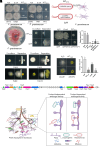
Pseudomonas strains 2P24 and FoE9 inhibit the growth of Fusarium in a contact-dependent manner mediated by T6SS. (A) P. fluorescens 2P24 demonstrated growth inhibition of F. graminearum PH-1. Lysobacter strains and Pseudomonas strains were cocultured with PH-1 for 48 h, respectively. OH13 is L. antibioticus; OH21 is L. brunescens; Pf01 is P. fluorescens; Pf-5 and CHA0 are P. protegens. Bacteria cells suspended in water were inoculated onto PDA media to assess antifungal activity. (B) P. fluorescens 2P24 secreted 2,4-diacetylphloroglucinol (2,4-DAPG) to inhibit F. graminearum PH-1, with the phl gene cluster being essential for 2,4-DAPG biosynthesis. (C) The 2,4-DAPG biosynthesis-defective mutant Δphl exhibited a loss of antagonistic activity against PH-1 compared to WT 2P24. Bacterial strains were cultured in LB media containing 2% glucose to induce 2,4-DAPG biosynthesis, after which the cultures were inoculated onto PDA media to evaluate antifungal activity. (D) P. fluorescens 2P24 inhibited the growth of PH-1 in a contact-dependent manner independent of 2,4-DAPG. Filter separation impeded intercellular contact, resulting in hindered antifungal activity of Δphl, but not WT 2P24. The blue dotted lines denote the 0.22-μM PVDF filter. Bacterial strains were cultured in LB media containing 2% glucose prior to being inoculated onto PDA media for antifungal testing. (E) The ΔphlΔhcp double mutant exhibited decreased antifungal activity compared to Δphl; however, the hcp complementation strain ΔphlΔhcp-c restored this defect. Bacteria cells, suspended in water, were inoculated onto PDA media to test antifungal activity. (F) Fungal colony sizes quantified from panel E. Three replicates of each sample were analyzed with a one-way ANOVA test. Different letters indicate the significant differences (P < 0.01). (G) The obstruction of intercellular contacts by filter separation resulted in hindered antifungal activity of FoE5, FoE9, and FoE13. The blue dotted lines indicate the 0.22-μM PVDF filter, with FoE4 and FoE9 cultured in LB plus 2% glucose media, while FoE5 and FoE13 were sustained in LB media. Cultures were then inoculated onto PDA media for antifungal assessment. (H) Mutation of tssM in FoE9 led to a partial reduction in antifungal activity on PDA media. (I) Fungal colony sizes quantified from panel H. Three replicates of each sample were analyzed with a one-way ANOVA test. Different letters indicate the significant differences (P < 0.01). (J) A T6SS gene cluster was identified in the genome of FoE9. (K) The working model depicts contact-dependent and contact-independent antifungal activities of bacteria. In the soil ecosystem, beneficial bacteria such as L. enzymogenes OH11 and P. fluorescence 2P24 secreted antibiotics to inhibit fungi in a contact-independent manner. Conversely, strains OH11, 2P24, and other soil microbiome bacteria, including FoE5 and FoE9, can also inhibit filamentous fungi through contact-dependent mechanisms, even in the absence of antibiotic metabolites. In the cases of strains OH11, 2P24, and FoE9, T6SS plays a crucial role in this contact-dependent antifungal activity, potentially by translocating T6SS effectors into fungal cells.

References

    1. Avery S. V., Singleton I., Magan N., Goldman G. H., The fungal threat to global food security. Fungal Biol. 123, 555–557 (2019). - PubMed
    1. Fisher M. C., Hawkins N. J., Sanglard D., Gurr S. J., Worldwide emergence of resistance to antifungal drugs challenges human health and food security. Science 360, 739–742 (2018). - PubMed
    1. Zhou Y., et al. , Bacterial-fungal interactions under agricultural settings: From physical to chemical interactions. Stress Biol. 2, 22 (2022). - PMC - PubMed
    1. Zhan C., Matsumoto H., Liu Y., Wang M., Pathways to engineering the phyllosphere microbiome for sustainable crop production. Nat. Food 3, 997–1004 (2022). - PubMed
    1. Fira D., Dimkic I., Beric T., Lozo J., Stankovic S., Biological control of plant pathogens by Bacillus species. J. Biotechnol. 285, 44–55 (2018). - PubMed

MeSH terms

Substances

LinkOut - more resources