. 2025 Jan 17;11(3):eadr7934.
doi: 10.1126/sciadv.adr7934. Epub 2025 Jan 15.

CD4+FOXP3Exon2+ regulatory T cell frequency predicts breast cancer prognosis and survival

Affiliations

CD4+FOXP3Exon2+ regulatory T cell frequency predicts breast cancer prognosis and survival

Clorinda Fusco et al. Sci Adv. .

Abstract

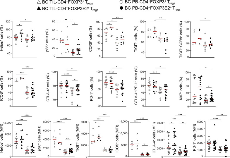
CD4+FOXP3+ regulatory T cells (Tregs) suppress immune responses to tumors, and their accumulation in the tumor microenvironment (TME) correlates with poor clinical outcome in several cancers, including breast cancer (BC). However, the properties of intratumoral Tregs remain largely unknown. Here, we found that a functionally distinct subpopulation of Tregs, expressing the FOXP3 Exon2 splicing variants, is prominent in patients with hormone receptor-positive BC with poor prognosis. Notably, a comprehensive examination of the TCGA validated FOXP3E2 as an independent prognostic marker in all other BC subtypes. We found that FOXP3E2 expression underlies BCs with defective mismatch repair and a stem-like signature and highlights pathways involved in tumor survival. Last, we found that the TME induces FOXP3E2 through the CXCL12/CXCR4 axis and confirmed the higher immunosuppressive capacity of FOXP3E2+ Tregs derived from patients with BC. Our study suggests that FOXP3E2+ Tregs might be used as an independent biomarker to predict BC prognosis and survival and to develop super-targeted immunotherapies.


Figures

Fig. 1.
Fig. 1.. Characterization of the immune infiltrate in PB and primary tissue from patients with BC and BF.
(A) Schematic representation of FOXP3+ and FOXP3E2+ Tregs in tumor immune escape. (B) CD8+/CD4+ ratio, % of (C) FOXP3+ and (D) FOXP3E2+ cells (gated on CD4+), (E) FOXP3E2+/FOXP3+ ratio (E2 ratio), and (F) CD8+/Treg ratio in peripheral blood (PB; dots) and tumor-infiltrating lymphocytes (TILs; triangles) from patients with BF (white empty) and BC (red empty). In (B) to (F), represented data are for BF at least n = 7 and n = 15 and for BC at least n = 15 and n = 24 (respectively, for TILs and PB). (G) Correlation between % of FOXP3E2+ and CD8+/Treg ratio in TILs from patients with BC (n = 24). (H) Representative immunohistochemical (IHC) staining of primary BC and BF tissue showing CD3+, CD8+, FOXP3+, and FOXP3E2+ cells. Immunohistochemistry-based quantification of (I) % of CD3+, (J) CD8+, (K) FOXP3+, (L) FOXP3E2+ cells, (M) CD8+/CD3+ ratio, (N) FOXP3+/CD3+ ratio, and (O) FOXP3E2+/CD3+ ratio [respectively, white dots (n = 6) for BF and red triangles (n = 23) for patients with BC]. Data are presented as median values. Each data point represents a different individual (i.e., independent biological samples) [(G) and (I) to (O)] or experimental replicates [(B) to (F)]. Statistical analyses were performed by using Mann-Whitney U test (two tails) [(B) to (F) and (I) to (O)] and Spearman r correlation test (G). *P ≤ 0.05; **P ≤ 0.01; ***P ≤ 0.005; ****P ≤ 0.0001.
Fig. 2.
Fig. 2.. FOXP3E2 transcript analysis from primary BC tissues delineates a subgroup of patients with poor prognosis and a distinctive gene expression profile.
(A) FOXP3 transcripts in normal (n = 112) and primary BC (n = 990) tissues. Data represent normalized RNA-seq by expectation maximization (RSEM) value obtained by RNA-seq analysis of datasets in the TCGA database. (B and C) Kaplan-Meier survival curve of patients with BC stratified into low– and high–FOXP3 expression levels within the primary tumor based on its Q2 (n = 495 and 495) or Q3 (n = 742 and 248) value. (D) Interquartile distribution of the FOXP3E2/FOXP3 ratio calculated in the primary BC tissue (n = 990). (E) Patients with BC were stratified into low (n = 741) and high (n = 249) FOXP3E2/FOXP3 ratio (E2 ratio) according to the Q3 value cutoff. (F) Hazard ratio (= 1.8, confidence interval of 1.1 to 2.8, Cox P = 0.014) and (G) Kaplan-Meier survival curve of patients with BC with low (n = 741) and high (n = 249) E2 ratios according to Q3 value cutoff. AIC, Akaike's Information Criterion. (H) Volcano plot of differentially expressed genes (DEGs) obtained by applying a threshold of log2 fold change > ±0.05 (x axis) and a P adj. < 0.001 (y axis) in the two groups of patients with BC. Dots represented single genes: 179 up-regulated (red) and 523 down-regulated (green) in the high-ratio BC group. (I) Circular composition overview plot for selected Gene Ontology (GO) pathways (represented in different colors) overrepresented among DEGs in high– versus low–FOXP3E2/FOXP3 ratio BC groups. GO analysis was performed by DAVID database. Gene color scale indicates the relevant fold change values (red, up-regulated; and green, down-regulated). Data are presented as median values [(A) and (E)]. Statistical analyses were performed by using Mann-Whitney U test (two tails) [(A) and (E)], Multivariate Cox regression model reference [(B), (C), and (G)] and log-rank test (F). ****P ≤ 0.0001.
Fig. 3.
Fig. 3.. Reduced co-mutation frequency and co-occurrence characterizes the BC group with a high FOXP3E2/FOXP3 ratio.
(A and B) Somatic interaction analysis between gene pairs showing that co-occurring mutations (green squares) and mutually exclusive mutations (brown squares) were detected using somaticInteractions function of Maftools (v.2.12.0), which performs pair-wise Fisher’s exact tests to detect significant [Benjamini-Hochberg false discovery rate (FDR) < 0.1] pairs of genes. The intensity of the color is proportionate to the −log10 (P value). (C and D) Diagram reporting the cancer network of patients with BC separated into low and high FOXP3E2/FOXP3 ratios, including nodes (driver genes) and edges (co-occurrences), obtained by WES analysis from TCGA. Red circles represent driver genes, and blue circles represent all occurrences found. The solid black links represent the key links found by the co-occurrence analysis. Genes were considered to have evidence of positive selection (driver gene) if the reported dN/dS ratio qglobal_cv was <0.05 for missense mutations, truncating variants, all substitutions, or indels. (E) Variant allele frequency (%) showing the mean of variant clusters across groups. ClonEvol was used to infer consensus clonal evolution models using the variant clusters generated by the Maftools R package. Statistical analyses were performed by using by Benjamini-Hochberg multiple testing correction.
Fig. 4.
Fig. 4.. dMMR and specific mutational signatures in patients with BC with a high FOXP3E2/FOXP3 ratio.
(A and B) Mutational signatures identified in patients with BC with low and high FOXP3E2/FOXP3 ratios, respectively. The y axis indicates exposure of 96 trinucleotide motifs to overall signature. In each plot, we report the best match against validated COSMIC signatures (COSMIC v.2) and cosine similarity value alongside the proposed etiology. (C) Supervised hierarchical clustering analysis of TCGA-BC reverse-phase protein array (RPPA) results using an analysis of variance (ANOVA) FDR P-value threshold lower than 0.05. On the basis of this threshold, 81 probes were differentially altered in the high–E2 ratio group, with 40 probes up-regulated (red bar) and 41 down-regulated (green bar). (D) Enrichment analysis of the differently expressed probes using Metascape. Statistical analyses were performed by using Cophenetic correlation. GTPases, guanosine triphosphatases.
Fig. 5.
Fig. 5.. Highly immunosuppressive FOXP3E2+ Tregs preferentially accumulate in TILs of patients with newly diagnosed HR+ BC.
Cumulative data of flow cytometry analysis showing cell percentage and mean fluorescence intensity (MFI) of Helios+, pS6+, CCR8+, TIGIT+, ICOS+, CTLA-4+, PD-1+, and Ki67+ cells (gated on CD4+FOXP3+ and CD4+FOXP3E2+) in freshly isolated TILs (at least n = 4, BC TIL) and PB (at least n = 9, BC PB) from patients with BC. Data are presented as median values. Statistical analysis was performed by using Wilcoxon and Mann-Whitney U test (two tails); *P ≤ 0.05; **P ≤ 0.01; ***P ≤ 0.005; ****P ≤ 0.0001.
Fig. 6.
Fig. 6.. Higher IC expression in Tregs from patients with HR+ BC correlates with increased FOXP3E2+/FOXP3+ ratio and peripheral Treg suppressive function.
(A) Percentage of suppression of Tregs in coculture with carboxyfluorescein diacetate succinimidyl ester (CFSE)–labeled Tconvs at different proportions of Tregs/Tconvs, purified from HDs (n = 20) and patients with BC (n = 16). (B) FOXP3E2 PB ratio (E2 PB ratio) calculated by flow cytometry quantification of the ratio of CD4+FOXP3E2+/CD4+FOXP3+ Tregs from PB of HDs (n = 38) and patients with BC (n = 33). (C) Percentage of TIGIT+, CCR8+, TIGIT+/CCR8+, CTLA-4+, CTLA-4+/PD-1+, Helios+, and ICOS+ Tregs and MFI of ICOS, CCR8, CTLA-4, and Helios on CD4+FOXP3+ and CD4+FOXP3E2+ Tregs from PB of HDs (at least n = 25) and patients with BC (at least n = 22). (D) Box plot representation of the E2 PB ratio (median, minimum to maximum values, and quartiles) from patients with BC (n = 33) and HDs (n = 38). Each symbol shows independent biological samples [(B) to (D)] or experimental replicates (A). Data are presented as median values. Statistical analysis was performed by using Wilcoxon and Mann-Whitney U test (two tails); *P ≤ 0.05; **P ≤ 0.01; ***P ≤ 0.005; ****P ≤ 0.0001.
Fig. 7.
Fig. 7.. Breast TS–derived CXCL12 induces FOXP3E2 in iTregs of HR+ BC.
(A) Representative immunoblot and (B and C) densitometry of FOXP3 (all splicing variants) and FOXP3E2 in Tconvs from HD (white columns) and BC (red columns) stimulated in vitro for 24 and 36 hours (h) with anti-CD3/anti-CD28 mAbs (0.1 bead per cell) to obtain inducible Tregs (iTregs). FOXP3 and FOXP3E2 were normalized to total extracellular signal–regulated kinase 1/2 (ERK 1/2) and presented relative to results obtained for 24-hour TCR-stimulated HD-Tconvs. Data were from n = 3 independent experiments from four HDs and six patients with HR+ BC [(B) and (C)]. (D) Representative immunoblot and (E to H) densitometry of FOXP3 (all splicing variants) and FOXP3E2 in Tconvs from patients with BC after 24 [(E) and (F)] and 36 [(G) and (H)] hours of in vitro TCR stimulation, in the presence of 5% of TS, alone or in combination with AMD3100 (50 μM). FOXP3 and FOXP3E2 were normalized to total ERK 1/2 and presented relative to results obtained for 24 [(E) and (F)]– and 36 [(G) and (H)]–hour TCR-stimulated BC-Tconvs (in medium alone). Data were from n = 5 independent experiments from at least four patients with BC, presented as means ± SEM. Statistical analyses were performed by using the Mann-Whitney U test (two tails). *P ≤ 0.05; **P ≤ 0.01; ****P ≤ 0.0001. h, hours.
Fig. 8.
Fig. 8.. Increased peripheral Treg suppressive function and IC expression in Tregs from patients with BC with a high FOXP3E2 TIL and PB ratio.
(A) Correlation between the FOXP3E2 TIL ratio (E2 TIL ratio, calculated by flow cytometry quantification of the ratio of CD4+FOXP3E2+/CD4+FOXP3+ Tregs from TILs of patients with BC) and the percentage of Treg peripheral suppression from patients with BC (n = 10). (B) Percentage of suppression of Tregs from patients with BC with high (n = 6) and low (n = 6) E2 TIL ratios at different proportions of Tregs/Tconvs. (C) E2 PB ratio from patients with BC with low (n = 24) and high (n = 18) E2 TIL ratios. (D) Percentage of Treg suppression in patients with BC divided into low (n = 10) and high (n = 7) E2 PB ratio. (E) Cumulative data calculated by flow cytometry quantification showing the percentage of Helios+, CTLA-4+, CCR8+, CTLA-4+PD-1+, TIGIT+CCR8+, and pS6+ cells and MFI (ICOS, CTLA-4, CCR8, pS6, and Helios) gated on CD4+FOXP3+ and CD4+FOXP3E2+ Tregs from PB of patients with BC with low (at least n = 13) and high (at least n = 11) E2 PB ratios. Each symbol shows independent biological samples [(A) to (C) and (E)] or experimental replicates [(B) and (D)]. Data are presented as median values. Statistical analysis was performed by using Wilcoxon and Mann-Whitney U test (two tails); *P ≤ 0.05; **P ≤ 0.01; ***P ≤ 0.005; ****P ≤ 0.0001.
Fig. 9.
Fig. 9.. High blood-derived FOXP3E2+/FOXP3+ ratio associates with worse prognosis and greater immune suppression in patients with HR+BC, and intratumoral FOXP3E2 predicts poor prognosis in KIRP-, CESC-, and LUAD cancer–affected patients.
(A) Intratumoral Ki67 evaluated in patients with BC with low (n = 19) and high (n = 21) FOXP3E2 PB ratios. (B) Luminal A and luminal B (average proportion) in the low– and high FOXP3E2 PB ratio BC groups. (C) FOXP3E2 PB ratio in luminal A (n = 57) and luminal B (n = 35) BC groups. (D) Percentage of suppression in PB-derived Tregs isolated from patients with luminal A (n = 12) and luminal B (n = 6) BC. (E) FOXP3E2 PB ratio from patients with BC with good (n = 54) and poor (n = 29) prognosis. (F) Percentage of suppression in PB-derived Tregs isolated from patients with BC with good (n = 13) and poor (n = 5) prognosis. (G to I) Kaplan-Meier survival curve of KIRP-, CESC-, and LUAD cancer–affected patients stratified into low and high FOXP3E2 ratios according to Q3 value cutoff. (J) Schematic summary of the results. Each symbol shows independent biological samples [(A) and (B)] or experimental replicates [(C) to (F)]. Data are presented as median values. Statistical analyses were performed by using Wilcoxon and Mann-Whitney U test (two tails) [(A) and (C) to (F)], Fisher’s exact test (B), and multivariate Cox regression model reference [(G) to (I)]; *P ≤ 0.05; **P ≤ 0.01; ***P ≤ 0.005; ****P ≤ 0.0001.

References

    1. Shan F., Somasundaram A., Bruno T. C., Workman C. J., Vignali D. A. A., Therapeutic targeting of regulatory T cells in cancer. Trends Cancer 8, 944–961 (2022). - PMC - PubMed
    1. Togashi Y., Shitara K., Nishikawa H., Regulatory T cells in cancer immunosuppression—Implications for anticancer therapy. Nat. Rev. Clin. Oncol. 16, 356–371 (2019). - PubMed
    1. Dunn G. P., Bruce A. T., Ikeda H., Old L. J., Schreiber R. D., Cancer immunoediting: From immunosurveillance to tumor escape. Nat. Immunol. 3, 991–998 (2002). - PubMed
    1. O’Donnell J. S., Teng M. W. L., Smyth M. J., Cancer immunoediting and resistance to T cell-based immunotherapy. Nat. Rev. Clin. Oncol. 16, 151–167 (2019). - PubMed
    1. Blanco-Heredia J., Souza C. A., Trincado J. L., Gonzalez-Cao M., Goncalves-Ribeiro S., Gil S. R., Pravdyvets D., Cedeno S., Callari M., Marra A., Gazzo A. M., Weigelt B., Pareja F., Vougiouklakis T., Jungbluth A. A., Rosell R., Brander C., Tresserra F., Reis-Filho J. S., Tiezzi D. G., de la Iglesia N., Heyn H., De Mattos-Arruda L., Converging and evolving immuno-genomic routes toward immune escape in breast cancer. Nat. Commun. 15, 1302 (2024). - PMC - PubMed

MeSH terms

LinkOut - more resources