. 2025 Jan 17;11(3):eadp9175.
doi: 10.1126/sciadv.adp9175. Epub 2025 Jan 15.

Revealing the reaction path of UVC bond rupture in cyclic disulfides with ultrafast x-ray scattering

Affiliations

Revealing the reaction path of UVC bond rupture in cyclic disulfides with ultrafast x-ray scattering

Lingyu Ma et al. Sci Adv. .

Abstract

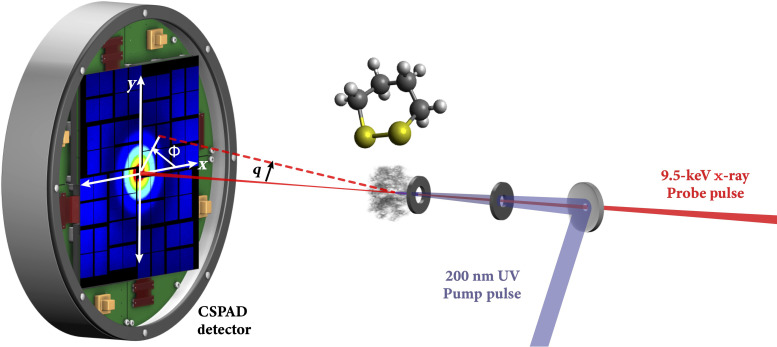
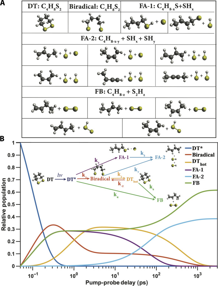
Disulfide bonds are ubiquitous molecular motifs that influence the tertiary structure and biological functions of many proteins. Yet, it is well known that the disulfide bond is photolabile when exposed to ultraviolet C (UVC) radiation. The deep-UV-induced S─S bond fragmentation kinetics on very fast timescales are especially pivotal to fully understand the photostability and photodamage repair mechanisms in proteins. In 1,2-dithiane, the smallest saturated cyclic molecule that mimics biologically active species with S─S bonds, we investigate the photochemistry upon 200-nm excitation by femtosecond time-resolved x-ray scattering in the gas phase using an x-ray free electron laser. In the femtosecond time domain, we find a very fast reaction that generates molecular fragments with one and two sulfur atoms. On picosecond and nanosecond timescales, a complex network of reactions unfolds that, ultimately, completes the sulfur dissociation from the parent molecule.


Figures

Fig. 1.
Fig. 1.. A schematic of the experiment.
The photoinduced reaction of DT is initiated by a 200-nm UV pump pulse, and the time-evolving molecular structures of reactants and photoproducts are probed by scattering using 9.5-keV x-ray pulses with a variable time delay. The scattering signals are recorded with a CSPAD detector. Created in Adobe Illustrator.
Fig. 2.
Fig. 2.. Comparison of experimental and simulated time-dependent percent difference scattering signals of the photoinduced chemical reaction of DT.
(A) The isotropic component of the time-dependent experimental percent difference scattering signal of DT as a function of pump-probe time delays. Plotted is the percent difference in scattering intensity (color bar) induced by the laser pulse as a function of the absolute value of the momentum transfer vector, q, and the pump-probe time delay. (B) Simulated time-dependent percent difference scattering signal of DT based on the proposed kinetic model and kinetic analysis fitting results. (C) Residuals of the experimental percent difference scattering signals with respect to the simulated percent difference scattering signals as a function of the absolute value of the momentum transfer vector q and the pump-probe time delay. (D) Experimental pump-probe x-ray scattering percent difference signals (black dots with 3σ experimental noise) and kinetic fit results (colored lines) at selected representative time delays.
Fig. 3.
Fig. 3.. The kinetic model of the photoinduced chemical reaction of DT upon 200-nm excitation.
(A) Illustrations of possible intermediate transients and photoproducts within the photoinduced chemical reaction network. The positions of hydrogen atoms remain undetermined because of the small x-ray scattering cross section for hydrogen. Several structures are possible for intermediate transient FA-1 and the final photoproducts FA-2 and FB as shown (see possible geometries in the “The structures of the photochemical reaction transients and photoproducts” section in Supplementary Text). (B) Time-dependent relative populations of transients and photoproducts. Inset: The photoinduced chemical reaction scheme.
Fig. 4.
Fig. 4.. Experimental extracted (data points) and computationally modeled (solid lines) percent difference scattering patterns of reaction transients and photoproducts of the photoinduced chemical reaction of DT.
The simulated scattering patterns are scaled by the optical excitation probability γ = 4.5%. (A) Sum of the biradical and DThot. (B) Intermediate fragment FA-1. (C and D) End photoproducts FB and FA-2, respectively. Shown in each panel are similar fragments for various placements of the hydrogen atoms. The insets show the molecular structures used for the simulations. The error bars of the experimental scattering patterns are from multiple iterative kinetic fitting analyses.
Fig. 5.
Fig. 5.. The conceptual potential energy surface and reaction scheme of DT upon excitation with 200-nm photons.
One reaction pathway (red dashed line) involves the rupture of the S─S disulfide bond, leading to biradical species. Two separate, fast dissociation pathways lead to the intermediates C4S + S (FA-1, purple dashed line) and end photoproducts C4 + S + S and C4 + S2 (blue and green dashed lines), with a distribution of 36 and 64%, respectively. The color coding matches Fig. 3.

References

    1. Creighton T. E., Disulphide bonds and protein stability. BioEssays 8, 57–63 (1988). - PubMed
    1. Sevier C. S., Kaiser C. A., Formation and transfer of disulphide bonds in living cells. Nat. Rev. Mol. Cell Biol. 3, 836–847 (2002). - PubMed
    1. Lange S. S., Takata K., Wood R. D., DNA polymerases and cancer. Nat. Rev. Cancer 11, 96–110 (2011). - PMC - PubMed
    1. Chen Y., Kamlet A. S., Steinman J. B., Liu D. R., A biomolecule-compatible visible-light-induced azide reduction from a DNA-encoded reaction-discovery system. Nat. Chem. 3, 146–153 (2011). - PMC - PubMed
    1. Crespo-Hernández C. E., Cohen B., Kohler B., Base stacking controls excited-state dynamics in A· T DNA. Nature 436, 1141–1144 (2005). - PubMed

LinkOut - more resources