Review
. 2025 Jul 1;105(3):1033-1074.
doi: 10.1152/physrev.00012.2024. Epub 2025 Jan 15.

The TRP channels serving as chemical-to-electrical signal converter

Affiliations
Review

The TRP channels serving as chemical-to-electrical signal converter

Yuhua Tian et al. Physiol Rev. .

Abstract

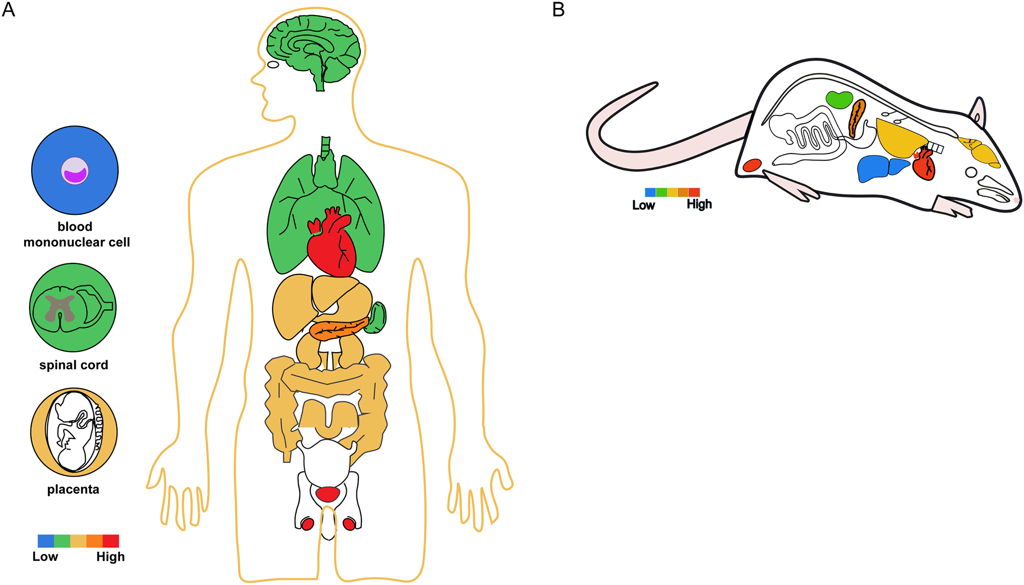
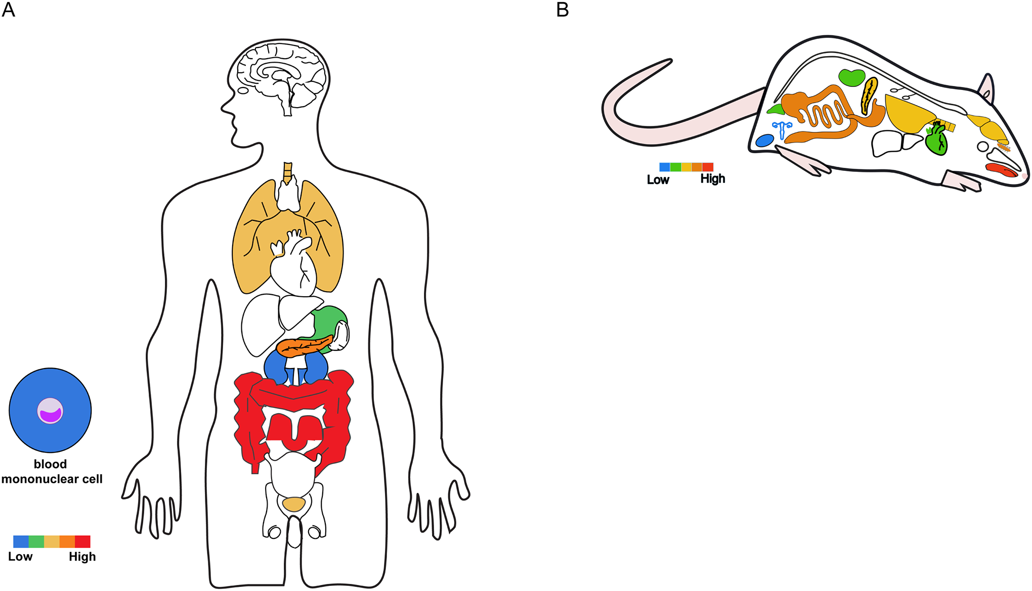
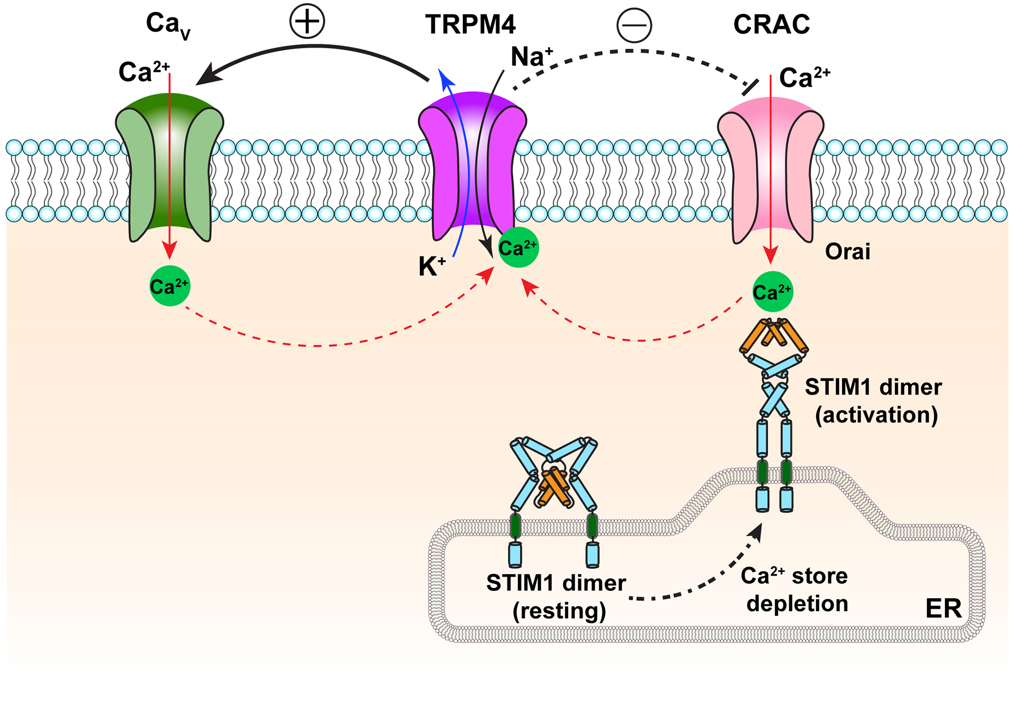
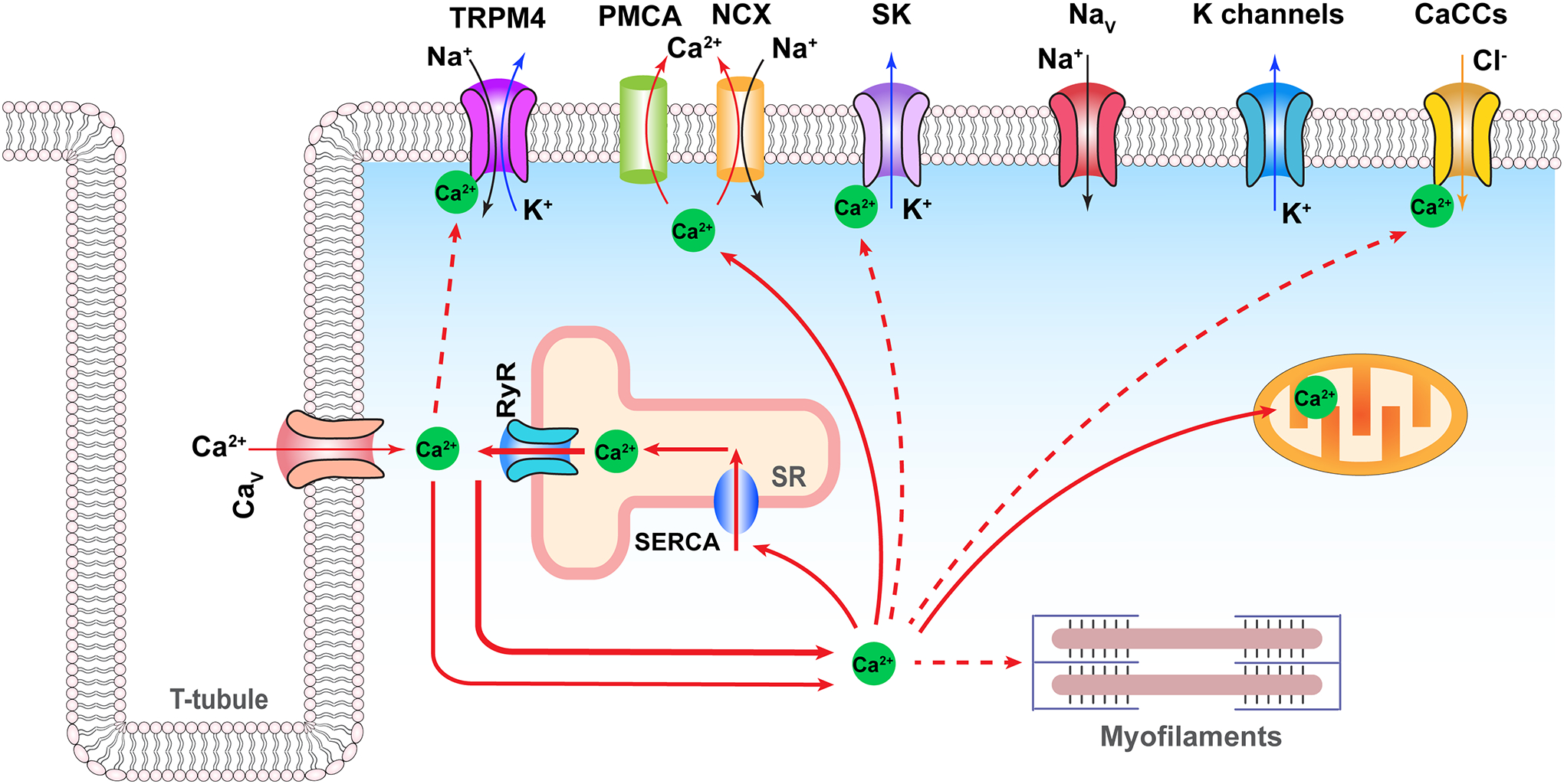
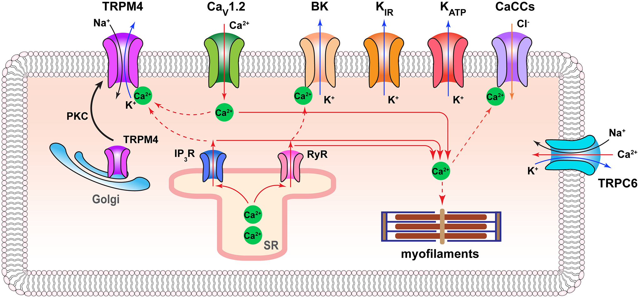
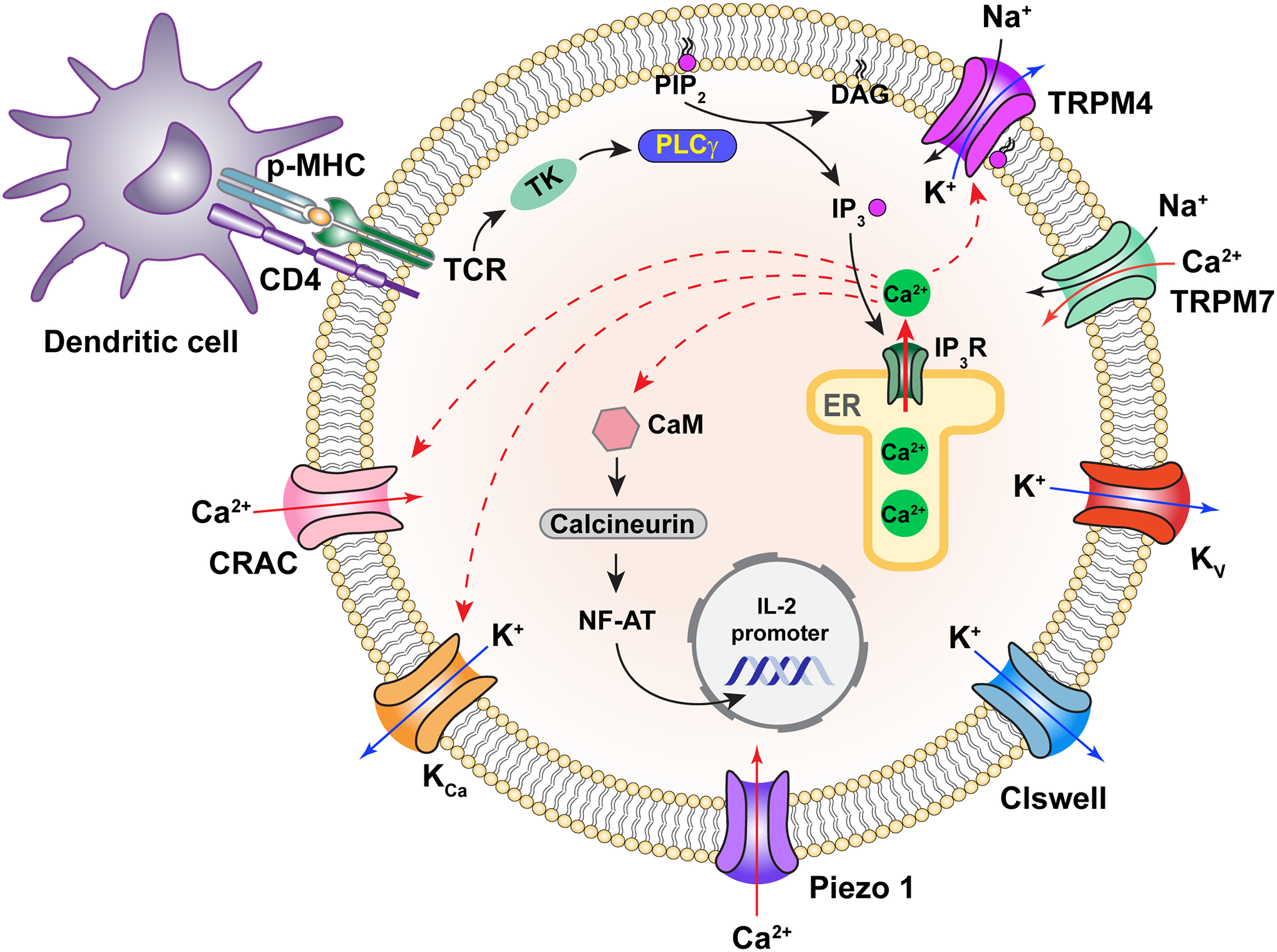
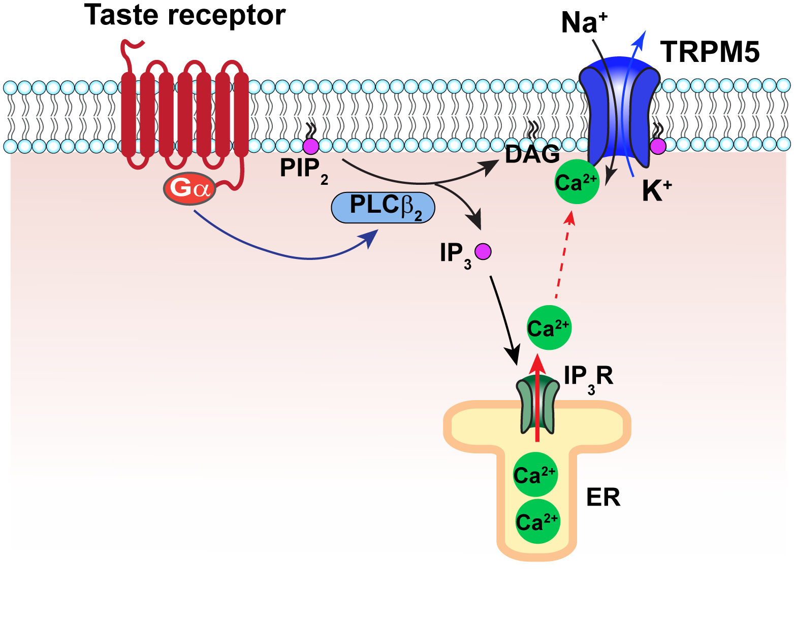
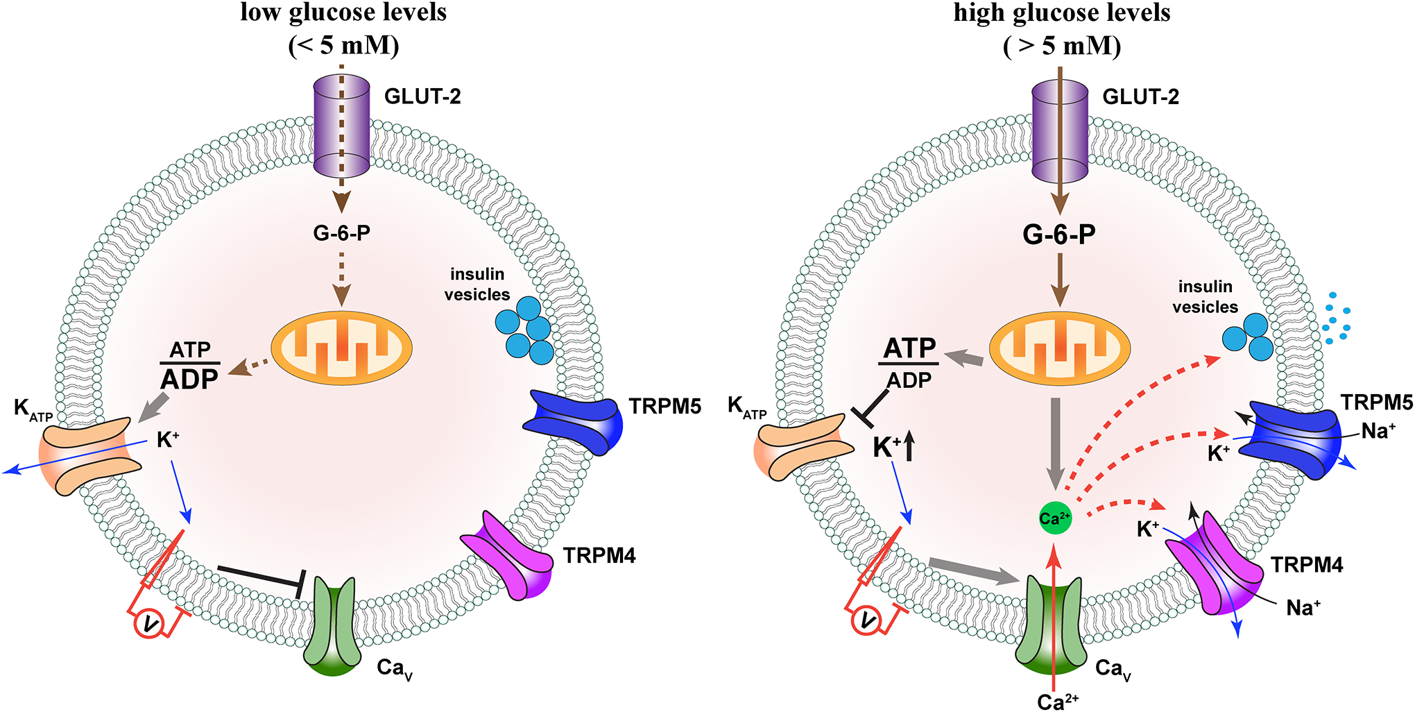
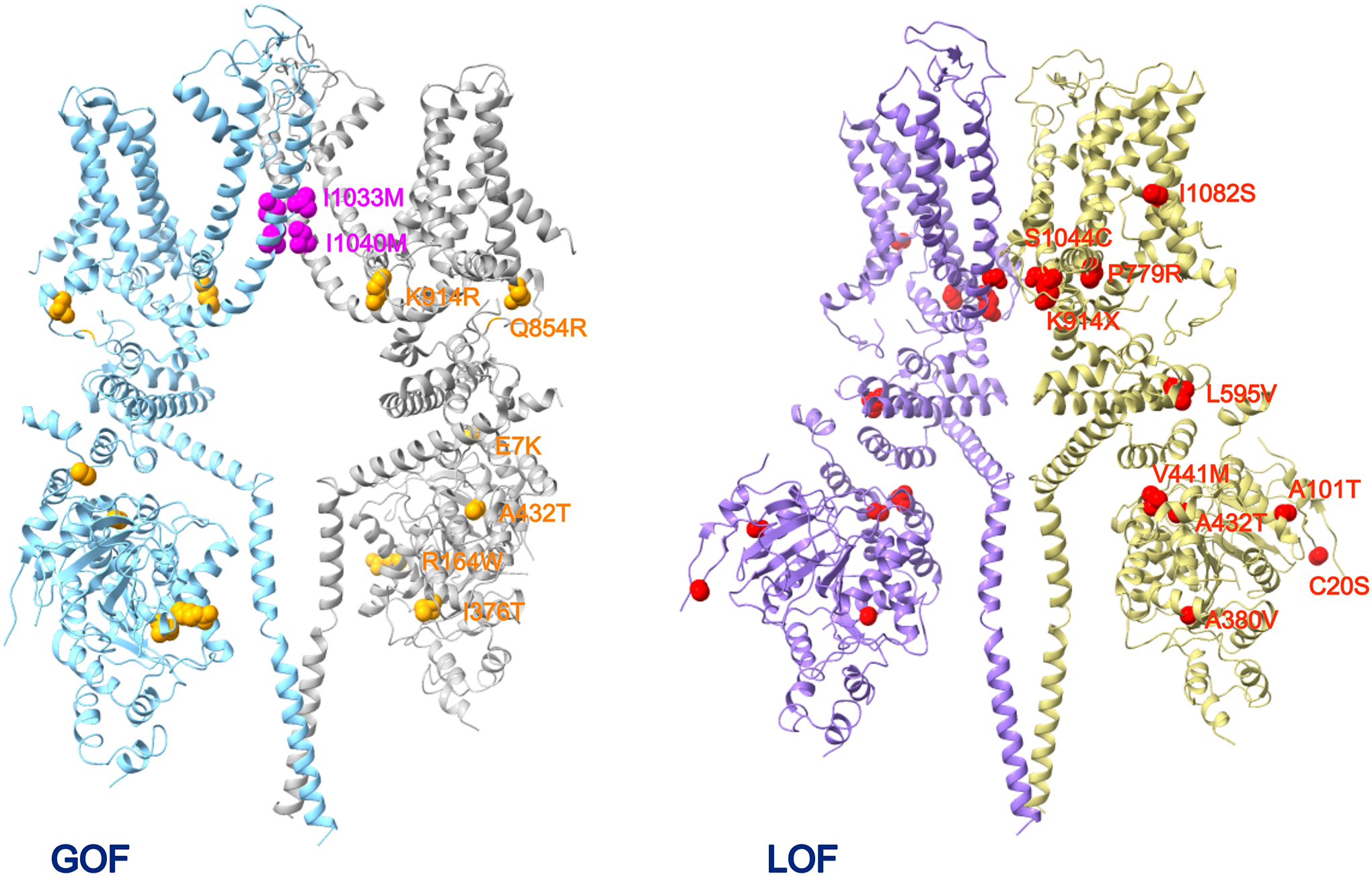
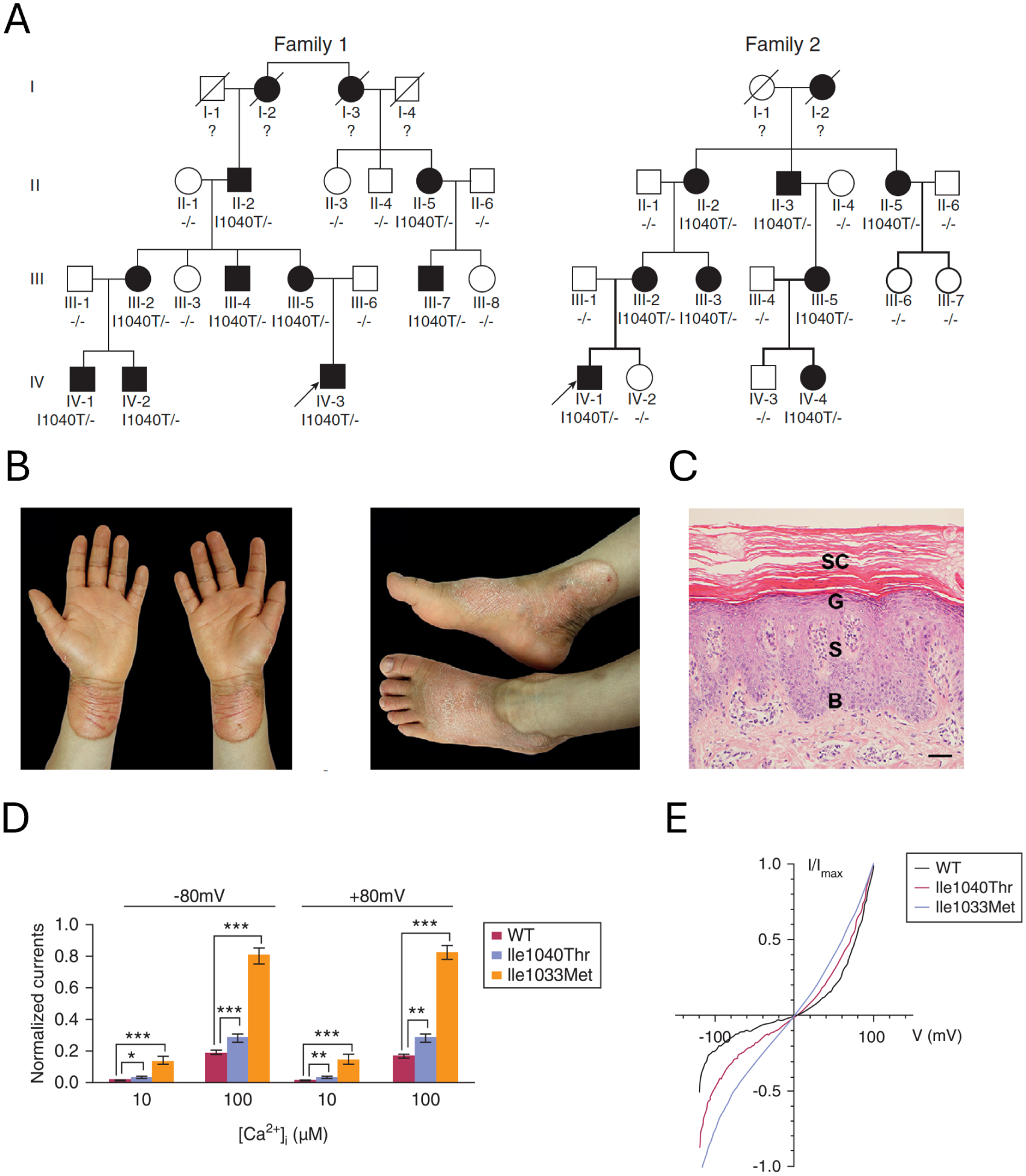
Biology uses many signaling mechanisms. Among them, calcium and membrane potential are two prominent mediators for cellular signaling. Transient receptor potential (TRP) melastatin 4 and 5 (TRPM4 and TRPM5), two calcium-activated monovalent cation-selective ion channels, offer a direct linkage between these two signals. Their activities convert a rise in the intracellular calcium level, a chemical signal, into depolarization of membrane potential, an electrical signal. Interestingly, membrane depolarization can in turn alter the electrical driving force or membrane permeability for calcium entry; hence, it offers feedback mechanisms for regulating calcium signaling. By converging two powerful cellular signals, TRPM4 and TRPM5 can contribute to many fundamental biological processes including cardiovascular biology, immunology, insulin release, chemosensation, and others. Numerous mutations in TRPM4 are linked to human hereditary cardiac and skin diseases, whereas knocking out TRPM5 in mice abolishes the perception of sweet, umami, and bitter tastes. This review summarizes what is currently known about the signaling roles of these unique TRP channels and what remains mysterious.

Keywords: calcium signaling; cardiac physiology; immune response; insulin release; membrane potential.


Figures

Figure 1.
Figure 1.
Representative current traces and I-V relationships of the CAN channels and cloned TRPM4 and TRPM5 channels. A&B, Example single-channel recordings of calcium-activated non-selective cation channels from cultured cardiac cells (A) and their I-V relationship (B). Recording voltage was −70 mV; intracellular calcium concentration was 6 μM (top), 1.5 μM (middle), and zero μM (bottom). Filled circles and open squares in B are data points with symmetrical sodium saline solutions and with intracellular potassium/extracellular sodium saline solutions, respectively, with reversal potentials around zero mV. Reproduced from Colquhoun et al. (5); used with permission under CC-BY license. C&D, TRPM4 single-channel currents activated by 0.3 μM calcium (C) and their I-V relationship with intracellular potassium/extracellular sodium saline solutions (D), with a reversal potential around zero mV. Reproduced from Launay et al. (13) with permission. E&F, TRPM5 currents activated by 12 μM calcium (E) and the I-V relationship (F). Reproduced from Liu and Liman (20) with permission.
Figure 2.
Figure 2.
Structural features of TRPM4 and TRPM5 revealed by cryo-EM. A. Overall structure of TRPM4 (PDB ID 9B8Y, left panel) and TRPM5 (7MBQ, right panel). The light green spheres represent bound Ca2+ ions. TMD, transmembrane domain; ICD, intracellular domain; MHR, melastatin homology region. B. The binding pocket of decavanadate (DVT, cyan) in TRPM4 (9B8Y). Two neighboring subunits are colored in magenta and gray, respectively. Key positively charged residues in direct contact with DVT are highlighted. C. The closed states and open states of TRPM4 and TRPM5 (PDB ID from left to right: 6BQR, 9B8Y, 7MBR, and 7MBQ), highlighting the ion permeation pore. S5 and S6 represent the fifth and sixth transmembrane helices, respectively.
Figure 3.
Figure 3.
Calcium binding sites of representative TRP channels. A. Amino acid sequence alignment of the S2-S3 regions of human (hs) TRPM4, zebrafish (dr) TRPM5, human TRPM2, mouse (mm) TRPM8, and human TRPC3, with the key conserved residues for calcium binding highlighted. B. Transmembrane calcium binding pockets of hsTRPM4 (magenta, 9B8Y), drTRPM5 (deep blue, 7MBQ), hsTRPM2 (purple, 6PUS), mmTRPM8 (light yellow, 7WRB), and hsTRPC3 (golden, 7DXB). S2 and S3 represent the second and third transmembrane helices, respectively. C. Amino acid sequence alignment of the intracellular N-terminal calcium binding sites of TRPM4 and TRPM5 of several species, with the key conserved residues highlighted. D. Intracellular calcium binding pockets of TRPM4 (magenta, 9B8Y) and TRPM5 (deep blue, 7MBQ). The light green spheres represent bound Ca2+ ions. Amino acid numbers are for human TRPM4.
Figure 4.
Figure 4.
Activation and regulation of TRPM4 and TRPM5. A. A representative current trace of human TRPM4 recorded at 80 mV from an inside-out patch in response to 3 mM Ca2+, 10 μg/mL poly-lysine, and 50 μM diC8-PIP2. Dotted blue trace is from another recording when diC8-PIP2 was applied before complete desensitization due to application of poly-lysine. Labels 1, 2, and 3 represent initial peak current, spontaneously desensitized current, and current recovered by PIP2, respectively. B. Simulated calcium-dependent activation of TRPM4 (solid curves) and TRPM5 (dash curves) at time point 1 (red), 2 (gray), and 3 (blue) in panel A. Naïve TRPM4 and TRPM5 channels have an apparent EC50 value in 100 μM and 10 μM, respectively, in inside-out patch recordings. C. Voltage-dependent activation of TRPM4 and TRPM5, using the same color scheme as B. D. Example mouse TRPM4 current traces in response to 100 μM Ca2+ over time at −100 mV and 100 mV (top left); co-expressing a calcium-insensitive calmodulin mutant CAM1,2,3,4 reduces the current amplitude (top right). The bottom panels show the corresponding I-V relationships. Reproduced with permission from Nilius et al. (2005) JBC. E. An example trace at −80 mV (left) and summary (right) of mouse TRPM4 current activated by 100 μM Ca2+ and recovery by 10 μM diC8-PIP2. Reproduced from Zhang et al. (44) with permission under CC-BY license.
Figure 5.
Figure 5.
Summary of reported TRPM4 distribution in human (A) and rodents (B). The color scale indicates relative levels of expression based on consensus of reports in the literature. The colored circles depict TRPM4-expressing specific cells, tissues or organs in humans.
Figure 6.
Figure 6.
Summary of reported TRPM5 distribution in human (A) and rodents (B). The color scale indicates relative levels of expression based on consensus of reports in the literature. The colored circles depict TRPM4-expressing specific cells, tissues or organs in humans.
Figure 7.
Figure 7.
Positive (left side) and negative (right side) feedback regulation of calcium signaling by TRPM4. Solid red arrows indicate calcium flow; dash red arrows indicate calcium regulation targets. Solid black and deep blue arrows indicate sodium and potassium flow, respectively. CRAC, calcium release-activated calcium channel; STIM, stromal-interaction molecule; ER, endoplasmic reticulum.
Figure 8.
Figure 8.
Physiological function of TRPM4 in cardiomyocytes. Solid red arrows indicate calcium flow; dash red arrows indicate calcium regulation targets. Solid black, deep blue, and orange arrows indicate sodium, potassium, and chloride flow, respectively. Only selected ion channels are highlighted in this illustration. PMCA, plasma membrane Ca2+ ATPase; NCX, sodium-calcium exchanger; CaCCs, calcium-activated chloride channels; SERCA, sarcoendoplasmic reticulum calcium ATPase; SR, sarcoplasmic reticulum; Nav, voltage-gated sodium channel subtype 1.5; Cav, voltage-gated calcium channel subtype 1.2; K channels represent a collection of potassium-selective channels.
Figure 9.
Figure 9.
Physiological function of TRPM4 in vascular smooth muscle cell. Solid red arrows indicate calcium flow; dash red arrows indicate calcium regulation targets. Solid black, deep blue, and orange arrows indicate sodium, potassium, and chloride flow, respectively. Only selected ion channels are shown. CaCCs, calcium-activated chloride channels. PKC, protein kinase C; SR, sarcoplasmic reticulum.
Figure 10.
Figure 10.
Physiological function of TRPM4 in T cells. Solid red arrows indicate calcium flow; dash arrows indicate calcium regulation targets. Solid black and deep blue arrows indicate sodium and potassium flow, respectively. Only selected ion channels are shown. CRAC, calcium release-activated calcium channel; CaM, calmodulin; p-MHC, peptide presented by major histocompatibility complex; CD4, cluster of differentiation 4; TCR, T-cell receptor; TK, tyrosine kinase; PLCγ, phosphoinositide phospholipase C γ subunit; IL-2, interleukin-2; ER, endoplasmic reticulum.
Figure 11.
Figure 11.
Physiological function of TRPM5 in taste cells. Solid red arrow indicates calcium flow; dash arrow indicates calcium regulation targets. Solid black and deep blue arrows indicate sodium and potassium flow, respectively. Gα, G protein α subunit; ER, endoplasmic reticulum.
Figure 12.
Figure 12.
Physiological function of TRPM4 and TRPM5 in pancreas beta-cells at low (A) and high (B) blood glucose levels. Solid red arrows indicate calcium flow; dash arrows indicate calcium regulation targets. Solid black, deep blue arrows indicate sodium and potassium flow, respectively. Endoplasmic reticulum serves as an important intracellular calcium store for beta-cells but is omitted due to space limitation. Only selected ion channels are shown. GLUT-2, glucose transporter 2; G-6-P, glucose 6-phosphate.
Figure 13.
Figure 13.
Location of human TRPM4 mutations linked to cardiac and skin diseases, shown on two subunits. (A) The gain-of-function (GOF) mutations associated with skin (magenta) and cardiac diseases (orange). (B) The loss-of-function (LOF) mutations associated with cardiac diseases (red).
Figure 14.
Figure 14.
TRPM4 gain-of-function mutations cause progress symmetric erythrokeratodermia (PSEK). A. Two example pedigrees containing the I1040T mutation. B. Erythematous hyperkeratotic plaques on the patient’s hands and feet. C. Affected skin shows psoriasiform hyperplasia with focal parakeratosis and mild perivascular lymphocytic infiltration in the superficial dermis. D. Increased current responses of TRPM4 mutants. E. Comparison of the I-V relationship between TRPM4 wildtype and mutants. Modified from Wang et al. (136) with permission.

Similar articles

References

    1. Boron WF, and Boulpaep EL. Human Physiology. Elsevier, 2016.
    1. Clapham DE. Calcium signaling. Cell 131: 1047–1058, 2007. - PubMed
    1. Berridge MJ, Lipp P, and Bootman MD. The versatility and universality of calcium signalling. Nat Rev Mol Cell Biol 1: 11–21, 2000. - PubMed
    1. Hu M, Feng X, Liu Q, Liu S, Huang F, and Xu H. The Ion Channels of Endomembranes. Physiol Rev 2024. - PMC - PubMed
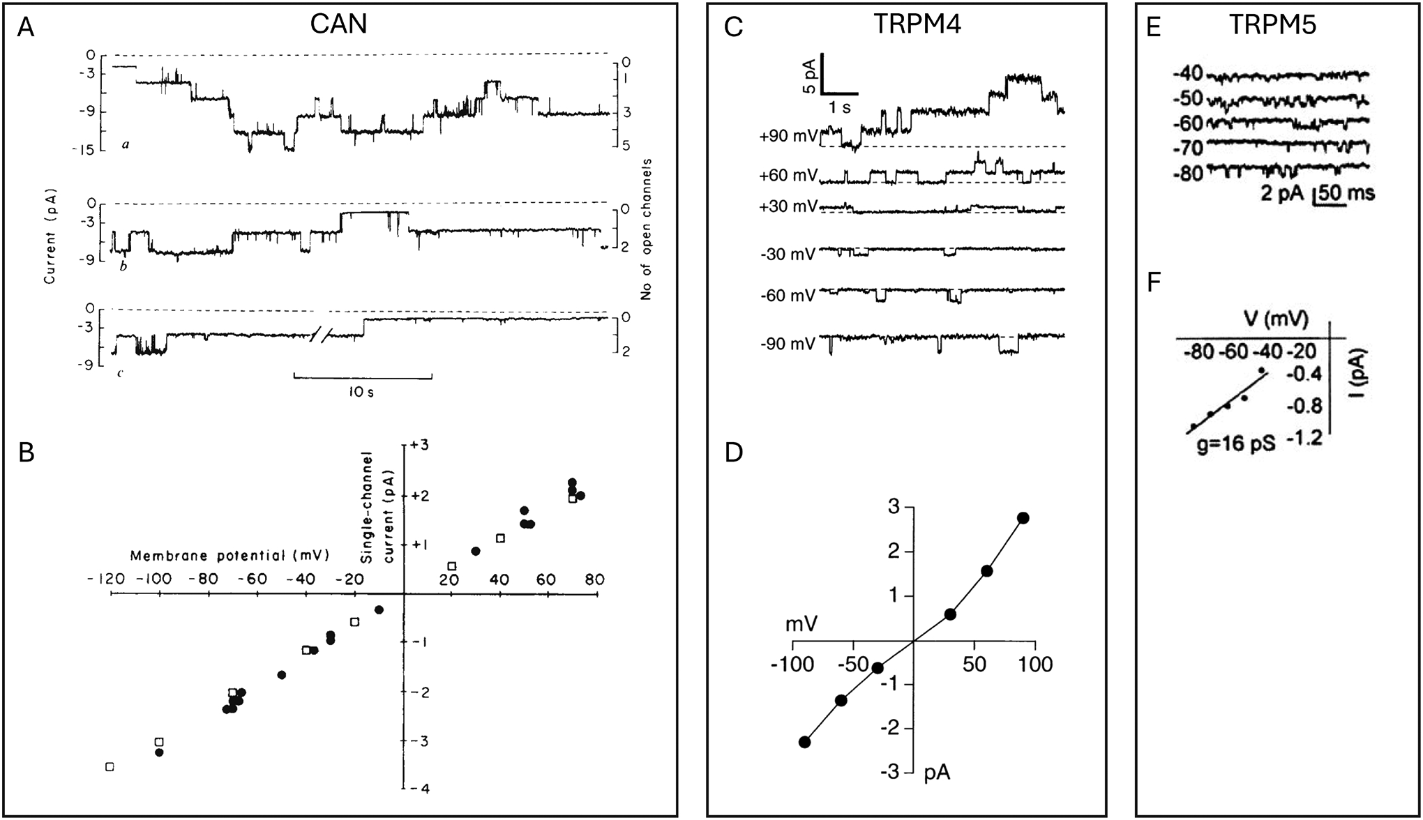
    1. Colquhoun D, Neher E, Reuter H, and Stevens CF. Inward current channels activated by intracellular Ca in cultured cardiac cells. Nature 294: 752–754, 1981. - PubMed

MeSH terms

LinkOut - more resources