Newborn with Refractory Seizures due to Hemimegalencephaly and Tuberous Sclerosis Complex: Case Report and Literature Review
- PMID: 39814050
- PMCID: PMC11932765
- DOI: 10.1055/a-2516-9103
Newborn with Refractory Seizures due to Hemimegalencephaly and Tuberous Sclerosis Complex: Case Report and Literature Review
Abstract
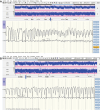
Hemimegalencephaly (HME) is a rare congenital disorder that is initiated during embryonic development with abnormal growth of one hemisphere. Tuberous sclerosis complex (TSC), a genetic disorder, is rarely associated with HME.We present a case of a newborn with HME with a confirmed mutation in the TSC-1 gene and describe the clinical course, findings on amplitude-integrated electroencephalography (aEEG), cranial ultrasound (CUS), MRI, and the postmortem evaluation. Furthermore, we conducted a comprehensive literature review of all reported newborns with HME and a genetically confirmed TSC mutation.This infant experienced therapy-resistant seizures after birth despite treatment with multiple antiseizure medications. CUS and MRI revealed HME of the left hemisphere. Early functional hemispherectomy, around the age of 3 months, was considered but dismissed after multidisciplinary evaluation, medical ethical consultation, and multiple discussions with the parents. Care was redirected due to worsening clinical and neurologic conditions, increasing respiratory insufficiency, and ongoing therapy-resistant seizures. Postmortem evaluation of the brain revealed hamartomatous brain changes and irregular gyration of the enlarged hemisphere. in addition, these changes were also present in the previously considered unaffected side, raising thoughts about the potential effectiveness of functional hemispherectomy.This case report illustrates that in cases with TSC abnormalities might not be confined solely to the initially considered affected side. This can have important therapeutic implications.
The Author(s). This is an open access article published by Thieme under the terms of the Creative Commons Attribution License, permitting unrestricted use, distribution, and reproduction so long as the original work is properly cited. (https://creativecommons.org/licenses/by/4.0/).
Conflict of interest statement
None declared.
Figures


References
-
- Di Rocco C, Battaglia D, Pietrini D, Piastra M, Massimi L. Hemimegalencephaly: clinical implications and surgical treatment. Childs Nerv Syst. 2006;22(08):852–866. - PubMed
-
- Shim S, Shin J E, Lee S M et al.A patient with tuberous sclerosis with hemimegalencephaly presenting with intractable epilepsy in the early neonatal period: a case report. Perinatology. 2022;33(04):201–207.
-
- Baek S T, Gibbs E M, Gleeson J G, Mathern G W. Hemimegalencephaly, a paradigm for somatic postzygotic neurodevelopmental disorders. Curr Opin Neurol. 2013;26(02):122–127. - PubMed
-
- Sidira C, Vargiami E, Dragoumi P, Zafeiriou D I. Hemimegalencephaly and tuberous sclerosis complex: a rare yet challenging association. Eur J Paediatr Neurol. 2021;30:58–65. - PubMed
-
- Serletis D, MacDonald C, Xu Q et al.Hemispherectomy for hemimegalencephaly in a 6.5-week-old infant with tuberous sclerosis complex. Childs Nerv Syst. 2022;38(07):1415–1419. - PubMed
Publication types
MeSH terms
Substances
Grants and funding
LinkOut - more resources
Full Text Sources
Medical

