. 2025 Mar;639(8053):225-231.
doi: 10.1038/s41586-024-08393-x. Epub 2025 Jan 15.

De novo designed proteins neutralize lethal snake venom toxins

Affiliations

De novo designed proteins neutralize lethal snake venom toxins

Susana Vázquez Torres et al. Nature. 2025 Mar.

Abstract

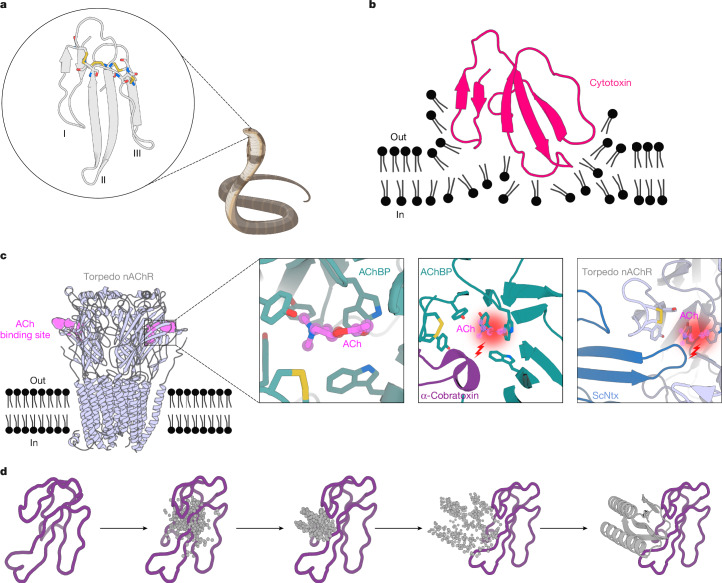
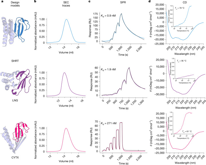
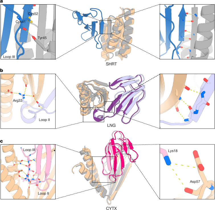
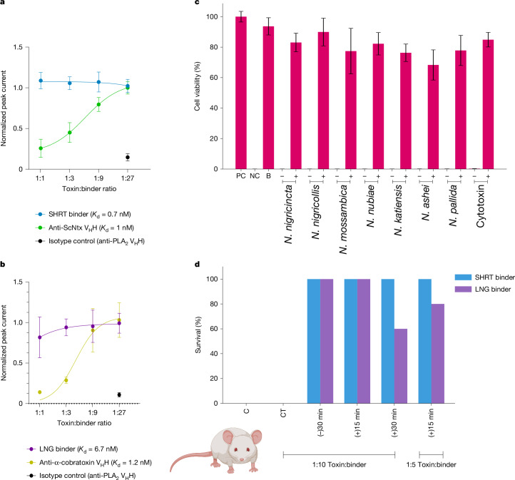
Snakebite envenoming remains a devastating and neglected tropical disease, claiming over 100,000 lives annually and causing severe complications and long-lasting disabilities for many more1,2. Three-finger toxins (3FTx) are highly toxic components of elapid snake venoms that can cause diverse pathologies, including severe tissue damage3 and inhibition of nicotinic acetylcholine receptors, resulting in life-threatening neurotoxicity4. At present, the only available treatments for snakebites consist of polyclonal antibodies derived from the plasma of immunized animals, which have high cost and limited efficacy against 3FTxs5-7. Here we used deep learning methods to de novo design proteins to bind short-chain and long-chain α-neurotoxins and cytotoxins from the 3FTx family. With limited experimental screening, we obtained protein designs with remarkable thermal stability, high binding affinity and near-atomic-level agreement with the computational models. The designed proteins effectively neutralized all three 3FTx subfamilies in vitro and protected mice from a lethal neurotoxin challenge. Such potent, stable and readily manufacturable toxin-neutralizing proteins could provide the basis for safer, cost-effective and widely accessible next-generation antivenom therapeutics. Beyond snakebite, our results highlight how computational design could help democratize therapeutic discovery, particularly in resource-limited settings, by substantially reducing costs and resource requirements for the development of therapies for neglected tropical diseases.


Conflict of interest statement

Competing interests: D.B., S.V.T., T.P.J., M.B.V., S.P.M., S.K.M., N.R.C., E.M., I.S., A.K.B. and A.K. are inventors on a provisional patent application submitted by the University of Washington for the design and composition of the proteins created in this study.

Figures

Fig. 1
Fig. 1. Targets of 3FTxs.
a, Structure of 3FTxs (Protein Data Bank (PDB) 1QKD). Highly conserved cysteine residues are highlighted in sticks, and each of the three fingers is indicated (I–III). b, Representation of type IA cytotoxin (dark pink) (PDB 5NQ4) interacting with a lipid bilayer. c, Muscle acetylcholine (ACh) (torpedo) receptor (light blue) (PDB 7Z14). The ACh-binding site is depicted in violet. Left inset: close-up of the acetylcholine-binding protein (AChBP) (teal) (PDB 3WIP) bound to ACh (violet). A set of aromatic residues form a cage around the neurotransmitter. Middle inset: close-up of α-cobratoxin (dark purple) blocking access to the ACh-binding site in AChBP (teal) (PDB 1YI5). A molecule of ACh is depicted to illustrate its binding site. Right inset: close-up of ScNtx (dark blue) blocking access to the ACh-binding site in the torpedo receptor (light blue) (PDB 7Z14). A molecule of ACh is depicted to illustrate its binding site. d, Schematic showing α-cobratoxin binder design using RFdiffusion. Starting from a random distribution of residues around the specified β-strands in the target toxin (dark purple), successive RFdiffusion denoising steps progressively removed the noise leading at the end of the trajectory to a folded structure interacting with α-cobratoxin β-strands. Schematics in ac were created using BioRender (https://biorender.com).
Fig. 2
Fig. 2. Experimental characterization of 3FTx-binding proteins.
a, Design models of protein binders (grey) bound to their 3FTx targets (dark blue, ScNtx; dark purple, α-cobratoxin; dark pink, consensus cytotoxin). b, SEC traces of purified proteins. mAU, milli-absorbance units. c, SPR-binding affinity measurements. Coloured solid lines represent fits using the heterogeneous ligand model, with the dissociation constant (Kd) values derived from these fits. RU, response units. d, CD data confirmed the presence of an αβ-secondary structure in the 3FTx-binding proteins and their thermal stability (inset). θ, molar ellipticity.
Fig. 3
Fig. 3. Crystal structures of 3FTx-binding proteins closely matched those of the design models.
a, Apo-state crystal structure of SHRT design. Left: hydrogen bonding between the carbonyl oxygen of Cys41 in ScNtx (dark blue) and the side chain of Tyr45 in the SHRT design model (grey). Middle: overlay of the SHRT design model (grey) with crystal structure (wheat). Right: backbone hydrogen bonding between the SHRT design model (grey) and ScNtx (dark blue) β-strands. b, Crystal structure of LNG design in complex with α-cobratoxin. Left: cross-interface hydrogen-bond network involving Arg33 in loop II of α-cobratoxin (light purple) and Glu69, Tyr40 and Tyr49 in the LNG crystal structure (wheat). Middle: overlay of the LNG design model (grey) bound to α-cobratoxin (dark purple) with crystal structure of binder (wheat) bound to toxin (light purple). Right: backbone hydrogen bonding between the crystal structure of the designed binder (wheat) and α-cobratoxin (light purple) β-strands. c, Crystal structure of CYTX_B10 design in complex with Naja pallida cytotoxin. Left: cross-interface electrostatic interaction network between loops III and II of N.pallida cytotoxin (light pink) and the binder crystal structure (wheat). Middle: overlay of the CYTX_B10 design model (grey) bound to the toxin (dark pink) with the crystal structure of the binder (wheat) bound to N.pallida cytotoxin (light pink). Right: salt bridge between positively charged Lys18 in cytotoxin (light pink) and Asp57 in the binder crystal structure (wheat).
Fig. 4
Fig. 4. In vitro and in vivo efficacy.
a, Concentration–response curves comparing SHRT binder and an anti-ScNtx nanobody (VHH) efficacy in preventing nAChR blocking by ScNtx at a dose corresponding to the 80% inhibitory concentration (IC80). The y axis corresponds to the ACh response in the presence of toxin and design, normalized to full ACh response, and averaged in each group (n = 16). b, Concentration–response curves comparing the efficacy of the LNG binder and anti-α-cobratoxin VHH in preventing nAChR blocking by one IC80 of α-cobratoxin. c, Neutralization of the cytolytic effects of whole venoms from seven different Naja species and isolated cytotoxin by the CYTX binder. Two IC50 values of the whole venom or toxin were preincubated with CYTX at a 1:5 molar ratio (toxin:binder). This ratio was estimated assuming that 70% of the whole venom consists of cytotoxins, on the basis of previous proteomic analyses. Keratinocyte medium was used as a positive control (PC). Triton X-100 was used as a negative control (NC). CYTX binder (B) was used as a PC. (−) denotes two IC50 values of the whole venom without a binder, and (+) denotes venom incubated with a binder. Experiments were performed in triplicate, and the results are expressed as mean ± s.d. d, Survival of mice following lethal neurotoxin challenge (n = 5). At a concentration corresponding to three times the LD50 value of ScNtx or α-cobratoxin, either were preincubated for 30 min (−30 min) with the corresponding protein binders at a 1:10 ratio and then administered intraperitoneally into groups of five mice. Toxins were administered intraperitoneally following IP administration of binders at 1:10 or 1:5 molar ratios (toxin:binder) either 15 min (+15 min) or 30 min (+30 min) post-toxin injection. Controls included mice that received the toxins alone (C). Specificity was assessed via cross-treatment (CT) experiments, in which non-target binders were preincubated with three LD50 values of ScNtx or α-cobratoxin and administered intraperitoneally. Signs of toxicity were observed, and deaths were recorded for a period of 24 h. Illustration in d created with BioRender (https://biorender.com).

Update of

  • De novo designed proteins neutralize lethal snake venom toxins.
    Torres SV, Valle MB, Mackessy SP, Menzies SK, Casewell NR, Ahmadi S, Burlet NJ, Muratspahić E, Sappington I, Overath MD, Rivera-de-Torre E, Ledergerber J, Laustsen AH, Boddum K, Bera AK, Kang A, Brackenbrough E, Cardoso IA, Crittenden EP, Edge RJ, Decarreau J, Ragotte RJ, Pillai AS, Abedi M, Han HL, Gerben SR, Murray A, Skotheim R, Stuart L, Stewart L, Fryer TJA, Jenkins TP, Baker D. Torres SV, et al. Res Sq [Preprint]. 2024 May 17:rs.3.rs-4402792. doi: 10.21203/rs.3.rs-4402792/v1. Res Sq. 2024. Update in: Nature. 2025 Mar;639(8053):225-231. doi: 10.1038/s41586-024-08393-x. PMID: 38798548 Free PMC article. Updated. Preprint.

References

    1. GBD 2019 Snakebite Envenomation Collaborators. Global mortality of snakebite envenoming between 1990 and 2019. Nat. Commun.13, 6160 (2022). - PMC - PubMed
    1. Gutiérrez, J. M. et al. Snakebite envenoming. Nat. Rev. Dis. Primer3, 17063 (2017). - PubMed
    1. Royte, L. & Sawarkar, A. Snake bite—cytotoxic effects of snake venom: a rare clinical image. Pan Afr. Med. J.44, 61 (2023). - PMC - PubMed
    1. Barber, C. M., Isbister, G. K. & Hodgson, W. C. Alpha neurotoxins. Toxicon66, 47–58 (2013). - PubMed
    1. Deka, A., Gogoi, A., Das, D., Purkayastha, J. & Doley, R. Proteomics of Naja kaouthia venom from North East India and assessment of Indian polyvalent antivenom by third generation antivenomics. J. Proteomics207, 103463 (2019). - PubMed

LinkOut - more resources