. 2025 Jan 15;8(1):57.
doi: 10.1038/s42003-025-07491-4.

Intranasally administrated fusion-inhibitory lipopeptides block SARS-CoV-2 infection in mice and enable long-term protective immunity

Affiliations

Intranasally administrated fusion-inhibitory lipopeptides block SARS-CoV-2 infection in mice and enable long-term protective immunity

Said Mougari et al. Commun Biol. .

Abstract

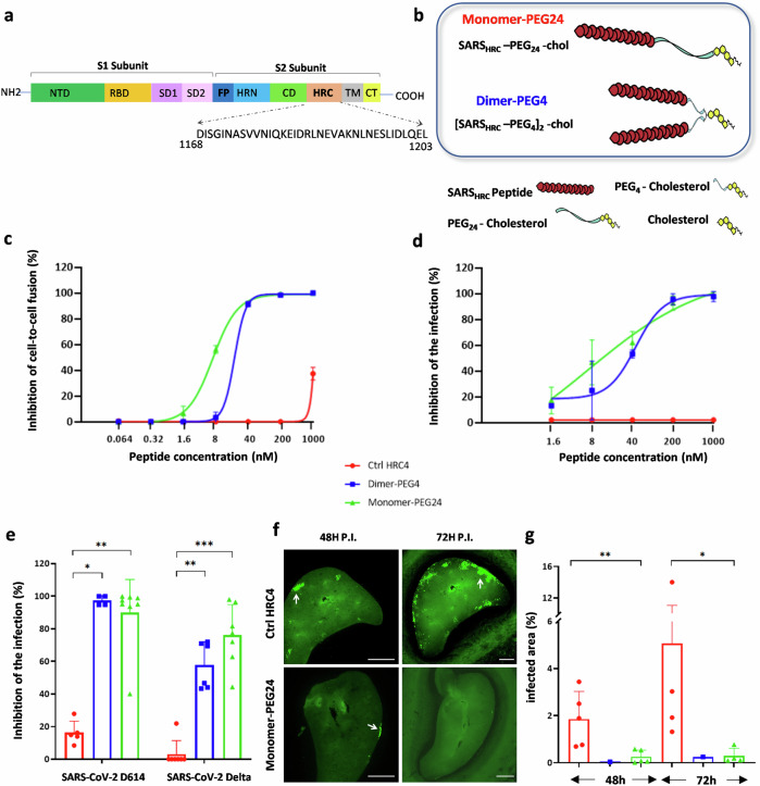
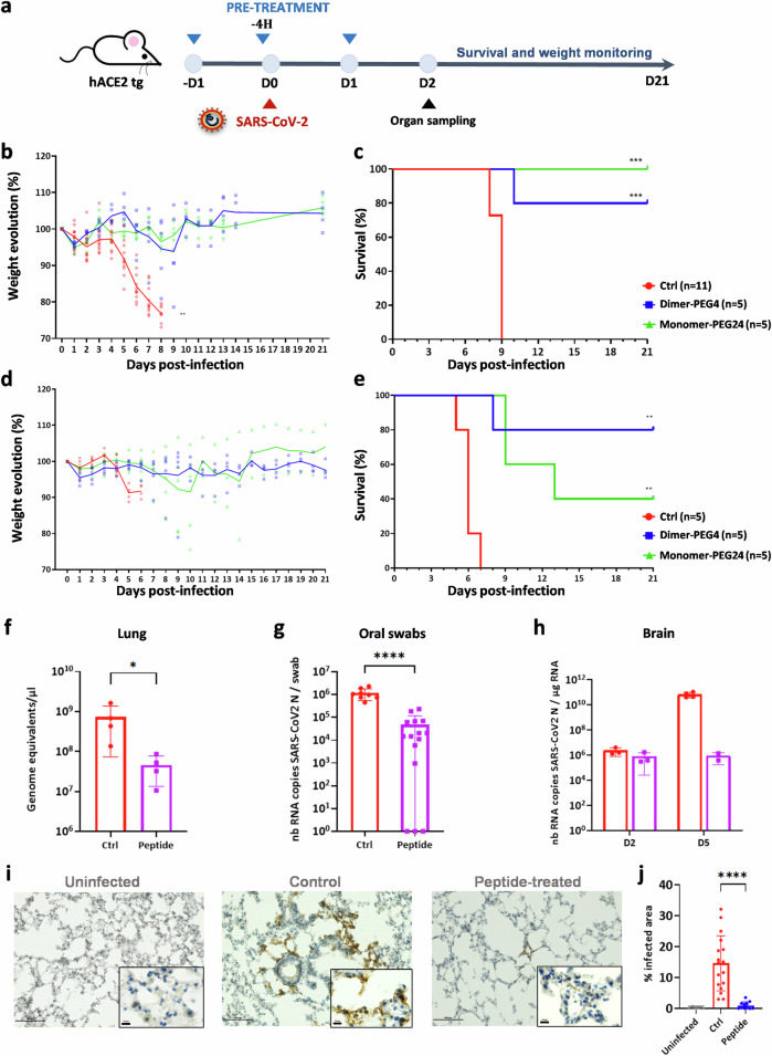
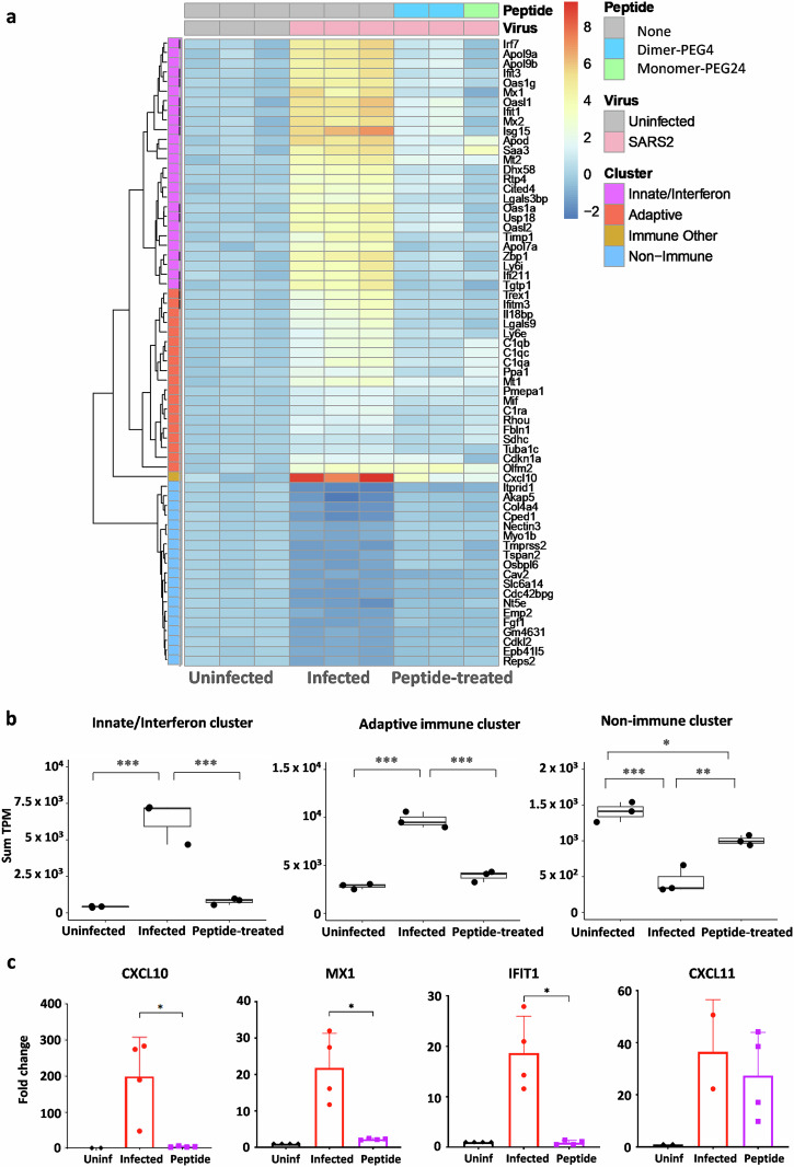
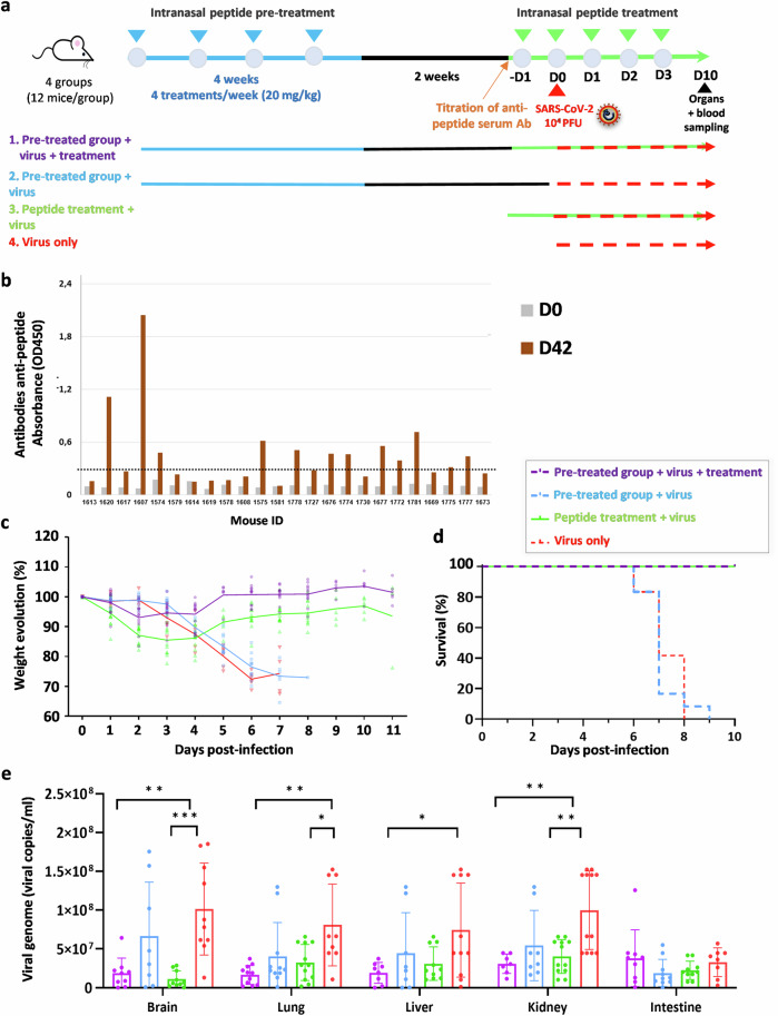
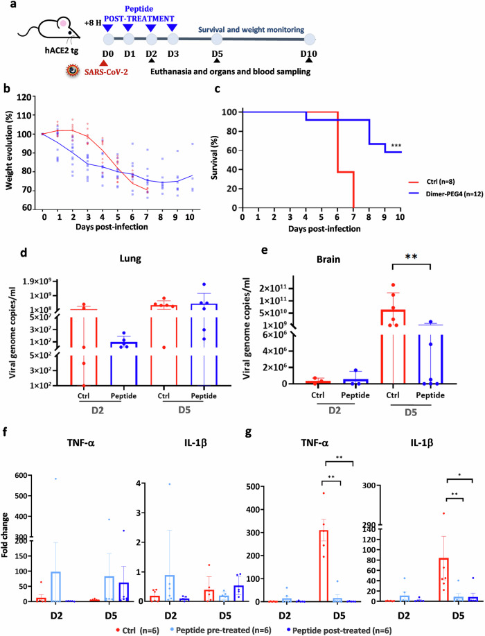
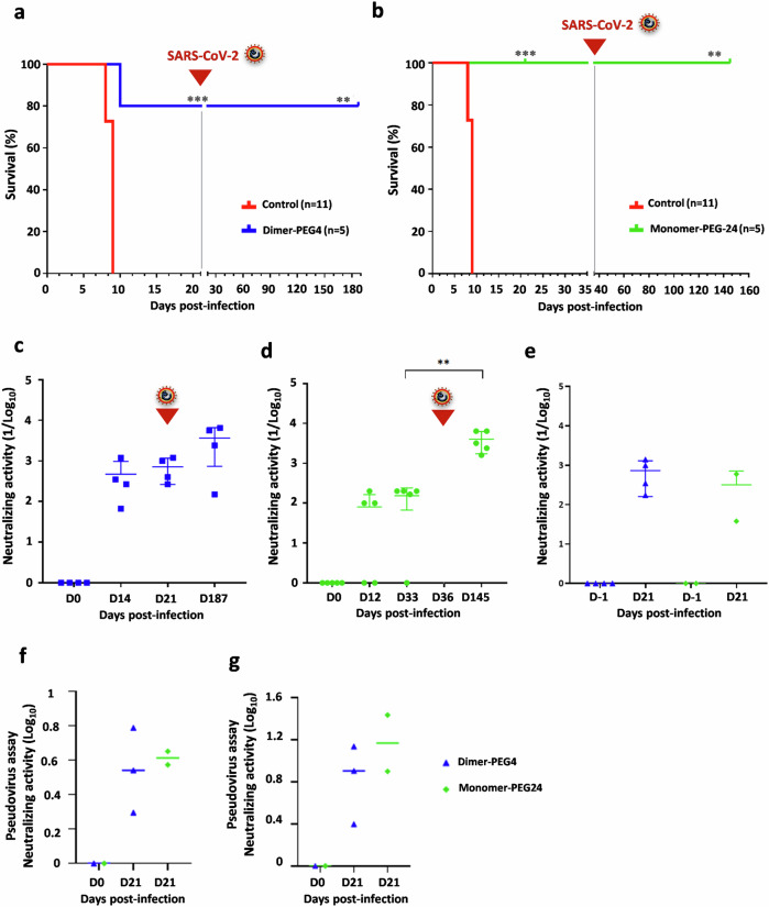
We have assessed antiviral activity and induction of protective immunity of fusion-inhibitory lipopeptides derived from the C-terminal heptad-repeat domain of SARS-CoV-2 spike glycoprotein in transgenic mice expressing human ACE2 (K18-hACE2). The lipopeptides block SARS-CoV-2 infection in cell lines and lung-derived organotypic cultures. Intranasal administration in mice allows the maintenance of homeostatic transcriptomic immune profile in lungs, prevents body-weight loss, decreases viral load and shedding, and protects mice from death caused by SARS-CoV-2 variants. Prolonged administration of high-dose lipopeptides has neither adverse effects nor impairs peptide efficacy in subsequent SARS-CoV-2 challenges. The peptide-protected mice develop cross-reactive neutralizing antibodies against both SARS-CoV-2 used for the initial infection and recently circulating variants, and are completely protected from a second lethal infection, suggesting that they developed SARS-CoV-2-specific immunity. This strategy provides an additional antiviral approach in the global effort against COVID-19 and may contribute to development of rapid responses against emerging pathogenic viruses.


Conflict of interest statement

Competing interests: M.P. and A.M. anticipate future financial interest in Thylacine Biotherapeutics, a company established to develop lipopeptide antiviral therapeutics. A.M., M.P., B.H. and C.M. are inventors of several provisional patent applications related to the use of antiviral lipopeptides.

Figures

Fig. 1
Fig. 1. Fusion inhibitory lipopeptides block SARS-CoV-2-Spike-mediated fusion in hACE2-transfected cells and SARS-CoV-2 replication in vitro and ex vivo.
a Schematic of SARS-CoV-2 Spike protein primary structure with peptide sequence. NTD N-terminal domain, RBD receptor-binding domain, SD1 subdomain-1, SD2 subdomain-2, FP fusion protein, HRN heptad repeat N-terminal, CD connector domain, HRC heptad repeat C-terminal, TM transmembrane domain, CT cytoplasmic tail. b Schematic presentation of the composition of the monomer-PEG24 (SARSHRC-PEG24-chol) and dimer-PEG4 ([SARSHRC-PEG4]2-chol) peptides. c Cell–cell fusion inhibition assay in 293T-ACE2 cells with 293T-Spike Wuhan D614. Data are means ± standard deviation (SD) (error bars) from three separate experiments with the curve representing a four-parameter dose–response model (Ctrl HRC4: control peptide, measles-specific). d Virus inhibition assay, measuring peptide-mediated inhibition of SARS-CoV-2 (Wuhan D614) infection (100 plaque-forming unit- PFU/well) in Vero cells. Percent inhibition was calculated as the ratio of PFU in the presence of a specific concentration of inhibitor and the PFU in the absence of inhibitor. Data are means ± SD from three separate experiments. e Inhibition of infection in organotypic cultures from lungs of K18-hACE2 mice with either recombinant SARS-CoV-2 neon-green or SARS-CoV-2 Delta (500 PFUs), with peptides added into the culture during 96 h, assessed using RT-qPCR (4–8 slices per condition, generated from 3 mice, results presented as mean ± SD). f Spread of fluorescent rSARS-CoV-2 neon-green virus in representative fluorescence micrographs of lung organotypic cultures, in the presence and absence of peptide (arrows indicate the sites of viral replication in cultures, (scale bars: 1 mm). g Quantification of the percentage of infected area, detected by immunofluorescence on scanned slides using QuPath, Open-source software for digital pathology image analysis, results presented as mean ± SD (*p < 0.05, **p < 0.01, ***p < 0.001, Mann–Whitney test).
Fig. 2
Fig. 2. Pretreatment with fusion inhibitory peptides protects K18-hACE2 mice from SARS-CoV-2-induced pathology.
a Experimental design: K18-hACE2 mice received either lipopeptide or vehicle (2% DMSO) intranasally daily for 3 days and were infected with SARS-CoV-2 intranasally 4 h after the second peptide administration or after receiving a vehicle solution (control), and followed for 21 days. b–e Body weight normalized to their initial weight on the day of infection and survival of mice following the infection with SARS-CoV-2 D614 (104 PFU) (b and c), or Delta variant (105 PFU) (d and e). Statistical significance of the effect of peptides on the evolution of weight was determined using a Mann-Whitney test and survival was measured using a Mantel–Cox test, **p < 0.01, ***p < 0.001). f Viral load in lungs (4 mice/group) determined by RT-qPCR, 2 dpi) with SARS-CoV-2 D614. g Viral load in oral swabs of mice (8 control mice and 15 peptide-treated mice) at 2 days post-infection (h) and in brains (7 control mice and 5 peptide-treated mice) at the indicated time points, using SARS-CoV-2 Delta, determined by RT-qPCR. Results obtained from two lipopeptide-treated series were grouped, results presented as mean ± SD, and statistical significance was determined using a Mann–Whitney test (*p < 0.05, ****p < 0.0001). i Immunohistological analysis of lung sections of mice (2 and 3 animals/condition) that received the indicated treatments 2 dpi with SARS-CoV-2 D614. Staining with anti-SARS-CoV-2 N-specific Ab is indicated with red arrows (scale bars: 500 µM for lower magnification and 10 µM for higher magnification). j Quantification of the percentage of infected cells, detected on scanned slides (infected controls, n = 17; uninfected, n = 5 and peptide-treated infected, n = 11), using QuPath, Open-source software for digital pathology image analysis (****p < 0.0001, Mann–Whitney test).
Fig. 3
Fig. 3. Intranasal administration of fusion inhibitory lipopeptides in SARS-CoV-2-infected mice modifies transcriptomic profile in lungs at 2 days post-infection.
a Heatmaps depicting the average log2 fold change of the 65 differentially expressed genes with the lowest adjusted p values in lungs of K18-hACE2 mice infected with SARS-CoV-2 D614 (n = 3), compared to noninfected mice (n = 3) and to mice pretreated with either dimer-PEG4 or monomer-PEG24 lipopeptides (n = 3), 2 days post-infection. Red denotes up-regulated and blue denotes down-regulated transcripts. b Differentially expressed genes with the lowest adjusted p values were placed into three groups, using their annotations and the clustering: Interferon/Innate-enriched, Adaptive-enriched, and Non-immune genes. Results are presented with adaptive/innate/interferon metadata bars, based on the non-redundant lists of genes from reactome+GO BioProcess. The sum of transcripts per million (TPM) of each gene category per sample was determined, and results are presented for each of the 3 clusters by box-plots showing individual values and the average sum TPM for each group. Comparisons between uninfected, infected-untreated, and infected-peptide-treated groups were performed using ANOVAs with Tukey’s Post Hoc test (**p < 0.01). c RT-qPCR analysis in analyzed lung samples from 3 mice/group of selected three up-regulated transcripts in the infected-untreated group, MX1, IFIT1, and CXCL10, and one transcript where differential expression was not observed (CXCL11). Results are expressed as fold change relative to the number of copies of mRNA in each group, compared to the uninfected group. Uninf: Uninfected, untreated group, Infected: Infected, untreated group; Peptide: infected, pretreated with either dimer or PEG-24 monomer group; results presented as mean ± SD (*p < 0.05, **p < 0.01, ***p < 0.001, ****p < 0.0001 Mann–Whitney test).
Fig. 4
Fig. 4. Multiple administrations of high-dose lipopeptides had no adverse effects and did not interfere with peptide-induced antiviral protection during subsequent SARS-CoV-2 challenges.
a Experimental design: 24 K18-hACE2 mice received 20 mg/kg of dimer-PEG4 peptide i.n. 4 times per week, for 4 weeks. After a 2-week break, mice were tested for the presence of peptide-specific antibodies in the serum and separated into 2 groups: one that received the additional peptides (group 1) and the other that remained without treatment (group 2). In addition, 2 groups of naive K18-ACE2 mice (12 mice in each group) were included in the experiment: one that received peptide treatment (group 3), and the other which remained without treatment (group 4). All mice were infected intranasally with SARS-CoV2 alpha-UK (104 PFU/mouse, in 40 µL) on day 0, and the additional peptide treatment was given intranasally for 5 days (20 mg/kg). Mice were monitored for 10 days and euthanized between days 6 and 9 if they presented clinical signs of infection, and all surviving mice were sacrificed at day 10, and RNA was isolated from indicated organs. b ELISA analysis for the presence of peptide-specific antibodies in the serum of mice from groups 1 and 2, showing the presence of antibodies in 50% of treated mice. c, d Weight and survival of mice, after challenge with a lethal dose of SARS-CoV-2 Alpha-UK (104 PFU). Mantel-Cox Log Rank test between groups shows a statistically significant difference between groups surviving 100% infection (groups 1 and 3) and groups succumbing to infection (groups 2 and 4) (****p < 0.0001). e Organ-specific viral load between 6 and 10 dpi, tested in different organs by RT-qPCR for the presence of SARS-CoV-2 N, and results are shown as average ± SD. The experiment was repeated in Fig. S5 with mice euthanized at the same dpi (one-way ANOVA with individual comparisons between groups analyzed via Tukey’s test (*p = 0.017–0.045, **p = 0.001-0.002, ***p = 0.0006).
Fig. 5
Fig. 5. Protective effect of fusion inhibitory lipopeptides given after SARS-CoV-2 infection.
a Experimental design: K18-hACE2 mice were infected i.n. with SARS-CoV-2 alpha-UK (104 PFU) and received dimer-PEG4 lipopeptide i.n. 8 h later (20 mg/kg), four times in a 24 h interval, and were followed for 10 days. b, c Follow up of weight and survival of mice treated with either vehicle (control, ctrl) or dimer-PEG4. Statistical significance of the effect of peptides on survival was measured using the Mantel–Cox test (***p < 0.0001). d Viral load in lungs in mice euthanatized at day 2 pi (n = 5) and 5 dpi (n = 6). e Viral load in the brain of mice euthanatized 2 dpi (n = 3) and 5 dpi (n = 6). Results are presented as mean ± SD. f, g RT-qPCR quantification of TNF-α and IL-1ß in lungs (f) and brain (g) of mice euthanized 2 and 5 dpi. Mice were either pretreated with dimer-PEG4 as described in Supplemental Fig. 5 or post-treated as in (a). Results are presented as fold-change (mean ± SD) in comparison to the cytokine expression in noninfected mice. Statistical significance was determined using one-way ANOVA with individual comparisons between groups analyzed via Tukey’s test (*p < 0.05; **p < 0.01).
Fig. 6
Fig. 6. Treatment with fusion-inhibitory lipopeptides allows protected mice to develop a long-term immune response against SARS-CoV-2 variants and resistance to subsequent viral reinfection.
Peptide-treated K18-hACE2 mice from the experiment in Fig. 2 were reinfected a 21 days or b 36 days after the first infection with the lethal dose of SARS-CoV-2 D614 (104 PFU) and followed for 6 months. Presence of serum neutralizing antibodies in either dimer-PEG4-treated (n = 4) (c) or monomer-PEG24-treated mice (n = 5) (d) at different time points after initial infection with SARS-CoV-2 D614 and reinfection (**p < 0.01, ***p < 0.001, Mantel–Cox test). e Presence of the serum-neutralizing antibodies in either dimer-PEG4-treated (blue, n = 4) or monomer-PEG24-treated mice (green, n = 2) at 21 days after infection with SARS-CoV-2 Delta. f, g Neutralizing capacity of mouse sera was analyzed using the pseudo-VSV system. f Ability of sera from mice infected with SARS-CoV-2 Delta (presented in e) to neutralize pseudotyped VSV-D614 and g to neutralize pseudotyped VSV-Omicron XBB (horizontal bars present mean values).

Similar articles

Cited by

References

    1. Düzgüneş, N., Fernandez-Fuentes, N. & Konopka, K. Inhibition of viral membrane fusion by peptides and approaches to peptide design. Pathogens10, 1599 (2021). - PMC - PubMed
    1. Schütz, D. et al. Peptide and peptide-based inhibitors of SARS-CoV-2 entry. Adv. Drug Deliv. Rev.167, 47–65 (2020). - PMC - PubMed
    1. Walls, A. C. et al. Structure, function, and antigenicity of the SARS-CoV-2 spike glycoprotein. Cell181, 281–292.e6 (2020). - PMC - PubMed
    1. Shang, J. et al. Cell entry mechanisms of SARS-CoV-2. Proc. Natl Acad. Sci. USA117, 11727–11734 (2020). - PMC - PubMed
    1. Bosch, B. J., van der Zee, R., de Haan, C. A. M. & Rottier, P. J. M. The coronavirus spike protein is a class I virus fusion protein: structural and functional characterization of the fusion core complex. J. Virol.77, 8801–8811 (2003). - PMC - PubMed

MeSH terms

LinkOut - more resources