

Clinical and radiological features of gastric and small intestinal anisakiasis: comparison with gastric ulcers and crohn's disease
- PMID: 39815123
- PMCID: PMC12053333
- DOI: 10.1007/s11604-024-01731-z
Clinical and radiological features of gastric and small intestinal anisakiasis: comparison with gastric ulcers and crohn's disease
Abstract
Purpose: To compare the clinical and radiological features of gastric and small intestinal anisakiasis with those of gastric ulcers and Crohn's disease.
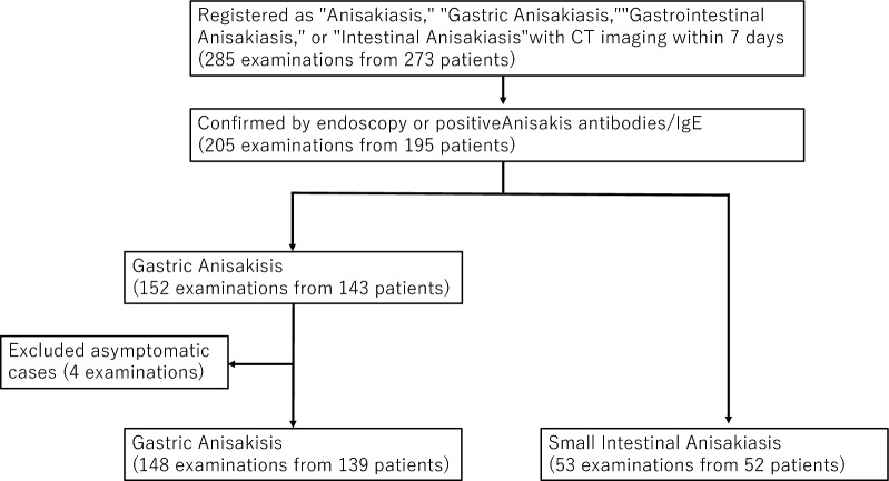
Materials and methods: In this retrospective cohort study, 205 cases of anisakiasis (148 gastric; 53 small intestinal) were identified between July 2003 and February 2022. The control groups included 130 and 31 patients with gastric ulcers and Crohn's disease, respectively. Clinical and imaging findings were compared between the groups using the chi-square test, Fisher's exact test, Mann-Whitney U test, and t-test.
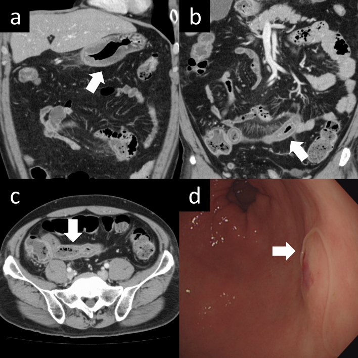
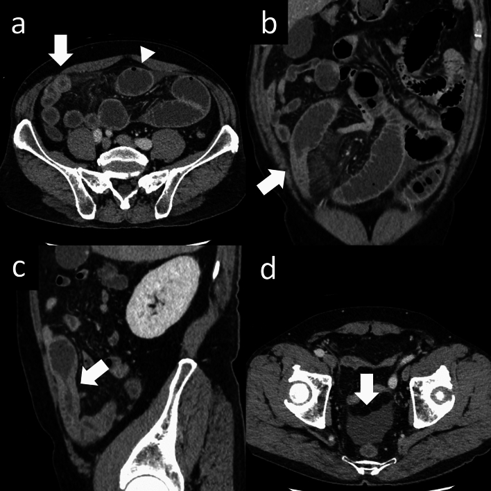
Results: Patients with gastric anisakiasis were younger (median age, 40 [21-85] years; 87 men) than those with gastric ulcers (median age, 64.5 [29-90] years; 101 men). Abdominal pain was common in the gastric anisakiasis group, whereas bleeding symptoms were frequent in the gastric ulcer group. Patients with small intestinal anisakiasis were older (mean age, 51.2 [38.6-63.7] years; 44 men) than those with Crohn's disease (mean age, 35.9 [21.6-50.3] years; 22 men). Patients with gastric anisakiasis exhibited more edematous wall thickening, increased surrounding fat density, ascites, and thickening of other intestinal walls than those with gastric ulcers. Patients with small intestinal anisakiasis showed greater wall edema, perienteric fat stranding, proximal dilatation, clamp sign, and ascites than those with Crohn's disease. Interobserver agreement was moderate to excellent, except for esophageal findings.
Conclusion: Anisakiasis demonstrates clinical and radiological features distinct from those of gastric ulcers and Crohn's disease. Recognizing these differences may aid in the differential diagnosis of gastrointestinal disorders, particularly in regions with high levels of raw fish consumption. This retrospective study compared CT findings of gastric and small intestinal anisakiasis with gastric ulcers and Crohn's disease. Anisakiasis exhibited distinct features, including edematous wall thickening, increased surrounding fat density, and ascites. These findings can aid in differential diagnosis, particularly in regions where raw fish consumption is common.
Keywords: Abdominal pain; Anisakiasis; Crohn’s disease; Gastric ulcer; Seafood.
© 2025. The Author(s).
Conflict of interest statement
Declarations. Conflict of interest: The authors of this manuscript declare no relationships with any companies whose products or services may be related to the subject matter of the article. Ethical approval: This retrospective cohort study was approved by the institutional review board of our tertiary hospital. Informed consent: The requirement for informed consent was waived because of the retrospective nature of the study (research number: 23-R001).
Figures

References
-
- Hochberg NS, Hamer DH. Anisakidosis: perils of the deep. Clin Infect Dis. 2010;51:806–12. 10.1086/656238. (PMID: 20804423). - PubMed
-
- Takabayashi T, Mochizuki T, Otani N, Nishiyama K, Ishimatsu S. Anisakiasis presenting to the ED: clinical manifestations, time course, hematologic tests, computed tomographic findings, and treatment. Am J Emerg Med. 2014;32:1485–9. 10.1016/j.ajem.2014.09.010. - PubMed
-
- Montalto M, Miele L, Marcheggiano A, Santoro L, Curigliano V, Vastola M, et al. Anisakis infestation: a case of acute abdomen mimicking Crohn’s disease and eosinophilic gastroenteritis. Dig Liver Dis. 2005;37:62–4. 10.1016/j.dld.2004.05.014. - PubMed
Publication types
MeSH terms
LinkOut - more resources
Full Text Sources
Medical
