

'The Stegosaurus Appearance' on ultrasound to assist in identifying the correct spinal level for primary lumbar spinal surgery
- PMID: 39816771
- PMCID: PMC11732314
- DOI: 10.21037/jss-24-61
'The Stegosaurus Appearance' on ultrasound to assist in identifying the correct spinal level for primary lumbar spinal surgery
Abstract
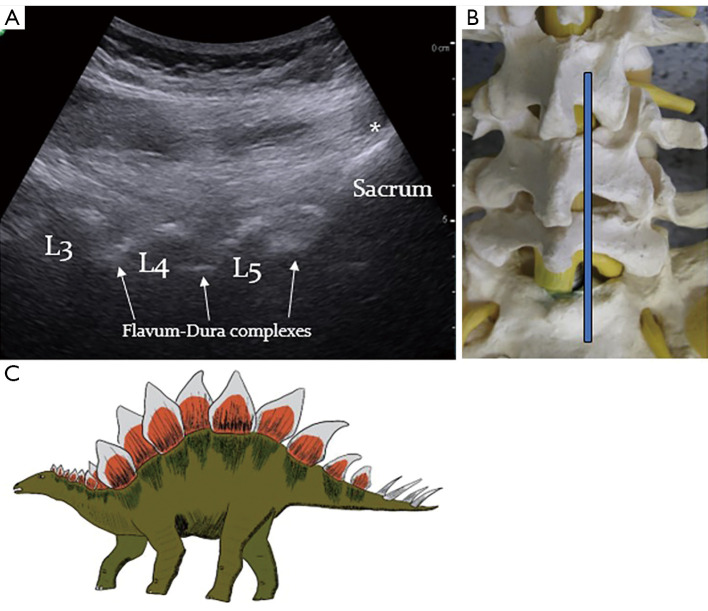
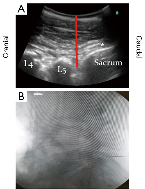
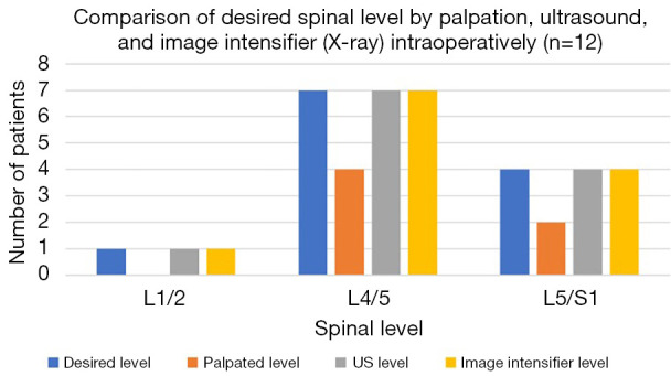
Lumbar spinal surgery relies on palpation of anatomical landmarks and X-ray imaging confirmation to identify the correct spinal level, therefore exposing patients and staff to radiation, and increasing intraoperative time and cost. Ultrasound (US) assistance is being used to visualise spinal anatomy by many specialities, such as neurology and anaesthetics, and can be used intraoperatively in selected spinal surgery cases. However, its potential use to check spinal levels prior to surgery remains understudied. This prospective, pilot study screened all patients requiring a primary elective or emergency lumbar discectomy, under the supervision of a single consultant neurosurgeon, over an 8-month period at a single neurosurgical unit. US assistance was used to identify and mark the proposed spinal level prior to skin incision. The resemblance of the parasagittal lumbar US images to the back of the dinosaur Stegosaurus aided users in identifying the relevant anatomical structures necessary to mark the desired spinal level, (e.g., lumbar laminae, intervertebral spaces, sacrum). This inspired our description of the US images of the lumbar spine as 'The Stegosaurus Appearance'. The spinal level marked by US was then confirmed in the standard fashion using intraoperative X-ray imaging. In 100% of cases (12/12), the desired spinal level was correctly identified using US, confirmed by the subsequent intraoperative X-ray images. US assistance appears to be a safe, quick, and accurate tool for identifying the correct lumbar spinal level prior to skin incision, and could therefore represent a useful adjunct to supplement level checking in lumbar spinal surgery.
Keywords: Ultrasound (US); check; discectomy; level; spinal.
2024 AME Publishing Company. All rights reserved.
Conflict of interest statement
Conflicts of Interest: All authors have completed the ICMJE uniform disclosure form (available at https://jss.amegroups.com/article/view/10.21037/jss-24-61/coif). The authors have no conflicts of interest to declare.
Figures
Similar articles
-
Intra-operative imaging for spinal level localisation in lumbar surgery.Br J Neurosurg. 2019 Jun;33(3):352-356. doi: 10.1080/02688697.2018.1562030. Epub 2019 Feb 11. Br J Neurosurg. 2019. PMID: 30741019
-
An evaluation of ultrasound imaging for identification of lumbar intervertebral level.Anaesthesia. 2002 Mar;57(3):277-80. doi: 10.1046/j.1365-2044.2002.2403_4.x. Anaesthesia. 2002. PMID: 11892638
-
Ability of pediatric emergency medicine physicians to identify anatomic landmarks with the assistance of ultrasound prior to lumbar puncture in a simulated obese model.Pediatr Emerg Care. 2015 Jan;31(1):15-9. doi: 10.1097/PEC.0000000000000330. Pediatr Emerg Care. 2015. PMID: 25526015
-
Ultrasonography for lumbar neuraxial block.Anesth Pain Med (Seoul). 2020 Oct 30;15(4):397-408. doi: 10.17085/apm.20065. Anesth Pain Med (Seoul). 2020. PMID: 33329842 Free PMC article. Review.
-
Intraoperative Ultrasound in Spine Decompression Surgery: A Systematic Review.Spine (Phila Pa 1976). 2022 Jan 15;47(2):E73-E85. doi: 10.1097/BRS.0000000000004111. Spine (Phila Pa 1976). 2022. PMID: 34474449
Cited by
-
Ultrasound measurements of lumbar spinous process movement during flexion distraction manipulation: a preliminary descriptive cross-sectional study with healthy participants.Chiropr Man Therap. 2025 Jul 25;33(1):29. doi: 10.1186/s12998-025-00593-0. Chiropr Man Therap. 2025. PMID: 40713659 Free PMC article.
References
LinkOut - more resources
Full Text Sources