. 2024 Dec 20;10(4):663-679.
doi: 10.21037/jss-24-71. Epub 2024 Dec 17.

Minimally invasive prone lateral retropleural or retroperitoneal antepsoas approach spinal surgery using the rotatable radiolucent Jackson table

Affiliations

Minimally invasive prone lateral retropleural or retroperitoneal antepsoas approach spinal surgery using the rotatable radiolucent Jackson table

Yu-Cheng Yeh et al. J Spine Surg. .

Abstract

Background: Prone lateral spinal surgery for simultaneous lateral and posterior approaches has recently been proposed to facilitate surgical room efficiency. The purpose of this study is to evaluate the feasibility and outcomes of minimally invasive prone lateral spinal surgery using a rotatable radiolucent Jackson table.

Methods: From July 2021 to June 2023, a consecutive series of patients who received minimally invasive prone lateral spinal surgery for various etiologies by the same surgical team were reviewed. A Mizuho Jackson Modular Table System was used for all prone lateral surgeries. All patients received combined lateral and posterior approach surgery on the same day. The lateral approaches were performed with the Jackson table rotated 30-40 degrees away from the surgical side. A table-mounted oblique lumbar interbody fusion (OLIF) retractor was applied in retropleural/retroperitoneal spaces. Minimally invasive lateral procedures such as discectomy or mini-open corpectomy were performed after adequate exposure. Posterior procedures were performed with the Jackson table rotated back horizontally. The disease etiologies, surgical levels, blood loss, operation time, and surgical procedures were collected and analyzed.

Results: The study included 64 patients with a mean age of 61.8 years (range, 26-88 years). The disease etiologies were 11 (17.2%) deformities, 15 (23.4%) degenerations, 25 (39.1%) infections, 9 (14.1%) traumas, and 4 (6.3%) tumors. The mean length of the surgical level was 4.1±2.0 (range, 2-10), with surgical levels ranging from T8 to L5 laterally and T6 to the ilium posteriorly. The mean blood loss was 863±843 mL (range, 50-4,600 mL) and the mean operation time was 314±148 minutes (range, 92-785 minutes). Of the lateral approaches, there were 25 retropleural and 39 retroperitoneal approaches (36 antepsoas approach). Surgical procedures performed included lateral discectomies, mini-open corpectomies, interbody reconstruction and fusion, and various posterior techniques such as pedicle screw instrumentation, cement augmentation, decompression, osteotomy, and spinal endoscopy. Patients who received both prone lateral retropleural and retroperitoneal approaches had significant improvement in the sagittal Cobb angle of the lateral surgical level, Visual Analogue Scale (VAS), and Oswestry Disability Index (ODI) at 1 year postoperatively.

Conclusions: Minimally invasive prone lateral spinal surgery is a feasible option for patients requiring combined lateral and posterior approach spinal surgery. Both lateral retropleural and retroperitoneal antepsoas approaches can be applied in combination with various posterior surgical procedures in the prone position using the rotatable radiolucent Jackson table.

Keywords: Prone lateral spinal surgery; retroperitoneal antepsoas approach; retropleural approach.


Conflict of interest statement

Conflicts of Interest: All authors have completed the ICMJE uniform disclosure form (available at https://jss.amegroups.com/article/view/10.21037/jss-24-71/coif). The authors have no conflicts of interest to declare.

Figures

Figure 1
Figure 1
Step by step preparation of prone lateral spinal surgery using rotatable radiolucent table. (A) The hip pad on the approach side was positioned downward to the position of the upper thigh as indicated by the red arrow. (B) Full exposure of the working space on the approach side from the ribs to the iliac crest with patient’s abdomen hanging freely. (C) Two lateral positioners are placed on the contralateral side ribs and femoral trochanteric area as support after rotation and backstop during graft placement. Tape is used to secure the thoracic and pelvic area of the patient while avoiding the sterilized surgical field. (D-F) Once the patient is secured by taping, the patient is manually rotated 30–40 degrees away from the approach side for a better approach to the surgical corridor. (G) The skin incision is marked anterior to the targeted vertebrae for lower lumbar segments in the lateral approach. (H) The skin incision mark is drawn centered over the closest rib to the targeted thoracic or thoracolumbar level, which is slightly more posterior than the lower lumbar segments. (I) The C-arm machine was positioned in the true anteroposterior view position before sterilization and draping. (J) True lateral view is obtained by rotating the C-arm over the patient. (K,L) Intraoperative true anteroposterior and lateral images obtained by the C-arm before incision of an L3–4 adjacent segment disease.
Figure 2
Figure 2
Comparisons of (A) traditional decubitus lateral antepsoas approach and (B) rotated prone lateral antepsoas approach.
Figure 3
Figure 3
A 49-year-old female with L4–5 degenerative spondylolisthesis received minimal invasive fusion surgery. (A-D) Anteroposterior, lateral standing and flexion-extension radiographs demonstrating L4–5 grade I degenerative spondylolisthesis. (E-G) The preoperative MRI revealed L4–5 grade B spinal stenosis. (H,I) The anteroposterior and lateral radiographs showed a L4–5 prone lateral OLIF and percutaneous posterior instrumentation. MRI, magnetic resonance imaging; OLIF, oblique lumbar interbody fusion.
Figure 4
Figure 4
A 74-year-old female with degenerative lumbar kyphosis received corrective surgery. (A-D) Anteroposterior and lateral standing radiographs demonstrating a L2-iliac posterior instrumentation, L5–S1 TLIF and prone lateral L2–5 OLIF. Prone lateral grade 1 ACR at L4–5 was performed to restore L4–S1 lordosis. TLIF, transforaminal lumbar interbody fusion; OLIF, oblique lumbar interbody fusion; ACR, anterior column realignment.
Figure 5
Figure 5
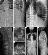
A 65-year-old man with T8–10 vertebral osteomyelitis of previous T9 vertebroplasty received debridement, cement removal and reconstruction surgery due to unresponsive to prolonged antibiotics treatment. (A-D) Anteroposterior and lateral radiographs demonstrating a T6–12 percutaneous posterior instrumentation and prone lateral T9 corpectomy and cement removal. (E-G) The preoperative MRI revealed T8–10 vertebral osteomyelitis with paraspinal abscess formation. (H,I) The patient recovered from septic shock with no recurrent infection and mobilized independently as shown in the anteroposterior and lateral standing radiographs. MRI, magnetic resonance imaging.
Figure 6
Figure 6
A 59-year-old woman suffered from pelvic fracture, L1 compression fracture and L3 comminuted burst fracture after falling from height received staged operations for multiple trauma. (A-E) The plain radiographs and CT showed L1 compression fracture and L3 comminuted burst fracture. The axial views revealed severe spinal canal compromised by the retropulsed fragments at L3 level. (F,G) The postoperative anteroposterior and lateral views demonstrating an open reduction and internal fixation for pelvic fracture, L1 vertebroplasty, L2–4 percutaneous posterior cemented instrumentation and anterior reconstruction after L3 corpectomy for direct decompression of the spinal canal. (H-J) The postoperative CT at 5 months showed stable implant construct with solid union of the fractured vertebra and adequate direct spinal canal decompression of the retropulsed fragments as compared to preoperative CT. CT, computed tomography.
Figure 7
Figure 7
A 50-year-old man with L1 single metastatic adenocarcinoma from rectal cancer received total en bloc spondylectomy. (A,B) The preoperative MRI showed L1 spinal tumor with right psoas muscle involvement. (C,D) The intraoperative fluoroscope and gross photo of the specimens. (E,F) The postoperative anteroposterior and lateral standing radiographs demonstrating a T11–L3 posterior instrumentation and anterior reconstruction after prone lateral simultaneous approached total en bloc spondylectomy. The vertebral body was removed via lateral window to avoid excessive traction of bilateral L1 roots. MRI, magnetic resonance imaging.

References

    1. White MD, Uribe JS. Transpsoas Approaches to the Lumbar Spine: Lateral and Prone. Neurosurg Clin N Am 2023;34:609-17. 10.1016/j.nec.2023.06.008 - DOI - PubMed
    1. DiGiorgio AM, Edwards CS, Virk MS, et al. Stereotactic navigation for the prepsoas oblique lateral lumbar interbody fusion: technical note and case series. Neurosurg Focus 2017;43:E14. 10.3171/2017.5.FOCUS17168 - DOI - PubMed
    1. Mehren C, Mayer HM, Zandanell C, et al. The Oblique Anterolateral Approach to the Lumbar Spine Provides Access to the Lumbar Spine With Few Early Complications. Clin Orthop Relat Res 2016;474:2020-7. 10.1007/s11999-016-4883-3 - DOI - PMC - PubMed
    1. Mobbs RJ, Phan K, Malham G, et al. Lumbar interbody fusion: techniques, indications and comparison of interbody fusion options including PLIF, TLIF, MI-TLIF, OLIF/ATP, LLIF and ALIF. J Spine Surg 2015;1:2-18. 10.3978/j.issn.2414-469X.2015.10.05 - DOI - PMC - PubMed
    1. Hung SF, Liao JC, Tsai TT, et al. Comparison of outcomes between indirect decompression of oblique lumbar interbody fusion and MIS-TLIF in one single-level lumbar spondylosis. Sci Rep 2021;11:12783. 10.1038/s41598-021-92330-9 - DOI - PMC - PubMed

LinkOut - more resources