. 2025 Jan 16:13:RP100060.
doi: 10.7554/eLife.100060.

Crispant analysis in zebrafish as a tool for rapid functional screening of disease-causing genes for bone fragility

Affiliations

Crispant analysis in zebrafish as a tool for rapid functional screening of disease-causing genes for bone fragility

Sophie Debaenst et al. Elife. .

Abstract

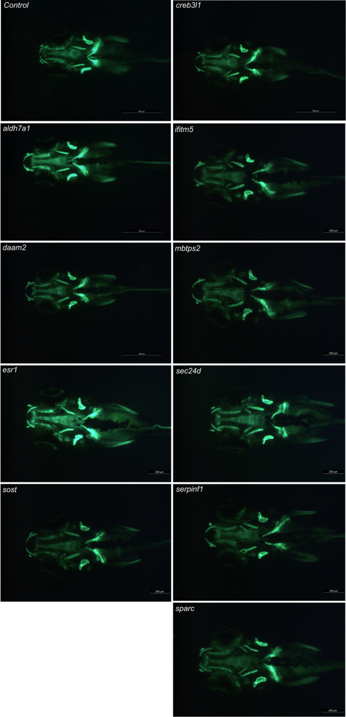
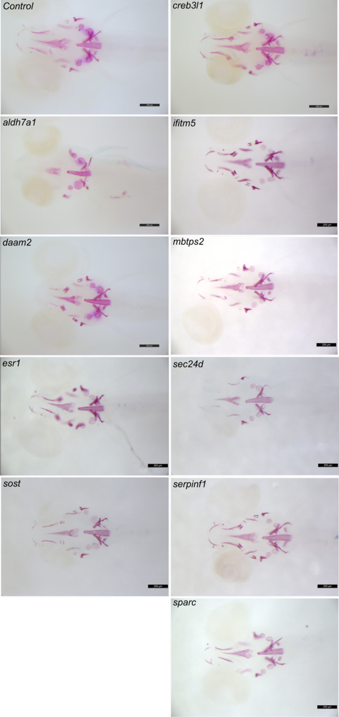
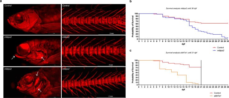
Heritable fragile bone disorders (FBDs), ranging from multifactorial to rare monogenic conditions, are characterized by an elevated fracture risk. Validating causative genes and understanding their mechanisms remain challenging. We assessed a semi-high throughput zebrafish screening platform for rapid in vivo functional testing of candidate FBD genes. Six genes linked to severe recessive osteogenesis imperfecta (OI) and four associated with bone mineral density (BMD) from genome-wide association studies were analyzed. Using CRISPR/Cas9-based crispant screening in F0 mosaic founder zebrafish, Next-generation sequencing confirmed high indel efficiency (mean 88%), mimicking stable knock-out models. Skeletal phenotyping at 7, 14, and 90 days post-fertilization (dpf) using microscopy, Alizarin Red S staining, and microCT was performed. Larval crispants showed variable osteoblast and mineralization phenotypes, while adult crispants displayed consistent skeletal defects, including malformed neural and haemal arches, vertebral fractures and fusions, and altered bone volume and density. In addition, aldh7a1 and mbtps2 crispants experienced increased mortality due to severe skeletal deformities. RT-qPCR revealed differential expression of osteogenic markers bglap and col1a1a, highlighting their biomarker potential. Our results establish zebrafish crispant screening as a robust tool for FBD gene validation, combining skeletal and molecular analyses across developmental stages to uncover novel insights into gene functions in bone biology.

Keywords: Crispants; Danio rerio; genetics; genomics; zebrafish.


Conflict of interest statement

SD, TJ, HD, JB, MB, IJ, PK, RK, PC, AW No competing interests declared

Figures

Figure 1.
Figure 1.. Measurements of osteoblast-positive surface area and mineralized surface area of the head skeleton in 7 and 14 dpf crispants.
The first four genes are associated with the pathogenesis of osteoporosis, while the last six are linked to osteogenesis imperfecta. (a) Schematic overview of the ventral and lateral perspective of the head of zebrafish larvae at 7 and 14 dpf. The notochord tip (n), the opercle (o) the mineralized vertebrae (v), the eyes (e) and parasphenoid (p) are shown. (b) Quantification of the osteoblast-positive surface area of the total head and the mineralized surface area of the total head and notochord tip at 7 dpf in comparison with their respective controls. (c) Quantification of the osteoblast-positive surface area of the total head and the mineralized surface area of the total head, opercle and notochord tip and number of vertebrae at 14 dpf in comparison with their respective controls. For easier visualization, obtained results were normalized to the respective controls (normalization = individual value crispant (or control) / mean control) (n=10). Statistical significance is evaluated using the Mann-Whitney U -test on non-normalized data and significant differences were visualized using an asterix (*=p ≤ 0,05; **=p ≤ 0,01; ***=p ≤ 0,001; ****=p ≤ 0,0001). Error bars show the standard deviation of non-normalized data.
Figure 1—figure supplement 1.
Figure 1—figure supplement 1.. InDelphi prediction and In-frame analysis.
The first four genes are associated with the pathogenesis of osteoporosis, while the last six are linked to osteogenesis imperfecta. (a) The predicted indel percentage using the InDelphi-mESC prediction tool. (b) Visualization of the in-frame analysis of the different crispants, showing the percentages of 9, 12, 15, 18, 21 and more than 21 base pair deletions in the crispants.
Figure 1—figure supplement 2.
Figure 1—figure supplement 2.. Osteoblast-positive head area at 7 dpf.
Visualization of the osteoblast using the osx:Kaede transgenic line. The four genes on the left are associated with the pathogenesis of osteoporosis, while the six genes on the right are linked to osteogenesis imperfecta. The presented image shows a representative image of the specific crispants. Images are taken with the Leica microscope and the osx:Kaede positive larvae are visualized from a ventral perspective. Scale bars = 500 μm and 200 μm.
Figure 1—figure supplement 3.
Figure 1—figure supplement 3.. Osteoblast-positive head area at 14 dpf.
Visualization of the osteoblast using the osx:Kaede transgenic line. The first four genes are associated with the pathogenesis of osteoporosis, while the last six are linked to osteogenesis imperfecta. The presented image shows a representative image of the specific crispants. Images are taken with the Leica microscope and the osx:Kaede-positive larvae are visualized from a ventral and lateral perspective. Scale bars = 1 mm.
Figure 1—figure supplement 4.
Figure 1—figure supplement 4.. Mineralization in the head area at 7 dpf.
Visualization of the mineralization after ARS staining. The four genes on the left are associated with the pathogenesis of osteoporosis, while the six genes on the right are linked to osteogenesis imperfecta. The presented image shows a representative image of the specific crispants. Images are taken with the Leica microscope and the stained larvae are visualized from a ventral perspective. Scale bars = 200 μm.
Figure 1—figure supplement 5.
Figure 1—figure supplement 5.. Mineralization in the head area at 14 dpf.
Visualization of the mineralization after ARS staining. The first four genes are associated with the pathogenesis of osteoporosis, while the last six are linked to osteogenesis imperfecta. The presented image shows a representative image of the specific crispants. Images are taken with the Leica microscope and the stained larvae are visualized from a ventral and lateral perspective. Scale bars = 1 mm.
Figure 2.
Figure 2.. RT-qPCR expression analysis of runx2, sp7, bglap and col1a1a in crispants at 7 (a) and 14 dpf (b) and their respective controls, normalized according to the controls (normalization = individual values crispant (or control) / mean control).
The first four genes are associated with the pathogenesis of osteoporosis, while the last six are linked to osteogenesis imperfecta. Statistical significance is evaluated using the Mann-Whitney U test on non-normalized data and significant differences were visualized using an asterix (*=p ≤ 0.05; **=p ≤ 0.01; ***=p ≤ 0.001; ****=p ≤ 0.0001). Error bars show the standard deviation of non-normalized data.
Figure 3.
Figure 3.. ARS images of mbtps2 crispants and survival analysis of aldh7a1 and mbtps2 crispants.
(a) ARS images of control fish and crispants for mbtps2, showing severe craniofacial abnormalities (arrows). (b) Survival curve of controls and crispants for mbtps2, showing a reduction in survival starting from 17 dpf. (c) Survival curve of controls and crispants for aldh7a1, showing a reduction in survival starting from 7 dpf.
Figure 4.
Figure 4.. Skeletal phenotyping of adult crispants and their respective controls.
The first three genes are associated with the pathogenesis of osteoporosis, while the last five are linked to osteogenesis imperfecta. (a) Pictures of ARS-stained vertebral column of a control and three crispants (from left to right: esr1, ifitm5 and creb3l1), showing fractures (arrow), fusions and compressions (squared) and malformations in the arches (arrowhead). (b) Measurements of the standard length, the head size and the eye diameter of the crispants compared to their control. The standard length was measured from the snout tip to the tail base. The head size was measured from the snout tip to the supraoccipital bone and the eye diameter was measured from the lateral ethmoid to the hyomandibula. The data was normalized for easier visualization (normalization = individual value crispants (or controls) / mean control (n=5)). (c) Quantification of the number of fractures, fusions or compressions and malformations in the arches of crispants and the controls. For this quantification, twelve vertebrae were selected per fish and a total of 5 crispants per assay was evaluated. Statistical analysis was evaluated using the Mann-Whitney U -test on non-normalized data and significant differences were visualized using an asterix (*=p ≤ 0.05; **=p ≤ 0.01; ***=p ≤ 0.001; ****=p ≤ 0.0001). Error bars showed the standard deviation of non-normalized data. (d) Quantification of skeletal parameters by quantitative micro-computed tomography (μCT) analysis. In the graphical representation, the different crispants were listed. Statistically significant differences from the control values for a given crispant were depicted as red bars. Significance levels were determined through the global test analysis (Figure 4—figure supplement 2).
Figure 4—figure supplement 1.
Figure 4—figure supplement 1.. Mineralization in the skeleton at 90 dpf.
Visualization of the mineralization after ARS staining. The first three genes are associated with the pathogenesis of osteoporosis, while the last five are linked to osteogenesis imperfecta. The presented image shows a representative image of the specific crispants. Images are taken with the Leica microscope and the stained adults are visualized from a lateral perspective. Scale bars = 1 mm.
Figure 4—figure supplement 2.
Figure 4—figure supplement 2.. Quantitative μCT-scanning analysis of the vertebral column of the crispants using FishCuT software.
Whole-body μCT-scanning was performed for the evaluation of skeletal structures in the vertebral column. Different bone related parameters are visualized: Tissue Mineral Density (TMD), Volume (Vol), Thickness (Th), and Length (Le). (a) Results of crispants for daam2. (b) Results for crispants for esr1. (c) Results for crispants for sost. (d) Results for crispants for creb3l1. (e) Results for crispants for ifitm5. (f) Results for crispants for sec24d. (g) Results for crispants for serpinf1. (h) Results for crispants for sparc. The first three genes are associated with the pathogenesis of osteoporosis, while the last five are linked to osteogenesis imperfecta. Statistically significant differences were represented with a lighter color scheme for easy visualization.
Figure 4—figure supplement 3.
Figure 4—figure supplement 3.. Mineralization in the skeleton at 90 dpf.
Representative images of skeletal mineralization following Alizarin Red S (ARS) staining in a second clutch, demonstrating the consistency of the observed skeletal phenotype. The three genes on the left are associated with the pathogenesis of osteoporosis, while the last five genes on the right are linked to osteogenesis imperfecta. Images show specific crispants from a lateral view, captured using a Leica microscope. Scale bars = 1 mm.

Update of

  • doi: 10.1101/2024.06.27.601074
  • doi: 10.7554/eLife.100060.1
  • doi: 10.7554/eLife.100060.2

References

    1. Amarasekara DS, Kim S, Rho J. Regulation of osteoblast differentiation by cytokine networks. International Journal of Molecular Sciences. 2021;22:2851. doi: 10.3390/ijms22062851. - DOI - PMC - PubMed
    1. Bek JW, Shochat C, De Clercq A, De Saffel H, Boel A, Metz J, Rodenburg F, Karasik D, Willaert A, Coucke PJ. Lrp5 mutant and crispant zebrafish faithfully model human osteoporosis, establishing the zebrafish as a platform for CRISPR-based functional screening of osteoporosis candidate genes. Journal of Bone and Mineral Research. 2020;36:1749–1764. doi: 10.1002/jbmr.4327. - DOI - PubMed
    1. Brett JO, Spring LM, Bardia A, Wander SA. ESR1 mutation as an emerging clinical biomarker in metastatic hormone receptor-positive breast cancer. Breast Cancer Research. 2021;23:85. doi: 10.1186/s13058-021-01462-3. - DOI - PMC - PubMed
    1. Charoenngam N, Rittiphairoj T, Ponvilawan B, Jaroenlapnopparat A, Waitayangkoon P, Suppakitjanusant P, Prasitsumrit V, Pongchaiyakul C, Holick MF. Bone fragility in hereditary connective tissue disorders: a systematic review and meta-analysis. Endocrine Practice. 2023;29:589–600. doi: 10.1016/j.eprac.2023.02.003. - DOI - PubMed
    1. Clement K, Rees H, Canver MC, Gehrke JM, Farouni R, Hsu JY, Cole MA, Liu DR, Joung JK, Bauer DE, Pinello L. CRISPResso2 provides accurate and rapid genome editing sequence analysis. Nature Biotechnology. 2019;37:224–226. doi: 10.1038/s41587-019-0032-3. - DOI - PMC - PubMed

LinkOut - more resources