. 2025 Jan 16;135(2):e183776.
doi: 10.1172/JCI183776.

Clonal analysis of SepSecS-specific B and T cells in autoimmune hepatitis

Affiliations

Clonal analysis of SepSecS-specific B and T cells in autoimmune hepatitis

Michael Kramer et al. J Clin Invest. .

Abstract

Autoimmune hepatitis (AIH) is a rare chronic inflammatory liver disease characterized by the presence of autoantibodies, including those targeting O-phosphoseryl-tRNA:selenocysteine-tRNA synthase (SepSecS), also known as soluble liver antigen (SLA). Anti-SepSecS antibodies have been associated with a more severe phenotype, suggesting a key role for the SepSecS autoantigen in AIH. To analyze the immune response to SepSecS in patients with AIH at the clonal level, we combined sensitive high-throughput screening assays with the isolation of monoclonal antibodies (mAbs) and T cell clones. The anti-SepSecS mAbs isolated were primarily IgG1, affinity-matured compared with their germline versions, and recognized at least 3 nonoverlapping epitopes. SepSecS-specific CD4+ T cell clones were found in patients with AIH who were anti-SLA-positive and anti-SLA-negative,and, to a lesser extent, in patients with non-AIH liver diseases and in healthy individuals. SepSecS-specific T cell clones from patients with AIH produced IFN-γ, IL-4, and IL-10, targeted multiple SepSecS epitopes, and, in one patient, were clonally expanded in both blood and liver biopsy. Finally, SepSecS-specific B cell clones, but not those of unrelated specificities, were able to present soluble SepSecS to specific T cells. Collectively, our study provides the first detailed analysis of B and T cell repertoires targeting SepSecS in patients with AIH, offering a rationale for improved targeted therapies.

Keywords: Autoimmunity; Hepatitis; Immunoglobulins; Immunology; T cells.


Figures

Figure 1
Figure 1. Characterization of anti-SLA positivity in AIH.
(A and B) Measurement of SepSecS-specific IgG in plasma from patients with AIH and individuals in the healthy control group by an in-house–developed flow cytometry assay based on SepSecS/eGFP-transfected EXPI-293 cells (A) or by an in-house–developed ELISA using SepSecS produced in EXPI-293 cells (B). Note that patient AIH18 was classified anti-SLA negative in the clinical laboratory but is anti-SLA positive in both of our assays. (C and D) Scatterplots showing the correlation between the EDF50 values of SepSecS-specific IgG measured using the flow cytometry assay and ELISA (C) and the ELISA and a commercially available ELISA (cELISA) (D). (E) SepSecS-specific IgG+ memory B cells in peripheral blood of patients with AIH and individuals in the healthy control group. PBMCs (3 × 10⁴; AIH11: 2 × 10⁴) were plated in 192 replicate wells (AIH11, 96) and stimulated with IL-2 and the TLR 7/8 agonist R848. After 12 days, the supernatant of each well was screened for the presence of secreted SepSecS-specific IgG using the flow cytometry assay. Reported is the mean fluorescence intensity (MFI) GFP-SepSecS+ / MFI GFP-SepSecS ratios for each individual supernatant (positive > 1.1). (F) Number of SepSecS-specific IgG+ memory B cells in 1 × 10⁶ PBMCs in patients with AIH and individulals in the healthy control group with positive cultures in the analysis in E and calculated according to the Poisson distribution.
Figure 2
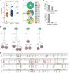
Figure 2. SepSecS-specific antibodies in patients with AIH are polyclonal and affinity matured.
(A) EC50 values of 155 anti-SepSecS mAbs isolated from EBV-immortalized IgG+ memory B cell clones from patients AIH11, AIH12, and AIH14, as calculated from curves reported in Supplemental Figure 3. (B) VH gene usage of sequenced SepSecS-specific B cell clones. Slices in the chart represent different VH genes, and their size is proportional to the number of clones using that particular gene. The total number of sequenced clones from each patient is reported at the center of the pie charts. (C) Percentages of sequenced SepSecS-specific B cell clones with variable identity to the germline VH and VL genes. (D) Genealogical trees of 4 B cell clonal families in patient AIH12. The number of somatic mutations at nucleotide and amino acid level (in parenthesis) is indicated on individual branches of the trees. Unmutated common ancestors (UCAs) are in green, intermediary development steps are in white, and sequenced clones are in purple. (E) Sequence alignments show heavy chain amino acid mutations of sequenced clones from the respective UCAs.
Figure 3
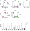
Figure 3. SepSecS-specific mAb recognize different epitopes and acquire high affinity through somatic mutations.
(A) Competition-ELISA identifying 3 groups of recombinantly produced mAbs with different binding regions. ELISA-plates were coated for 24 hours at 4°C with 5 μg/mL SepSecS. First, mAbs were added in excess (20 μg/mL) to block mAb-specific epitopes completely. Biotinylated mAbs were added as second mAbs, and binding was assessed using streptavidin-HRP. The 12 recombinant mAbs were analyzed in 2 independent experiments of 7 (upper 3 plots) and 5 mAbs (lower 2 plots). MAb codes start with number of the patient with AIH from whom the mAb is derived. (B) EC50 values of SepSecS binding of recombinantly produced mAbs carrying different combinations of mutated (M) and germline (GL) heavy and light chain of the indicated mAbs as measured by flow cytometry assay. MAb codes start with number of the patient with AIH from whom the mAb is derived.
Figure 4
Figure 4. SepSecS proliferative response of memory CD4+ T cells in the blood of patients with AIH and individuals in the control groups.
Total memory CD4+ T cells from the blood of patients with AIH, patients with non-AIH liver diseases in the PC group and individuals in the HC group were labeled with CFSE and stimulated with autologous monocytes in the presence or absence of the SepSecS peptide pool. On day 7, cells were collected and stained with anti–CD25-PE and anti–ICOS-APC mouse mAbs. (A) Histograms represent CFSE profile and contour plots represent CD25 and ICOS staining on gated CFSElo cells from AIH12 (anti-SLA positive), PC3 and HC2. Red lines, no SepSecS; gray lines, SepSecSpp. (B) Pooled data from all study participants shown as the percentage of CFSEloCD25+ICOS+ cells in the SepSecS stimulated cultures. Lines represent the median values. Blue dots represent anti-SLA positive patients with AIH. *P < 0.05 when determined by 2-tailed Mann-Whitney test and < 0.01 when determined by Spearman Partial correlation considering age as a covariable. (C) Number of total CD4+ T cell clones (left) isolated by limiting dilution from sorted CFSEloCD25+ICOS+ cells from ex vivo cultures of memory CD4+ T cells from patients with AIH, and individuals in the PC and HC groups. The right plot shows the frequency of SepSecS-specific CD4+ T cell clones among the isolated clones for each donor. Specificity was tested by stimulation with irradiated autologous B cells untreated or pulsed with SepSecS peptide pool. Proliferation was measured on day 3 after a 16 hour incubation with 1 μCi/mL methyl-3H-thymidine. (D) Epitope mapping of SepSecS-specific CD4+ T cell clones from patients with AIH and individuals in the control groups. Epitopes were identified by stimulating the clones with individual SepSecS peptides. Patients with AIH are represented in blue, patients in the PC group are in orange and individuals in the HC group are in black. Predicted binding peptides are shown as shaded blue (DRB1*03:01) and shaded green (DRB1*0401) squares in patients carrying the AIH predisposing HLA alleles DRB1*0301 and DRB1*0401, respectively. Anti-SLA status and disease activity are shown on the left-hand side; number of CD4+ T cell autoreactive clones are shown on the right-hand side.
Figure 5
Figure 5. SepSecS-specific CD4+ T cells are clonally expanded in the blood and liver of patient AIH12.
(A) Proliferative response to SepSecS of CD4+ T cell clones from the liver biopsy of patient AIH12. Cpm, counts per minute. Autologous EBV-B cells were either left untreated (No Ag) or pulsed with SepSecSpp. (B) TCR Vα and TCR Vβ chain sequencing of the 87 SepSecS-specific CD4+ T cell clones identify 4 unique clonotypes. Reported are the T cell receptor α variable (TRAV) gene and the T cell receptor β variable (TRBV) gene used as well as the CDR3 amino acid (aa) sequences of the TCRα and TCRβ chains. The number of identical sequences per each clone and the SepSecS epitopes, as identified by epitope mapping, are shown. aa, amino acids. (C) Pairwise comparison of TCR Vβ clonotype frequency distribution in samples of total memory CD4+ T cells sorted from PBMCs (x-axis) and in vitro-expanded total memory CD4+ T cells from a liver biopsy taken on the same day as the PBMCs (y-axis). The 4 SepSecS-specific clonotypes, which were identified by TCR Vβ chain sequencing of the 87 CD4+ T cell clones isolated from liver-infiltrating T cells are highlighted in color.
Figure 6
Figure 6. Specific B cells effectively present SepSecS protein to specific CD4+ T cells.
(A and B) Proliferative response of CD4+ T cell clones derived from blood (A) or liver (B) of patient AIH12 to different concentrations of SepSecS protein. The T cell clones targeted different SepSecS epitopes (C3 and A7: aa 341-370; B3: aa 71-90; LB A11 and LB C10: 181-200; LB A2*: 261-280). A SepSecS-specific (blue or orange symbols) EBV-B cell clone and an EBV-B cell clone of unrelated-specificity (white symbols) derived from the same patient were used as APCs. Cpm, counts per minute.

Comment in

  • Elucidating the role of autoreactive T cells and B cells in autoimmune hepatitis

References

    1. Mieli-Vergani G, et al. Autoimmune hepatitis. Nat Rev Dis Primers. 2018;4:18017. doi: 10.1038/nrdp.2018.17. - DOI - PubMed
    1. Terziroli Beretta-Piccoli B, et al. Autoimmmune hepatitis. Cell Mol Immunol. 2022;19(2):158–176. doi: 10.1038/s41423-021-00768-8. - DOI - PMC - PubMed
    1. Floreani A, et al. Etiopathogenesis of autoimmune hepatitis. J Autoimmun. 2018;95:133–143. doi: 10.1016/j.jaut.2018.10.020. - DOI - PubMed
    1. Ma Y, et al. Human leukocyte antigen profile predicts severity of autoimmune liver disease in children of European Ancestry. Hepatology. 2021;74(4):2032–2046. doi: 10.1002/hep.31893. - DOI - PMC - PubMed
    1. Costa M, et al. Isolation and characterization of cDNA encoding the antigenic protein of the human tRNP(Ser)Sec complex recognized by autoantibodies from patients withtype-1 autoimmune hepatitis. Clin Exp Immunol. 2000;121(2):364–374. doi: 10.1046/j.1365-2249.2000.01280.x. - DOI - PMC - PubMed