. 2025 May;49(5):778-789.
doi: 10.1111/aor.14947. Epub 2025 Jan 16.

Isogenic Transplantation of Hybrid Artificial Pleural Tissue Consisting of Rat Cells and Polyglycolic Acid Nanofiber Sheet Induces Restoration of Mesothelial Defects in Rat Model

Affiliations

Isogenic Transplantation of Hybrid Artificial Pleural Tissue Consisting of Rat Cells and Polyglycolic Acid Nanofiber Sheet Induces Restoration of Mesothelial Defects in Rat Model

Kengo Tani et al. Artif Organs. 2025 May.

Abstract

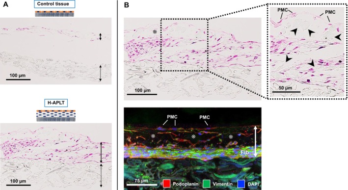
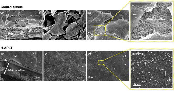
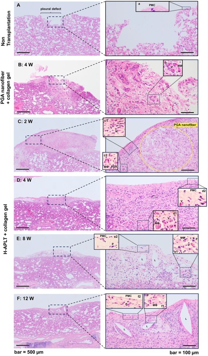
Background: Impairment of the visceral pleura following thoracic surgery often leads to air leaks and intrathoracic adhesions. For preventing such complications, mesothelial cell proliferation at the pleural defects can be effective. To develop new materials for pleural defects restoration, we constructed a hybrid artificial pleural tissue (H-APLT) combining polyglycolic acid (PGA) nanofiber sheets with a three-dimensional culture of mesothelial cells and fibroblasts and evaluated its therapeutic efficacy in a rat pleural defect model.

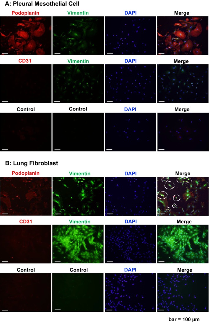
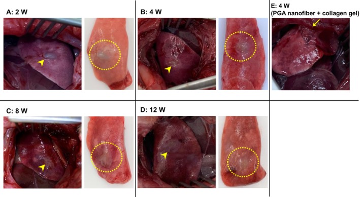
Methods: After rat lungs were harvested, pleural mesothelial cells and lung fibroblasts were cultured separately. To construct H-APLT, the cells were then coated with multiple layers of fibronectin and gelatin, followed by a single layer of mesothelial cells on top of multiple layers of fibroblasts accumulated onto a collagen-coated PGA nanofiber sheet. Left lateral thoracotomy was performed, and H-APLTs were transplanted into a rat model with pleural defects (N = 8). After 2-12 weeks of transplantation, lung resection and histological analyses were performed.

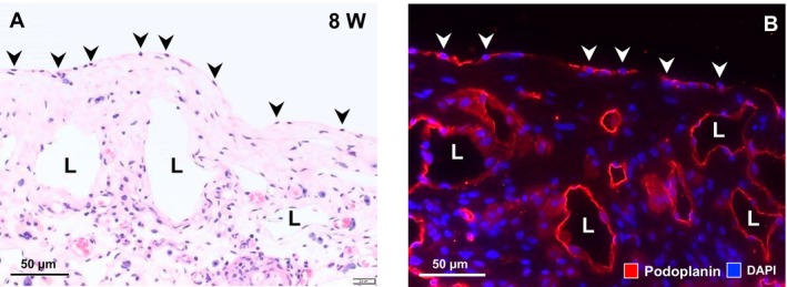
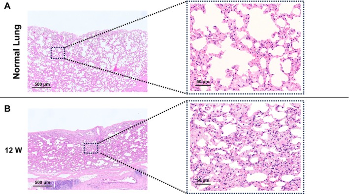
Results: H-APLTs exhibited a pleural structure with a highly integrated mesothelial layer in vitro. After transplantation, all eight rats survived until sacrifice. At 12 weeks post-transplantation, the mesothelial layer on the lung surface was observed to be without defects with no intrathoracic adhesions detected.

Conclusion: Successful isogenic engraftment of H-APLTs was achieved in a rat model of pleural defects. The combination of accumulated fibroblasts and collagen-coated PGA nanofiber sheets contributed to the maintenance of the mesothelial layer's structure and function, potentially preventing air leaks and intrathoracic adhesions.

Keywords: artificial pleural tissue; cell‐accumulation method; mesothelial cells; polyglycolic acid nanofiber sheet; tissue engineering.


Conflict of interest statement

The authors declare no conflicts of interest.

Figures

FIGURE 1
FIGURE 1
Construction of H‐APLT and transplantation of H‐APLT into a rat lung air leak model. (A) Isolation and culture of pleural mesothelial cells and lung fibroblasts. Rat lungs were treated with 0.05% trypsin–EDTA, and pleural mesothelial cells were isolated. Lungs were then minced and treated with 0.15% collagenase type V, the lung fibroblasts were isolated. Pleural mesothelial cells were characterized as having a cobblestone‐like arrangement, whereas fibroblasts were spindle‐shaped. (B) Construction of artificial tissues by cell accumulation technique. Lung fibroblasts with fibronectin‐gelatin nanofilms accumulated on the collagen‐coated PGA nanofiber sheet. A 3D tissue comprising four layers of lung fibroblasts was formed. Pleural mesothelial cells were cultured on top of these layers to generate H‐APLTs. The cells were cultured in Transwell inserts to create H‐APLT. The control tissue consisted of a single layer of pleural mesothelial cells on a collagen‐coated PGA nanofiber sheet. (C) The timeframe for implanting the H‐APLT into the rat lung air leak models. For immunosuppression, cyclosporin was added to drinking water (100 mg/L), 1 week before and throughout the transplantation period. Left lateral thoracotomy was performed. An air leak was generated via cutting the lung parenchyma into an approximately 5 mm long incision with a depth of 3 mm using surgical scissors (yellow dotted line). H‐APLTs (6 mm in diameter, yellow arrowhead) were transplanted into the air‐leak site. A collagen solution was instilled over the H‐APLT to prevent dislodgement. At 2, 4, 8, and 12 weeks post‐transplantation, the rats were euthanized. At each time point post transplantation, two rats were assessed (total N = 8). ECM, extracellular matrix; FN, fibronectin; G, gelatin; H‐APLT, hybrid artificial pleural tissue; PGA, polyglycolic acid. [Color figure can be viewed at wileyonlinelibrary.com]
FIGURE 2
FIGURE 2
Immunofluorescence analysis of pleural mesothelial cells and lung fibroblasts. (A) Immunostaining findings of pleural mesothelial cells. Podoplanin was strongly positive and vimentin was weakly positive (upper panel). CD31 was negative (middle panel). The negative control without primary antibodies is shown in the lower panel. (B) Immunostaining findings of lung fibroblasts. Vimentin was strongly positive (upper and middle panels). CD31 was negative (middle panel). Some cells were weakly positive for podoplanin (white dotted line in the upper panel); these were thought to be pleural mesothelial cells. These staining results likely occurred because lung fibroblasts were collected after pleural mesothelial cells from the same lung. The negative control without primary antibodies is shown in the lower panel. [Color figure can be viewed at wileyonlinelibrary.com]
FIGURE 3
FIGURE 3
Construct findings of artificial tissue. (A) Hematoxylin and eosin staining of artificial tissues. The H‐APLT and the control tissue was observed 5 days after construction. Solid and dotted lines represent cells and PGA nanofiber sheets, respectively. In the control tissue, the cells detached from the PGA nanofiber sheet (upper panel). H‐APLT had a thicker cellular component, without separation between the cellular components and collagen‐coated PGA nanofiber sheet (lower panel). (B) In the H‐APLT, PGA nanofibers were identified within the cellular components (upper panel, arrowheads), demonstrating the integration of tissue structure with PGA nanofiber. Immunofluorescence staining of H‐APLT is shown in the lower panel. The solid line outlines the cellular components. Podoplanin‐positive cells were observed on the surface of H‐APLT, indicating the presence of pleural mesothelial cells (PMC). Numerous vimentin‐positive cells were observed on the substrate PGA nanofiber, suggesting the presence of lung fibroblasts (Fib). Podoplanin‐positive cells were also detected within the lung fibroblast layer, indicating a mixture of pleural mesothelial cells. These staining results likely occurred because lung fibroblasts were collected after pleural mesothelial cells from the same lung. The gaps between mesothelial cells and the fibroblast layer are considered artifactual spaces related to the preparation process for histological sections (asterisk). H‐APLT, hybrid artificial pleural tissue; PGA, polyglycolic acid. [Color figure can be viewed at wileyonlinelibrary.com]
FIGURE 4
FIGURE 4
Scanning electron microscopy findings. (A, B) Are control tissues from different specimens. (C, D) are H‐APLT from different specimens. (A1) Uncomplete sheet structures. (A2, B) In the control tissue, the pleural mesothelial cells were detached from each other. (C1) In the H‐APLT, pleural mesothelial cells and fibroblasts formed sheet‐like structures. (C2, D) Pleural mesothelial cells were arranged in a cobblestone‐like pattern with no gaps between them. Numerous microvilli were identified in the high‐power field. Reproducibility has been demonstrated, confirming that these findings are not artifacts related to the SEM preparation process. Fib, fibroblasts; H‐APLT, hybrid artificial pleural tissue; PGA, polyglycolic acid; PMC, pleural mesothelial cells. [Color figure can be viewed at wileyonlinelibrary.com]
FIGURE 5
FIGURE 5
Macroscopic findings in the thoracic cavity and lungs after H‐APLT transplantation. H‐APLTs grew into the tissue, and the engrafted sites gradually became obscured (yellow arrowhead). The engrafted site is marked with a yellow dotted line. The H‐APLT was identified as a thick transplant at 2 weeks after surgery (A), but gradually integrated into the host tissue along the postoperative course (B–D). No intrathoracic adhesions were observed at any time point. However, 4 weeks after the attachment of a rat with only a PGA nanofiber sheet and collagen gel, adhesion between the parietal pleura and lung parenchyma was observed (E, yellow arrow). H‐APLT, hybrid artificial pleural tissue; PGA, polyglycolic acid. [Color figure can be viewed at wileyonlinelibrary.com]
FIGURE 6
FIGURE 6
Microscopic findings of engrafted site of the H‐APLT (Hematoxylin–eosin staining). The right‐hand images are magnified images of the black dotted boxes in the left‐hand images. (A) A pleural defect model. The alveolar structures were exposed. Only a few pleural mesothelial cells are present (a). (B) Four weeks after the attachment of the PGA nanofiber sheet and collagen gel. Although no remnants of the PGA nanofiber were seen, numerous instances of neovascularization were observed, suggesting a strong inflammatory response. Multinucleated macrophage (MΦ) and neovascular vessels (V) are shown in (6b). (C) Two weeks post‐transplantation, engraftment of H‐APLT was confirmed. Remnants of the PGA nanofibers were observed with abundant surrounding connective tissues (yellow dotted line). Immune cells including neutrophils (c1) and multinucleated macrophages (c2) were also identified. (D) After 4 weeks, the pleural defect was thoroughly replaced by thinned transplanted tissue. The PGA nanofibers were completely degraded, although multinucleated macrophages were still present (c1). Neovascularization was observed (d1 and d2, V). The surface of the graft was covered by flattened pleural mesothelial cells with fibrous connective tissue (d2). (E) After 8 weeks, macrophages, neovascularization and plasma cells were observed (e1). Dilated luminal structure (L) developed in the connective tissue. Pleural mesothelial cells were identified on the surface (e2). (F) After 12 weeks, the transplanted tissue showed development of luminal structures (L) with stromal fibrosis. Multinucleated macrophages were observed around the luminal structures (f1). Pleural mesothelial cells were identified on the surface (f2). H‐APLT, hybrid artificial pleural tissue; L, luminal structure; MΦ, macrophage; P, plasma cell; PGA, polyglycolic acid; PMC, pleural mesothelial cell; V, neovascularization. [Color figure can be viewed at wileyonlinelibrary.com]
FIGURE 7
FIGURE 7
High‐magnification findings of the engrafted site at 8 weeks. (A) A single layer of flattened cells covers the pleura‐defective area (arrowheads). (B) Immunohistochemical findings. Single flat surface cells (arrowheads) and luminal structures (L) stained positive for podoplanin. Pleural mesothelial cells and lymphatic vessels were suggested, respectively. [Color figure can be viewed at wileyonlinelibrary.com]
FIGURE 8
FIGURE 8
Lung parenchyma findings beneath the H‐APLT‐engrafted site. (A) The normal lung parenchyma and alveolar tissue. The right image is a magnification of the boxed area in the left image. (B) Lung parenchyma beneath the H‐APLT–engrafted site. The alveolar septa appeared to be thickened with fibrosis as compared with normal lungs. H‐APLT, hybrid artificial pleural tissue. [Color figure can be viewed at wileyonlinelibrary.com]

Similar articles

References

    1. Mutsaers S. E., Prele C. M., Brody A. R., and Idell S., “Pathogenesis of Pleural Fibrosis,” Respirology 9, no. 4 (2004): 428–440. - PubMed
    1. Mutsaers S. E., Prêle C. M., Pengelly S., and Herrick S. E., “Mesothelial Cells and Peritoneal Homeostasis,” Fertility and Sterility 106, no. 5 (2016): 1018–1024. - PubMed
    1. Mutsaers S. E., “Mesothelial Cells: Their Structure, Function and Role in Serosal Repair,” Respirology 7, no. 3 (2002): 171–191. - PubMed
    1. Mutsaers S. E., Birnie K., Lansley S., Herrick S. E., Lim C. B., and Prêle C. M., “Mesothelial Cells in Tissue Repair and Fibrosis,” Frontiers in Pharmacology 6 (2015): 113, 10.3389/fphar.2015.00113. - DOI - PMC - PubMed
    1. Attaar A., Luketich J. D., Schuchert M. J., Winger D. G., Sarkaria I. S., and Nason K. S., “Prolonged Air Leak After Pulmonary Resection Increases Risk of Noncardiac Complications, Readmission, and Delayed Hospital Discharge: A Propensity Score‐Adjusted Analysis,” Annals of Surgery 273, no. 1 (2021): 163–172. - PubMed

Substances

Grants and funding

LinkOut - more resources