. 2025 Jan 28;44(1):115205.
doi: 10.1016/j.celrep.2024.115205. Epub 2025 Jan 14.

Opposing roles of p38α-mediated phosphorylation and PRMT1-mediated arginine methylation in driving TDP-43 proteinopathy

Affiliations

Opposing roles of p38α-mediated phosphorylation and PRMT1-mediated arginine methylation in driving TDP-43 proteinopathy

Mari Aikio et al. Cell Rep. .

Erratum in

  • Opposing roles of p38α-mediated phosphorylation and PRMT1-mediated arginine methylation in driving TDP-43 proteinopathy.
    Aikio M, Odeh HM, Wobst HJ, Lee BL, Chan Ú, Mauna JC, Mack KL, Class B, Ollerhead TA, Ford AF, Barbieri EM, Cupo RR, Drake LE, Smalley JL, Lin YT, Lam S, Thomas R, Castello N, Baral A, Beyer JN, Najar MA, Dunlop J, Gitler AD, Javaherian A, Kaye JA, Burslem GM, Brown DG, Donnelly CJ, Finkbeiner S, Moss SJ, Brandon NJ, Shorter J. Aikio M, et al. Cell Rep. 2025 Mar 25;44(3):115386. doi: 10.1016/j.celrep.2025.115386. Epub 2025 Feb 19. Cell Rep. 2025. PMID: 39977265 Free PMC article. No abstract available.

Abstract

Amyotrophic lateral sclerosis (ALS) is a devastating neurodegenerative disorder typically characterized by insoluble inclusions of hyperphosphorylated TDP-43. The mechanisms underlying toxic TDP-43 accumulation are not understood. Persistent activation of p38 mitogen-activated protein kinase (MAPK) is implicated in ALS. However, it is unclear how p38 MAPK affects TDP-43 proteinopathy. Here, we show that p38α MAPK inhibition reduces pathological TDP-43 phosphorylation, aggregation, cytoplasmic mislocalization, and neurotoxicity. Remarkably, p38α MAPK inhibition mitigates aberrant TDP-43 phenotypes in diverse ALS patient-derived motor neurons. p38α MAPK phosphorylates TDP-43 at pathological S409/S410 and S292, which reduces TDP-43 liquid-liquid phase separation (LLPS) but allows pathological TDP-43 aggregation. Moreover, we establish that PRMT1 methylates TDP-43 at R293. Importantly, S292 phosphorylation reduces R293 methylation, and R293 methylation reduces S409/S410 phosphorylation. Notably, R293 methylation permits TDP-43 LLPS and reduces pathological TDP-43 aggregation. Thus, strategies to reduce p38α-mediated TDP-43 phosphorylation and promote PRMT1-mediated R293 methylation could have therapeutic utility for ALS and related TDP-43 proteinopathies.

Keywords: ALS; CP: Molecular biology; CP: Neuroscience; PRMT1; TDP-43; arginine methylation; p38α; phosphorylation.


Conflict of interest statement

Declaration of interests H.J.W., D.G.B., and N.J.B. were full-time employees and shareholders of AstraZeneca at the time these studies were conducted. S.J.M. is a consultant for SAGE Therapeutics and AstraZeneca, relationships that are regulated by Tufts University.

Figures

Figure 1.
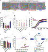
Figure 1.. Genetic and pharmacological inhibition of p38α MAPK reduces TDP-43 aggregation, S409/S410 phosphorylation, and neurotoxicity
(A) TDP-43 domain architecture with location of ALS-linked mutations and phosphorylation sites (P) detected after p38α treatment in vitro. (B) Western blot of total and pTDP-43M337V in RIPA and urea fractions of SH-SY5Y cells with siRNA-induced p38α knockdown. GAPDH serves as a loading control. SH-SY5Y cells were treated with siRNA for 24 h and then transfected with TDP-43M337V (myc tagged). Positions of myc-tagged TDP-43M337V (exo) and endogenous TDP-43 (endo) are indicated. (C) Quantification of urea/RIPA ratio of total TDP-43 (exogenously expressed) normalized to levels in scrambled siRNA (si-scr; mean ± SD, two-way ANOVA with Sidak’s multiple comparison test, n = 3). **p < 0.01, ****p < 0.0001. (D) Quantification of pTDP-43/total TDP-43 ratio (exogenously expressed) in urea fraction normalized to levels in si-scr (mean ± SD, two-way ANOVA with Sidak’s multiple comparison test, n = 3). *p < 0.05, **p < 0.01, ***p < 0.001. (E) Chemical structures of compound 1 and VX-745 (neflamapimod). (F) Western blot of total and pTDP-43M337V in RIPA and urea fractions of SH-SY5Y cells with pharmacological p38α inhibition with compound 1. GAPDH serves as a loading control. Positions of myc-tagged TDP-43M337V (exo) and endogenous TDP-43 (endo) are indicated. (G and H) Quantification of urea/RIPA ratio of total TDP-43 at 24 h (G) and 48 h (H) after transfection (mean ± SD, one-way ANOVA with Dunnett’s multiple comparison test, n = 3). **p < 0.01. (I) Quantification of pTDP-43/total TDP-43 ratio in urea fraction at 48 h after transfection normalized to levels in DMSO-treated cells (mean band signal ± SD, one-way ANOVA with Dunnett’s multiple comparison test, n = 3). **p < 0.01, ***p < 0.001. (J) Quantification of lactate dehydrogenase (LDH) activity in conditioned medium normalized to levels in DMSO-treated TDP-43WT-transfected NSC-34 cells (one-way ANOVA with Dunnett’s multiple comparison test, n = 4 with 6 replicates in each). **p < 0.01, ****p < 0.0001. (K) Hazard ratios of primary neurons expressing mApple and TDP-43M337V-EGFP treated with p38α inhibitor VX-745 at 0.3, 1, 3, and 9 μM compared with DMSO control (reference, set at 1.0) were 0.6296, 0.6378, 0.4596, and 0.6869, respectively. Reduction of hazard ratio was most significant at 3 μM (Cox proportional hazard, p < 0.01). The number of neurons tracked per treatment group were 82, 76, 54, and 58, for each concentration of the drug, respectively; and 246 for the DMSO control. For comparison, the hazard ratio for control neurons expressing only mApple and GFP (without TDP43M337V) and treated with DMSO was 0.4149 (N = 555 neurons) (p < 0.001). See also Figures S1–S4.
Figure 2.
Figure 2.. CA-p38α induces aggregation, phosphorylation, and cytoplasmic accumulation of TDP-43
(A) Western blot of total and pTDP-43 in RIPA and urea fractions of SH-SY5Y cells co-transfected with TDP-43M337V and empty control plasmid (ctrl), WT-p38α, CA-p38α or DN-p38α. GAPDH serves as a loading control. The positions of myc-tagged TDP-43M337V (exo) and endogenous TDP-43 (endo) are indicated. Quantification of urea/RIPA ratio of total TDP-43 at time points 24 h (B) or 48 h (C) after transfection, and pTDP-43/total TDP-43 ratio in urea fraction at time point 48 h after transfection (D), normalized to levels in ctrl-plasmid transfected cells (mean ± SD, one-way ANOVA with Dunnett’s multiple comparison test, n = 3). *p < 0.05, ***p < 0.001. (E) Representative confocal images of SH-SY5Y cells co-expressing TDP-43M337V (green, detected using an anti-TDP-43 antibody) and WT-p38α, CA-p38α or DN-p38α (red, detected using an anti-FLAG antibody) with quantification of the number of cells with nuclear TDP-43 puncta/granules (F) (one-way ANOVA with Dunnett’s multiple comparison test, n = 4 and 10 fields each; scale bar, 20 μm). **p < 0.01. (G) Western blot of TDP-43 in different cellular compartments of SH-SY5Y cells co-transfected with TDP-43M337V and ctrl-plasmid or CA-p38α. Exo denotes exogenous TDP-43 and endo denotes endogenous TDP-43. GAPDH serves as a loading control for cytoplasmic fraction (CP), COX IV for the membrane fraction (Mem), and histone H3 for the soluble nuclear (NS) and chromatin-bound (CB) fractions. The positions of myc-tagged TDP-43M337V (exo) and endogenous TDP-43 (endo) are indicated. (H) Quantification of total TDP-43 in cytoplasmic, membrane, soluble nuclear and chromatin-bound fractions (mean ± SD, two-way ANOVA with Sidak’s multiple comparison test, n = 3). ****p < 0.0001. See also Figure S5.
Figure 3.
Figure 3.. TDP-43 directly interacts with and is phosphorylated by p38α at S292 and S409/S410, with S292 regulating phosphorylation at S409/S410
(A) Western blot of immunoprecipitated TDP-43WT and co-immunoprecipitated WT-p38α and immunoprecipitated WT-p38α (FLAG tagged) and co-immunoprecipitated TDP-43WT in SH-SY5Y cells. Inputs are shown on the right. GAPDH serves as a loading control. (B) Western blot of in vitro kinase assay (30°C for 30 min) of recombinant human TDP-43WT (750ng) and recombinant active p38α (300 ng) with and without the p38α inhibitor compound 1. (C) Western blot of total and pTDP-43 in RIPA and urea fractions of SH-SY5Y cells transfected with TDP-43WT and with S292 and S409/S410 mutant constructs. The positions of myc-tagged TDP-43 variants (exo) and endogenous TDP-43 (endo) are indicated. An arrowhead shows the position of TDP-43 CTF. (D and E) Quantification of urea/RIPA ratio of total TDP-43 (D), and CTF/full-length ratio of TDP-43 in RIPA fraction (E) at time point 24 h after transfection normalized to levels in TDP-43WT (mean ± SD, one-way ANOVA with Dunnett’s multiple comparison test, n = 3). *p < 0.05. See also Figure S5.
Figure 4.
Figure 4.. TDP-43 is methylated by PRMT1 at R293
(A) Sequence alignment of amino acids 287–307 of TDP-43 from diverse vertebrates. Conserved 292–293 sites are bolded. S292 phosphorylation site and R293-G295 RGG-motif are highlighted in yellow and green, respectively. (B) Western blot of immunoprecipitated endogenous TDP-43 from SH-SY5Y-cells probed with antibodies against TDP-43, ADMA, MMA, and SDMA. TDP-43 with arginine methylation is marked by arrowheads. (C) Western blot of immunoprecipitated endogenous TDP-43 from SH-SY5Y-cells with siRNA-induced PRMT1 knockdown probed with antibodies against TDP-43 or MMA. A PRMT1 blot was run separately to confirm siRNA-mediated knockdown of PRMT1. (D) Western blot of expressed and immunoprecipitated TDP-43WT using anti-FLAG antibody from SH-SY5Y-cells treated with DMSO (D) or with methyltransferase inhibitor AdOx (A) at a final concentration of 20 μM for 24 h. FL, full-length TDP-43; CTF35, C-terminal 35-kDa fragment of TDP-43. (E) Western blot of expressed and immunoprecipitated TDP-43WT and TDP-43R293K using anti-FLAG antibody from SH-SY5Y cells probed with antibodies against TDP-43 and MMA. See also Figures S6 and S7.
Figure 5.
Figure 5.. TDP-43 phosphorylation favors aggregation, whereas arginine methylation favors LLPS
(A) Representative DIC microscopy images of liquid-like droplets of 10μM TDP-43-MBP WT and variants. Purified recombinant proteins were incubated for 30min with phase separation buffer prior to imaging. Scale bar, 10 μm. (B) Bar graph showing the total droplet area for each protein. Mean ± SEM, one-way ANOVA with Dunnett’s multiple comparison test (n = 6, *p < 0.05). (C) Vertical scatterplot displaying the size distribution of droplets for each protein. Each data point corresponds with a single droplet. Bolded bars represent the average droplet area for each variant. (D) Turbidity measurements of 5 μM TDP-43-MBP co-incubated with TEV protease (1 μg/mL). Turbidity was measured at an absorbance of 395 nm. Values represent the normalized mean ± SEM (n = 4). (E) Aggregation data from (D) was quantified by calculating the area under the curve. Values represent means ± SEM (n = 4). One-way ANOVA with Dunnett’s multiple comparison test was performed (*p < 0.05). (F) Aggregation versus LLPS plot shows the phospho-mimetics cluster below the dotted line, which represents the behavior of TDP-43WT, while methylation-mimic appears above the line. The y axis represents normalized LLPS propensity relative to WT based on the average area of droplets from analysis in (C). The x axis represents normalized aggregation relative to WT from (D). Error bars represent SEM with n = 4–6. (G) Schematic diagram describing the dichotomy in outcomes between TDP-43 phosphorylation and arginine methylation. Phospho-mimicking mutants favor aberrant aggregation over LLPS, whereas the arginine methylation-mimic favors LLPS over aberrant aggregation. See also Figures S6 and S7.
Figure 6.
Figure 6.. Arginine methylation of TDP-43 regulates its aggregation
(A) Western blot of total and pTDP-43 in RIPA and urea fractions of TDP-43WT-transfected SH-SY5Y-cells treated with DMSO (D) or with methyltransferase inhibitor AdOx (A) at a final concentration of 20 μM for 24 h. The positions of FLAG-tagged TDP-43 (exo) and endogenous TDP-43 (endo) are indicated. (B) Quantification of urea/RIPA ratio of total TDP-43 normalized to levels in DMSO-treated cells (mean ± SD, unpaired t test, n = 4). *p < 0.05. (C) Quantification of pTDP-43 in the urea fraction normalized to levels in DMSO-treated cells (mean ± SD, unpaired t test, n = 4). (D) Western blot of total and pTDP-43 in RIPA and urea fractions of TDP-43WT-transfected SH-SY5Y-cells co-transfected with empty control plasmid (Ctrl) or PRMT1. The positions of FLAG-tagged TDP-43 (exo) and endogenous TDP-43 (endo) are indicated. The positions of Myc-tagged PRMT1 (exo) and endogenous PRMT1 (endo) are also indicated. (E) Quantification of urea/RIPA ratio of total TDP-43 normalized to levels in Ctrl-plasmid co-transfected cells (mean ± SD, unpaired t test, n = 5). ***p < 0.001. (F) Quantification of pTDP-43 in the urea fraction normalized to levels in control cells (mean ± SD, unpaired t test, n = 5). ****p < 0.0001. See also Figure S7.
Figure 7.
Figure 7.. Crosstalk between PRMT1-catalyzed arginine methylation and p38α-mediated phosphorylation of TDP-43
(A) Western blot of immunoprecipitated FLAG-tagged TDP-43 from SH-SY5Y cells probed with antibodies against TDP-43, pTDP-43, and MMA. GAPDH serves as a loading control. The positions of FLAG-tagged TDP-43 (exo) and endogenous TDP-43 (endo) are indicated. (B) Western blot of immunoprecipitated FLAG-tagged TDP-43WT from SH-SY5Y cells with or without siRNA-induced p38α knockdown probed with antibodies against TDP-43, p38α, pTDP-43, and MMA. GAPDH serves as a loading control. (C) Quantification of TDP-43-CTF/full length-ratio (mean ± SD, unpaired t test, n = 3). **p < 0.01. (D) Quantification of mono-methylated TDP-43-CTF normalized to total TDP-43-CTF (mean ± SD, unpaired t test, n = 3). *p < 0.05. (E) Western blot of immunoprecipitated FLAG-tagged TDP-43WT from SH-SY5Y cells with or without PRMT1 overexpression probed with antibodies against TDP-43, pTDP-43, and MMA. GAPDH serves as a loading control. The positions of FLAG-tagged TDP-43 (exo) and endogenous TDP-43 (endo) are indicated. The positions of Myc-tagged PRMT1 (exo) and endogenous PRMT1 (endo) are also indicated. See also Figure S7.

Similar articles

Cited by

References

    1. Wobst HJ, Mack KL, Brown DG, Brandon NJ, and Shorter J (2020). The clinical trial landscape in amyotrophic lateral sclerosis-Past, present, and future. Med. Res. Rev. 40, 1352–1384. 10.1002/med.21661. - DOI - PMC - PubMed
    1. Rowland LP, and Shneider NA (2001). Amyotrophic lateral sclerosis. N. Engl. J. Med. 344, 1688–1700. 10.1056/NEJM200105313442207. - DOI - PubMed
    1. Valdmanis PN, and Rouleau GA (2008). Genetics of familial amyotrophic lateral sclerosis. Neurology 70, 144–152. 10.1212/01.wnl.0000296811.19811.db. - DOI - PubMed
    1. Nguyen HP, Van Broeckhoven C, and van der Zee J (2018). ALS Genes in the Genomic Era and their Implications for FTD. Trends Genet. 34, 404–423. 10.1016/j.tig.2018.03.001. - DOI - PubMed
    1. Arai T, Hasegawa M, Akiyama H, Ikeda K, Nonaka T, Mori H, Mann D, Tsuchiya K, Yoshida M, Hashizume Y, and Oda T (2006). TDP-43 is a component of ubiquitin-positive tau-negative inclusions in frontotemporal lobar degeneration and amyotrophic lateral sclerosis. Biochem. Biophys. Res. Commun. 351, 602–611. 10.1016/j.bbrc.2006.10.093. - DOI - PubMed

Publication types

MeSH terms