. 2025 Jan 17;387(6731):322-329.
doi: 10.1126/science.adq3586. Epub 2025 Jan 16.

Reconstitution of synaptic junctions orchestrated by teneurin-latrophilin complexes

Affiliations

Reconstitution of synaptic junctions orchestrated by teneurin-latrophilin complexes

Xuchen Zhang et al. Science. .

Abstract

Synapses are organized by trans-synaptic adhesion molecules that coordinate assembly of pre- and postsynaptic specializations, which, in turn, are composed of scaffolding proteins forming liquid-liquid phase-separated condensates. Presynaptic teneurins mediate excitatory synapse organization by binding to postsynaptic latrophilins; however, the mechanism of action of teneurins, driven by extracellular domains evolutionarily derived from bacterial toxins, remains unclear. In this work, we show that only the intracellular sequence, a dimerization sequence, and extracellular bacterial toxin-derived latrophilin-binding domains of Teneurin-3 are required for synapse organization, suggesting that teneurin-induced latrophilin clustering mediates synaptogenesis. Intracellular Teneurin-3 sequences capture liquid-liquid phase-separated presynaptic active zone scaffolds, enabling us to reconstitute an entire synaptic junction from purified proteins in which trans-synaptic teneurin-latrophilin complexes recruit phase-separated pre- and postsynaptic specializations.


Conflict of interest statement

Competing interests: Authors declare that they have no competing interests.

Figures

Fig. 1.
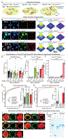
Fig. 1.. A minimal teneurin-3 (Tenm3) protein fully mediates synapse formation as long as latrophilin-binding is maintained
(A) Domain structures of wild-type and mutant Tenm3 proteins. (B and C) STED super-resolution imaging of the synaptic localization of wild-type and mutant Tenm3 proteins expressed in cultured cortical neurons lacking Tenm3 and Tenm4 (B, representative images of dendrites stained for surface HA-tagged Tenm3 and the pre- and postsynaptic markers vGluT1 and Homer1; C, summary graphs of the synaptic Tenm3 levels, with synapses defined as puncta with coincident vGluT1- and Homer1-staining). (D and E) STED super-resolution imaging of rescue experiments of the synapse loss in Tenm3/4 double-deficient neurons. Cortical cultures from Tenm3/4 double cKO mice were infected with lentiviruses expressing the indicated Tenm3 proteins (ΔCre = mutant Cre) and analyzed after 14 (DIV14) or 21 days in culture (DIV21) (D, representative images; E, summary graphs of the synapse density and size at DIV14 and DIV21). For additional data including spine analyses, see figs. S1 to S9). (F to H) mEPSC measurements of the synapse loss rescue by minimal Tenm3 proteins in Tenm3/4 double-deficient cultured cortical neurons. mEPSCs were recorded at DIV14-16 in the presence of tetrodotoxin (1 μM) and picrotoxin (50 μM) (F, representative traces; G, cumulative plots of the interevent intervals and summary graphs of the mEPSC frequency; H, cumulative plots and summary graphs of the mEPSC amplitude). (I to K) In vivo trans-synaptic tracing experiments using pseudo-typed rabies viruses documenting that minimal Tenm3 proteins rescue the loss of entorhinal cortex→CA1 region synapses induced by the Tenm3/4 double deletion (I, representative images of postsynaptic starter neurons in the CA1 region (top) and of presynaptic ipsilateral entorhinal cortex input neurons (bottom); J and K, quantification of synaptic input neurons in the ipsilateral entorhinal cortex (J), ipsilateral CA3 region (K, left; negative control), and contralateral CA3 region (K, right; additional negative control)). Numerical data are means ± SEM (numbers of cells, experiments, and mice are indicated in bars). ***P < 0.001, **P < 0.01, *P < 0.05 [One-way ANOVA with post hoc Tukey/Dunnett tests; G and H, Kolmogorov–Smirnov t test for cumulative distributions]; a.u., arbitrary units. For additional data, see figs. S1 to S10.
Fig. 2.
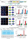
Fig. 2.. The intracellular domain (ICD) of Tenm3 incorporates into active zone liquid-liquid phase-separated (LLPS) condensates and recruits the Tenm3 extracellular domains (ECDs) to the surface shell of active zone LLPS condensates
(A) Experimental design of LLPS experiments. RIM1 and RIM-BP2 forming presynaptic active zone LLPS condensate via phase separation (40) were analyzed with addition of the Tenm3 ECD (Tenm3ECD), ICD (Tenm3ICD), or full-length Tenm3 (Tenm3ECD-ICD; fig. S12). (B) Representative images illustrating the recruitment of Tenm3ICD and Tenm3ECD-ICD but not of Tenm3ECD (all 1 μM) to RIM1 (10 μM) and RIM-BP2 (10 μM) active zone LLPS condensate (RIM-BP2, Tenm3ECD, and Tenm3ICD were labeled by iFluor-405, iFluor-546 and iFluor-488, respectively, while RIM1 was unlabeled). (C) Representative heatmaps of the enrichment of RIM-BP2, Tenm3ICD, and Tenm3ECD signals in active zone LLPS condensates (examples indicated by dashed circles in B). (D to F) Quantification of RIM-BP2, Tenm3ICD, and Tenm3ECD signals across phase-separated active zone LLPS condensates illustrating that addition of Tenm3 has no effect on the distribution of RIM-BP2 in the condensates (D), that Tenm3ICD localizes to the condensate core in the absence of the ECDs but to the condensate shell when coupled to ECDs in Tenm3ECD-ICD proteins (E), and that the Tenm3 ECDs are only present on the condensate surface when coupled to the Tenm3 ICD in Tenm3ECD-ICD protein (F). (G) Quantification of the size of LLPS condensates formed by active zone proteins RIM1 and RIM-BP2 in the presence of Tenm3ECD, Tenm3ICD, and Tenm3ICD-ECD. (H) Quantification of Tenm3ECD and Tenm3ICD-ECD shell cluster sizes on the surface of RIM1/RIM-BP2 active zone LLPS condensates. (I) Imaging of transfected HEK293T cells co-expressing Flag-tagged RIM1 or RIM-BP2 constructs with HA-tagged Tenm3WT or Tenm3ΔICD (red, Flag-epitope; green, HA-epitope). (J) Co-immunoprecipitation experiments of purified Tenm3ICD, RIM1, and RIM-BP2 confirm that the Tenm3ICD directly binds to RIM1 but not RIM-BP (Coomassie-stained SDS-gel (input (In), 5% of total; asterisk, co-immunoprecipitated RIM1). Numerical data are means ± SEM (numbers of condensates and experiments are indicated in bars). ***P < 0.001, **P < 0.01, *P < 0.05 [D, E, and F: Two-way ANOVA with post hoc Tukey tests; G and H left: Kolmogorov–Smirnov t test; H right: One-way ANOVA with post hoc Tukey tests]; a.u., arbitrary units.
Fig. 3.
Fig. 3.. Recruitment of the Tenm3 ICD into active zone LLPS condensates is sequence-specific
(A) Design of mutations in the Tenm3 ICD (see figs. S11 and S16 for sequence information). (B and C) Representative images (left) and heatmaps (right, corresponding to the circled LLPS active zone condensates on the left) demonstrating the effect of the ΔRKΦ and ΔPRM mutations on the recruitment of the Tenm3ECD-ICD to the shell of active zone LLPS condensates. Condensates composed of RIM1 and RIM-BP2 (both 20 μM, RIM-BP2 is labeled with iFluor-405) were incubated without or with wild-type or mutant Tenm3ICD-ECD (1 μM; ECD is labeled with iFluor-546). (D and E) Quantification of Tenm3ICD-ECD recruitment to the shell of presynaptic active zone condensates as a function of ICD mutations (left, absolute signal intensity; right, ratio of shell to core signal with ‘control’ signals constituting background). (F and G) Representative images (left) and heatmaps (right, corresponding to circled active zone LLPS condensates on the left) demonstrating that the effect of the small ΔRKΦ3 deletion on the Tenm3ECD-ICD protein recruitment to active zone LLPS condensates. Conditions were the same as in B and C. (H and I) Quantification of Tenm3ICD-ECD recruitment to the shell of presynaptic active zone condensates as a function of the RKΦ3 mutation (left, absolute signal intensity; right, ratio of shell to core signal with ‘control’ signals constituting background). (J) Sedimentation assay of LLPS active zone condensates documenting that the RKΦ3 mutation blocks Tenm3 recruitment to condensates (top, Coomassie-stained gel; bottom, immunoblot [yellow arrowheads, RIM1 and RIM-BP2; red arrowheads, Tenm3ICD; asterisks, Tenm3ICD-ECD]). Active zone LLPS condensates composed of RIM1 and RIM-BP2 (both 20 μM) were incubated without or with wild-type or mutant Tenm3ICD or Tenm3ICD-ECD (1 μM) and pelleted by centrifugation (P, pellets; S, supernatants). (K) Quantification of the recruitment of Tenm3ICD and Tenm3ICD-ECD to active zone LLPS condensates as measured by the sedimentation assay. Summary graph depicts the percentage of Tenm3 in the pellet as measured by quantitative immunoblotting. Numerical data are means ± SEM (numbers of condensates and experiments are indicated in bars). ***P < 0.001, **P < 0.01, *P < 0.05 [D, H, and K: Two-way ANOVA with post hoc Tukey tests; E and I: One-way ANOVA with post hoc Tukey tests]; a.u., arbitrary units.
Fig. 4.
Fig. 4.. Tenm3 ICD mutations that disrupt recruitment of the ICD to active zone LLPS condensates abolish Tenm3 function in synapse formation.
(A to C) STED super-resolution imaging demonstrating that mutations of Tenm3 that delete its intracellular domain (Tenm3ΔICD), its RKΦ regions (Tenm3ΔRKΦ), or its RKΦ3 sequence (Tenm3ΔRKΦ3) abolish the ability of Tenm3 to rescue decreased excitatory synapse numbers and sizes in Tenm3/4-deficient neurons (A, representative images; B and C, summary graphs of the synapse density and size, respectively). (D to F) The same mutations as analyzed in A-C also abolish the ability of Tenm3 to rescue the decreased mEPSC frequency of Tenm3/4-deficient cultured neurons at DIV14-16 (D, representative traces; E, summary graphs of the mEPSC frequency and cumulative plots of the interevent intervals; F, summary graphs and cumulative plots of the mEPSC amplitude). Numerical data are means ± SEM (numbers of cells and experiments are indicated in bars). ***P < 0.001, **P < 0.01, *P < 0.05 [One-way ANOVA with post hoc Tukey/Dunnett tests; E and F, Kolmogorov–Smirnov t test for cumulative distributions].
Fig. 5.
Fig. 5.. Reconstitution of synaptic junctions from purified proteins enabled by the Lphn3-Tenm3 complex that selectively recruits presynaptic active zone and postsynaptic LLPS condensates
(A) Representative images of phase-separated active zone and postsynaptic density LLPS condensates with addition of full-length Tenm3ICD-ECD and/or Lphn3 (both at 0.5 μM). Active zone condensates were formed with RIM1 and RIM-BP2 (both 20 μM) and PSD condensates with PSD-95, Homer3, truncated GKAP with a DLS sequence at the GK binding region (a phospho-mimicking mutation to enhance PSD-95 binding (38), SynGAP, and Shank3 (all 2.5 μM) (32, 42). (B) High-magnification images of representative reconstituted synaptic junctions that are formed by phase-separated active zone and postsynaptic density LLPS condensates containing Tenm3ICD-ECD (red) and Lphn3 (green), respectively (left, STED images; right, 3D-rendering heatmaps of Tenm3ICD-ECD and Lphn3 levels). (C) Quantification of the co-localization of Tenm3ICD-ECD and Lphn3 with active zone or postsynaptic LLPS condensates documenting that Tenm3ECD-ICD and Lphn3 are selectively recruited to active zone or postsynaptic density condensates, respectively, even when these condensates are mixed. (D) Cumulative frequency plots of the sizes of the Tenm3ICD-ECD and Lphn3 puncta on the LLPS condensate surfaces (inset, summary graph of mean sizes). (E) Quantifications of active zone and postsynaptic LLPS condensate contacts. Summary graphs show the percentages of active zone condensates contacting PSD condensates (left), of PSD condensates contacting active zone condensates (middle), and of total condensates forming contacts (right). (F) Sedimentation assays confirming reconstitution of synaptic junctions. Tenm3ICD-ECD and Lphn3 were incubated in the absence and presence of mixtures of pre- and postsynaptic LLPS condensates that were formed as in panel A and centrifuged. The supernatants and pellets were analyzed by SDS-PAGE and Coomassie staining (left) or immunoblotting (right). (G) Quantification of the pellet enrichment of Tenm3ICD-ECD and Lphn3 in the absence and presence of pre- and postsynaptic LLPS condensates. (H) Quantification of the fold enrichment of Tenm3ICD-ECD and Lphn3 in the pellet in the absence and presence of pre- and postsynaptic scaffold protein LLPS condensates. Numerical data are means ± SEM (numbers of condensates and experiments are indicated in bars). ***P < 0.001, **P < 0.01, *P < 0.05 [C, G, H, and D inset: two-tailed t test; D: Kolmogorov–Smirnov t test; E: One-way ANOVA with post hoc Tukey tests]. a.u., arbitrary units.

Similar articles

Cited by

References

    1. Oohashi T et al., Mouse ten-m/Odz is a new family of dimeric type II transmembrane proteins expressed in many tissues. J Cell Biol 145, 563–577 (1999). - PMC - PubMed
    1. Tucker RP, Teneurins: Domain Architecture, Evolutionary Origins, and Patterns of Expression. Front Neurosci 12, 938 (2018). - PMC - PubMed
    1. Leamey CA, Sawatari A, Teneurins: Mediators of Complex Neural Circuit Assembly in Mammals. Front Neurosci 13, 580 (2019). - PMC - PubMed
    1. Jackson VA, Busby JN, Janssen BJC, Lott JS, Seiradake E, Teneurin Structures Are Composed of Ancient Bacterial Protein Domains. Front Neurosci 13, 183 (2019). - PMC - PubMed
    1. Arac D, Li J, Teneurin Structure: Splice Variants of a Bacterial Toxin Homolog Specifies Synaptic Connections. Front Neurosci 13, 838 (2019). - PMC - PubMed

Publication types

LinkOut - more resources