. 2025 Mar;56(3):692-704.
doi: 10.1161/STROKEAHA.124.049079. Epub 2025 Jan 17.

Proteins Involved in Endothelial Function and Inflammation Are Implicated in Cerebral Small Vessel Disease

Affiliations

Proteins Involved in Endothelial Function and Inflammation Are Implicated in Cerebral Small Vessel Disease

Zihan Sun et al. Stroke. 2025 Mar.

Abstract

Background: Endothelial dysfunction and inflammation have been implicated in the pathophysiology of cerebral small vessel disease (SVD). However, whether they are causal, and if so which components of the pathways represent potential treatment targets, remains uncertain.

Methods: Two-sample Mendelian randomization (MR) was used to test the association between the circulating abundance of 996 proteins involved in endothelial dysfunction and inflammation and SVD. The genetic instruments predicting protein levels were obtained from the Iceland 36K (n=35 892) and the UK Biobank Proteomics (n=34 557) cohorts, both of which were longitudinal studies with follow-up from 2000 to 2023 and 2006 to 2023, respectively. SVD was represented by lacunar stroke (n=6030 cases) and 5 neuroimaging features (white matter hyperintensities [n=55 291], diffusion tensor imaging metrics: mean diffusivity [n=36 460] and fractional anisotropy [n=36 533], extensive white matter perivascular space burden [n=9324 cases], and cerebral microbleeds [n=3556 cases]). Among the proteins supported by causal evidence from the MR, cross-sectional analysis was performed to assess their associations with cognitive performance; survival analysis with Fine-Gray models was applied to examine their associations with incident all-cause dementia and stroke within the UK Biobank Proteomics cohort.

Results: MR suggested COL2A1 (collagen type II α-1 chain) was associated with lacunar stroke (odds ratio, 0.89 [95% CI, 0.86-0.91]; P=5×10⁻⁵). Moreover, 12 proteins related to endothelial function and inflammation were associated with neuroimaging features of SVD. Cross-sectional analyses showed 5 of the 13 proteins (EPHA2 [ephrin type-A receptor 2], METAP1D [methionine aminopeptidase 1D, mitochondrial], FLT4 [vascular endothelial growth factor receptor 3], COL2A1, and TIMD4 [T-cell immunoglobulin and mucin domain-containing protein 4]) were associated with cognitive performance with effects concordant with their MR findings. Survival analyses with the Fine-Gray models indicated that 5 of the 13 proteins (EPHA2, METAP1D, FLT4, APOE [apolipoprotein E], and PDE5A [cGMP-specific 3',5'-cyclic phosphodiesterase]) were associated with the risk of all-cause dementia or stroke independent of age and sex, consistent with their MR evidence.

Conclusions: Our findings suggest that endothelial-platelet activation and complement-mediated regulation of inflammation play roles in SVD and identify potential therapeutic targets and pathways.

Keywords: Mendelian randomization analysis; cerebral small vessel diseases; dementia; inflammation; stroke.


Conflict of interest statement

None.

Figures

Figure 1.
Figure 1.
Study workflow. CMB indicates cerebral microbleeds; EPVS, enlarged perivascular space; FA, fractional anisotropy; MD, mean diffusivity; MR, Mendelian randomization; pQTL, protein quantitative trait loci; SVD, small vessel disease; and WMH, white matter hyperintensity.
Figure 2.
Figure 2.
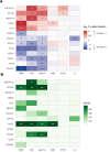
Heatmaps showing the proteins that were associated with ≥1 small vessel disease (SVD)–related outcomes. A, Mendelian randomization results from the primary analysis. Red color indicates an increased risk while blue color suggests a decreased risk per 1-unit increase in the normalized expression level of the circulating protein. The color shade corresponds to the strength of the P values, with darker color indicating stronger evidence for a causal association. False discovery rate (FDR) was calculated via the Benjamin-Hochberg method to account for multiple testing across the 6 outcomes. A total of 17 protein-outcome pairs, covering 13 unique proteins, demonstrated significant associations below an FDR of 5%. **FDR-corrected P<0.05,* unadjusted P<0.05. All genetic instruments were identified in cis association with the protein below a genome-wide P threshold of 5×10⁻⁸. B, Pairwise colocalization results. Color shade indicates the posterior probability (PP) of both traits sharing a single causal variant (ie, PP.H4). **PP.H4>0.8; *PP.H4>PP of any other hypothesis. CD46, membrane cofactor protein; CMB, cerebral microbleeds; COL2A1, collagen type II α-1 chain; EPHA2, ephrin type-A receptor 2; EPVS, enlarged perivascular space; FA, fractional anisotropy; FLT4, vascular endothelial growth factor receptor 3; HEXIM1, hexamethylene bis-acetamide-inducible protein 1; LS, lacunar stroke; MD, mean diffusivity; MEGF10, multiple epidermal growth factor-like domains protein 10; MERTK, tyrosine-protein kinase Mer; METAP1D, methionine aminopeptidase 1D, mitochondrial; NPTX1, neuronal pentraxin-1; PDE5A, cGMP-specific 3',5'-cyclic phosphodiesterase; PEAR1, platelet endothelial aggregation receptor 1; TIMD4, T-cell immunoglobulin and mucin domain–containing protein 4; and WMH, white matter hyperintensity.
Figure 3.
Figure 3.
Plots showing estimates for the association between each candidate protein and cognitive functions as measured by reaction time and pairs matching tests at the UK Biobank baseline. Multivariable linear regression and negative binomial regression were used to estimate the percent difference in reaction time and matching errors per 1-unit increase in the inverse-normal transformed normalized protein expression value of each protein, respectively. For both cognitive outcomes, the models were adjusted for sex and age at baseline. A, The % change in reaction time. B, The % change in matching errors. *False discovery rate–corrected P<0.05. CD46 indicates membrane cofactor protein; COL2A1, collagen type II α-1 chain; EPHA2, ephrin type-A receptor 2; FLT4, vascular endothelial growth factor receptor 3; HEXIM1, hexamethylene bis-acetamide-inducible protein 1; MEGF10, multiple epidermal growth factor-like domains protein 10; MERTK, tyrosine-protein kinase Mer; METAP1D, methionine aminopeptidase 1D, mitochondrial; NPTX1, neuronal pentraxin-1; PDE5A, cGMP-specific 3',5'-cyclic phosphodiesterase; PEAR1, platelet endothelial aggregation receptor 1; and TIMD4, T-cell immunoglobulin and mucin domain–containing protein 4.
Figure 4.
Figure 4.
Plots showing estimates for the association between candidate proteins and all-cause dementia and all-cause stroke. Fine-Gray model estimated the hazard ratios (HR) of incident dementia or stroke per 1-unit increase in the inverse-normal transformed normalized protein expression value of each protein after competing risk was accounted for. For dementia, the models were adjusted for age, sex, education in years, and APOE ε4 carrier status. For stroke, the model was adjusted for age and sex. A, All-cause dementia as the outcome. B, All-cause stroke as the outcome. *False discovery rate–corrected P<0.05. CD46 indicates membrane cofactor protein; COL2A1, collagen type II α-1 chain; EPHA2, ephrin type-A receptor 2; FLT4, vascular endothelial growth factor receptor 3; HEXIM1, hexamethylene bis-acetamide-inducible protein 1; MEGF10, multiple epidermal growth factor-like domains protein 10; MERTK, tyrosine-protein kinase Mer; METAP1D, methionine aminopeptidase 1D, mitochondrial; MR, Mendelian randomization; NPTX1, neuronal pentraxin-1; PDE5A, cGMP-specific 3',5'-cyclic phosphodiesterase; PEAR1, platelet endothelial aggregation receptor 1; and TIMD4, T-cell immunoglobulin and mucin domain–containing protein 4.
Figure 5.
Figure 5.
A summary of evidence among the 13 protein candidates. A, Analysis results across multiple methods. *The results were referenced from prior studies. B, Functional categorization of the 13 protein candidates. CD46, membrane cofactor protein; COL2A1, collagen type II α-1 chain; EPHA2, ephrin type-A receptor 2; eQTL, expression quantitative trait loci; FLT4, vascular endothelial growth factor receptor 3; HEXIM1, hexamethylene bis-acetamide-inducible protein 1; MEGF10, multiple epidermal growth factor-like domains protein 10; MERTK, tyrosine-protein kinase Mer; METAP1D, methionine aminopeptidase 1D, mitochondrial; NPTX1, neuronal pentraxin-1; PBMC, peripheral blood mononuclear cell; PDE5A, cGMP-specific 3',5'-cyclic phosphodiesterase; PEAR1, platelet endothelial aggregation receptor 1; pQTL, protein quantitative trait loci; TIMD4, T-cell immunoglobulin and mucin domain–containing protein 4.

References

    1. Markus HS, de Leeuw FE. Cerebral small vessel disease: recent advances and future directions. Int J Stroke. 2023;18:4–14. doi: 10.1177/17474930221144911 - PMC - PubMed
    1. Duering M, Biessels GJ, Brodtmann A, Chen C, Cordonnier C, de Leeuw FE, Debette S, Frayne R, Jouvent E, Rost NS, et al. . Neuroimaging standards for research into small vessel disease—advances since 2013. Lancet Neurol. 2023;22:602–618. doi: 10.1016/S1474-4422(23)00131-X - PubMed
    1. Debette S, Schilling S, Duperron MG, Larsson SC, Markus HS. Clinical significance of magnetic resonance imaging markers of vascular brain injury: a systematic review and meta-analysis. JAMA Neurol. 2019;76:81–94. doi: 10.1001/jamaneurol.2018.3122 - PMC - PubMed
    1. Rajani RM, Quick S, Ruigrok SR, Graham D, Harris SE, Verhaaren BFJ, Fornage M, Seshadri S, Atanur SS, Dominiczak AF, et al. . Reversal of endothelial dysfunction reduces white matter vulnerability in cerebral small vessel disease in rats. Sci Transl Med. 2018;10:eaam9507. doi: 10.1126/scitranslmed.aam9507 - PubMed
    1. Low A, Mak E, Rowe JB, Markus HS, O’Brien JT. Inflammation and cerebral small vessel disease: a systematic review. Ageing Res Rev. 2019;53:100916. doi: 10.1016/j.arr.2019.100916 - PubMed