

Multidisciplinary, multicenter consensus for the care of patients affected with Sturge-Weber syndrome
- PMID: 39819452
- PMCID: PMC11740666
- DOI: 10.1186/s13023-024-03527-w
Multidisciplinary, multicenter consensus for the care of patients affected with Sturge-Weber syndrome
Abstract
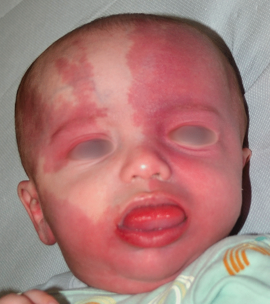
Background: Sturge-Weber Syndrome (SWS) is a rare, sporadic neurocutaneous disorder affecting the skin, brain, and eyes, due to somatic activating mutations in GNAQ or, less commonly, GNA11 gene. It is characterized by at least two of the following features: a facial capillary malformation, leptomeningeal vascular malformation, and ocular involvement. The spectrum of clinical manifestations includes headache, seizures, stroke-like events, intellectual disability, glaucoma, facial asymmetry, gingival hyperplasia, etc. An early diagnosis is crucial to guarantee an appropriate care, which is best performed in reference centres by multidisciplinary teams. The aim of this study was to develop a multidisciplinary expert consensus for diagnosis, treatment, and follow-up of all disease manifestations, according to the recommendations of the Italian Law on Rare Disease Care.
Results: Through a Delphi consensus methodology, 28 recommendations have been developed concerning (i) dermatological SWS manifestations and related treatment timing and modalities, (ii) neurological referral, diagnosis, pharmacological treatment of neurological signs and symptoms, neurosurgical indications, neurocognitive evaluation and related treatment, psychosocial support and patient follow-up, (iii) diagnosis of ophthalmological manifestations, medical and surgical treatment, and follow-up, (iv) maxillofacial surgical treatment, (v) oral cavity assessment, care and follow-up, and (vi) primary care paediatrician/general practitioner involvement.
Conclusions: The present consensus developed by a multidisciplinary group of experts from Italian reference centres comprises practical recommendations for SWS global management, including currently controversial issues. Specific statements for all disease aspects, from skin manifestations and neurological and ocular signs and symptoms to oral and maxillofacial care, are provided. They can be exploited to uniform clinical practice in reference centres, but also in other hospitals and outpatient settings. Though this consensus has been developed taking primarily into account the Italian National Health System organization and rules on rare disorders, it could be translated also to other countries.
Keywords: Anaesthesia; Capillary malformation; Epilepsy; Glaucoma; Intellectual disability; Medical and surgical treatment; Neuroimaging; Psychosocial care; Pulsed dye laser; Sturge–Weber syndrome.
© 2025. The Author(s).
Conflict of interest statement
Declarations. Ethics approval and consent to participate: This study did not need the Ethical Committee approval according to Italian law, since no human participants, human material, or human data were involved. We obtained informed consent from patients for publication of the pictures. Consent for publication: Consent for publication of the clinical images was obtained from the patients or their parents in case of a minor. Competing interests: The Authors have no competing interests to declare.
Figures


References
-
- Poliner A, Fernandez Faith E, Blieden L, Kelly KM, Metry D. Port-wine birthmarks: update on diagnosis, risk assessment for Sturge–Weber syndrome, and management. Pediatr Rev. 2022;43:507–16. - PubMed
-
- Geronemus RG, Ashinoff R. The medical necessity of evaluation and treatment of port-wine stains. J Dermatol Surg Oncol. 1991;17:76–9. - PubMed
Publication types
MeSH terms
LinkOut - more resources
Full Text Sources
