. 2025 Jan 16;29(1):29.
doi: 10.1186/s13054-025-05257-2.

Inhibition of Interleukin-40 prevents multi-organ damage during sepsis by blocking NETosis

Affiliations

Inhibition of Interleukin-40 prevents multi-organ damage during sepsis by blocking NETosis

Shijie Cai et al. Crit Care. .

Abstract

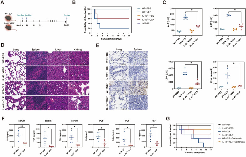
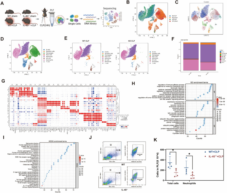
Despite intensive clinical and scientific efforts, the mortality rate of sepsis remains high due to the lack of precise biomarkers for patient stratification and therapeutic guidance. Interleukin 40 (IL-40), a novel cytokine with immune regulatory functions in human diseases, was elevated at admission in two independent cohorts of patients with sepsis. High levels of secreted IL-40 in septic patients were positively correlated with PCT, CRP, lactate (LDH), and Sequential Organ Failure Assessment (SOFA) scores, in which IL-40 levels were used to stratify the early death of critically ill patients with sepsis. Moreover, genetic knockout of IL-40 (IL-40-/-) improved outcomes in mice with experimental sepsis, as evidenced by attenuated cytokine storm, multiple-organ failure, and early mortality, compared with those of wild-type (WT) mice. Mechanistically, single-cell RNA sequencing (scRNA-seq) and bulk RNA sequencing (RNA-seq) have revealed that S100A8/9hi neutrophil influx into the peritoneal cavity along with neutrophil extracellular trap (NETs) formation accounts predominantly for the IL-40-mediated worsening of sepsis outcomes. Clinically, the IL-40 level was positively correlated with the NET-related MPO/dsDNA ratio in septic patients. Finally, with antibiotics (gentamycin), genetic knockout of IL-40 prevented polymicrobial sepsis fatalities more efficiently than without gentamycin treatment. In summary, these data reveal a novel prognostic strategy for sepsis and that IL-40 may serve as a novel therapeutic target for sepsis.

Keywords: IL-40; NETs; Prognostic; Sepsis; Theranostic.


Conflict of interest statement

Declarations. Ethics approval and consent to participate: The human study was approved by the affiliated Zhongda Hospital of Southeast University Clinical Research Ethics Committee (Registration No. 2023ZDSYLL463-P01), and written informed consent was obtained from patients or their legally authorized representatives before enrollment, according to the Declaration of Helsinki. All animal experiments complied with the guidelines of the Animal Experimentation Ethics Committee (AEEC) Guide for the Care and Use of Laboratory Animals and were approved by the AEEC, Southeast University. Competing interests: The authors declare no competing interests.

Figures

Fig. 1
Fig. 1
Elevated IL-40 levels correlate with multiple organ damage and mortality in critically ill patients with sepsis. A Plasma levels of IL-40 in healthy controls (n = 50), patient control (n = 50) as well as patients with sepsis (n = 116). B Dynamic changes in plasma IL-40 levels in adult sepsis patients at days 0, 3, 7, or 10. C Plasma levels of IL-40 in septic shock patients (n = 63) and non-shock patients (n = 53). D Plasma levels of IL-40 in survivors (n = 85) and non-survivors (n = 31). E Correlations between IL-40 levels and PCT (n = 111), CRP (n = 102) and LDH (n = 98) levels as well as SOFA score (n = 80) in adult septic patients. F Plasma levels of IL-40 in healthy controls (n = 90), patient controls (n = 50) and pediatric patients with sepsis (n = 106). G Dynamic changes in plasma IL-40 levels at admission and 3, 7, and 10 days after admission in pediatric patients with sepsis, when appropriate. H Plasma levels of IL-40 in septic shock pediatric patients (n = 18) and non-shock pediatric septic patients (n = 88). I Plasma levels of IL-40 in survivors (n = 98) and non-survivors of pediatric septic patients (n = 8). J Correlations between IL-40 levels and PCT levels (n = 100), CRP levels (n = 101), LDH levels (n = 105) and the SOFA score (n = 99) in pediatric patients with sepsis. The data are expressed as the means with 95% confidence intervals (means ± SEMs). Statistical analysis was performed with the Mann–Whitney U test (A, C-D, F, H-I), nonparametric Kruskal–Wallis test (B, G) and Spearman’s correlation coefficient test (E, J). *P < 0.05, **P < 0.01, ***P < 0.001 and ****P < 0.0001
Fig. 2
Fig. 2
IL-40 promotes hyperinflammation and organ dysfunction in polymicrobial sepsis. A Experimental design for establishing a polymicrobial sepsis model via CLP. B Circulating IL-40 concentrations in the serum, PLF, liver, and kidney at 6 h and 24 h after CLP, as measured via ELISA (n = 4–5). C Study design for establishing a polymicrobial sepsis model in WT mice treated with rmIL-40. rmIL-40 (800 ng/100 μL) or vehicle control was intraperitoneally (i.p.) administered to C57BL/6 mice at the time of CLP and were sacrificed 24 h after CLP for analysis, or rmIL-40 (800 ng/100 μL) was injected i.p. into WT mice immediately, 24 h and 48 h after CLP, and survival was monitored for 14 consecutive days. D Monitoring of the 14-day survival of septic mice treated with sterile PBS (100 μL) or rmIL-40 (800 ng/100 μL) (n = 5). E Representative images of hematoxylin and eosin (H&E)-stained lung, liver, spleen, and kidney tissues from vehicle- and rmIL-40-treated septic mice at 24 h after CLP (n = 5). Scale bars, 50 μm at X400 magnification. F Serological parameters of organ damage, including ALT, AST, LDH, and urea, in vehicle- and rmIL-40-treated septic mice at 24 h after CLP (n = 3–6). G Bar chart showing the total number of inflammatory cells in the PLF and corresponding CD11b+Ly6G+ neutrophils determined by flow cytometry in vehicle- and rmIL-40-treated septic mice 24 h after CLP (n = 4). H Concentrations of the proinflammatory mediators IL-6, TNF-α, and CCL2 in the blood and PLF of vehicle- and rmIL-40-treated septic mice 24 h after CLP, as detected via the Milliplex system (n = 5). I Representative images of apoptotic lung and spleen cells measured by TUNEL in vehicle- and rmIL-40-treated septic mice at 24 h after CLP (n = 5). The red arrow indicates apoptotic cells. The values are expressed as the means ± SEMs. Statistical analysis was performed with the Mann–Whitney U test (B, FH) and log-rank test (D). *P < 0.05 and **P < 0.01
Fig. 3
Fig. 3
Deficiency in IL-40 prevents the progression and outcome of polymicrobial sepsis. A Experimental design for establishing a polymicrobial sepsis model via CLP in WT and IL-40−/− mice. The mice were sacrificed 24 h after CLP for analysis or monitored for survival for 14 days. B 14-day survival of the WT and IL-40−/− septic mice (n = 5). C Serological markers of organ damage, including ALT, AST, LDH, and urea, were measured in WT and IL-40−/− septic mice 24 h after CLP (n = 4). D Representative examples of H&E-stained lung, liver, spleen, and kidney sections from WT and IL-40−/− septic mice 24 h after CLP (n = 5). Scale bars, 100 μm. E Representative examples of TUNEL-stained lungs and spleens from WT and IL-40−/− septic mice 24 h after CLP showing apoptotic cells (n = 5). Scale bars, 50 μm. F Multiplex detection of IL-6, TNF-α, and CCL2 in the blood and PLF of WT and IL-40−/− septic mice 24 h after CLP (n = 5). G Gentamicin (3 mg/kg) was injected i.p. two hours before CLP and at 2, 24, 48, and 72 h after CLP, and survival was monitored for 14 consecutive days (n = 5). The data are expressed as the means ± SEMs and represent three independent tests. Statistical analysis was performed with the Mann–Whitney U test (C, F) and log-rank test (B, G). *P < 0.05 and **P < 0.01
Fig. 4
Fig. 4
IL-40 promotes sepsis by promoting S1100A8/9hi neutrophil recruitment and activation. A Schematic workflow of scRNA-seq of PLFs from IL-40−/− and WT mice 24 h after CLP. B Uniform manifold approximation and projection (UMAP) representation of 42,896 single peritoneal cells from WT sham, WT + CLP, IL-40−/− sham, and IL-40−/− + CLP mice colored according to inferred cluster identity and sample origin. C The clusters were annotated into cell populations based on the marker genes and were distributed in UMAP colored by cell type. D UMAP plot showing the annotated cell types from the clusters in C based on the marker genes and colored by cell type. E UMAP plot showing the distributions of annotated cell types in WT and IL-40−/− septic mice. F Proportions of annotated cell populations in WT and IL-40−/− septic mice. G Dot plot showing the top three marker genes differentially expressed per cluster. The dot size represents the percentage of cells in each cluster with more than one read of the corresponding gene. H and I The top 10 enriched GO terms are categorized by biological process, molecular function, and cellular component, and the top 30 enriched KEGG terms are between those of WT and IL-40−/− neutrophils. KO: genetic knockout of IL-40 (IL-40−/−). J and K Gating strategies for counting peritoneal neutrophils via flow cytometry according to CD11b and Ly6G expression. Neutrophils were identified as CD11b+Ly6G+ populations. Bar chart showing the total number of inflammatory cells and CD11b+Ly6G+ neutrophils in the PLF (n = 4). The data are expressed as the means ± SEMs and represent three independent tests. Statistical analysis was performed with the Mann–Whitney U test (K). *P < 0.05
Fig. 5
Fig. 5
IL-40 deficiency prevents NETosis during sepsis both in vivo and in vitro. A UMAP plot showing the 11 clusters from WT and IL-40−/− neutrophils classified based on marker genes. B The clusters in (A) were annotated into G1-G5a cell populations and were distributed in UMAP and colored by cell type. C Dot plot showing the scaled expression of three selected signature genes for each cluster, colored according to the average gene expression in each cluster scaled across all clusters. The dot size represents the percentage of cells in each cluster with more than one read of the corresponding gene. D The proportions of G1-G5a neutrophil populations in WT and IL-40−/− septic mice. E The top 24 enriched GO terms were categorized by biological process, molecular function, and cellular component between WT and IL-40−/− G1-G5a neutrophils. F Representative UMAP plot of proinflammatory CXCL2, CXCL1, CCL6, TNF-α, S100A8 and S100A9 in WT and IL-40−/− G1-G5a neutrophils. G Relative expression of NETs associated markers and pathways in WT and IL-40−/− G1-G5a neutrophils. H and I Bown marrow-derived neutrophils were isolated from WT and IL-40−/− mice and stimulated with LPS to induce NETs; the cells were collected for electron microscopy visualization of NETs, while the supernatants were collected for MPO and H3Cit detection via ELISA (n = 3). J Experimental scheme for bulk RNA-seq of bone marrow-derived neutrophils from septic WT and IL-40−/− mice stimulated with LPS. K GO enriched terms categorized by biological process, molecular functions, and cellular components and KEGG enriched terms between WT and IL-40−/− neutrophils based on the DEG. L Representative images of IF staining for the NET-related MPO and H3Cit in peritoneal cells from WT and IL-40−/− septic mice in vivo. The nuclei were stained with DAPI (blue). MPO: green; H3Cit: red. M WT and IL-40.−/− mice were sacrificed 24 h after CLP, and serum and PLF were collected for MPO and H3Cit detection via ELISA (n = 4–6). The data are representative of three individual experiments. The values are expressed as the means ± SEMs. Statistical analysis was performed with the Mann–Whitney U test (I, M). *P < 0.05, and **P < 0.01
Fig. 6
Fig. 6
IL-40 was associated with NETs release in septic patients. A Adult septic patients (n = 18) and healthy controls (n = 20) were enrolled, and serum was collected for NETs-related dsDNA and MPO detection. B and C Correlations between circulating IL-40 levels and dsDNA and MPO in septic patients (n = 18) and healthy controls (n = 20). D Representative images showing the colocalization of IL-40 (orange), MPO (green), and H3Cit (red) in neutrophils from septic patients and healthy controls; the nuclei were stained with DAPI (blue). E Neutrophils from septic patients and healthy controls were isolated and induced with LPS for 4 h, with or without rhIL-40 treatment (500 ng/mL). The cells were collected for MPO (green) and H3Cit (red) staining; nuclei were stained with DAPI (blue). The data are representative of at least two individual experiments. The values are expressed as the means ± SEMs and were analyzed with the Mann–Whitney U test (A) and Spearman’s correlation coefficient test (B and C). ***P < 0.001

References

    1. Singer M, Deutschman CS, Seymour CW, Shankar-Hari M, Annane D, Bauer M, et al. The third international consensus definitions for sepsis and septic shock (Sepsis-3). JAMA. 2016;8:801–10. - PMC - PubMed
    1. van der Poll T, Shankar-Hari M, Wiersinga WJ. The immunology of sepsis. Immunity. 2021;11:2450–64. - PubMed
    1. Evans L, Rhodes A, Alhazzani W, Antonelli M, Coopersmith CM, French C, et al. Surviving sepsis campaign: international guidelines for management of sepsis and septic shock 2021. Intensive Care Med. 2021;11:1181–247. - PMC - PubMed
    1. Rudd KE, Johnson SC, Agesa KM, Shackelford KA, Tsoi D, Kievlan DR, et al. Global, regional, and national sepsis incidence and mortality, 1990–2017: analysis for the global burden of disease study. Lancet. 2020;10219:200–11. - PMC - PubMed
    1. Liu D, Huang SY, Sun JH, Zhang HC, Cai QL, Gao C, et al. Sepsis-induced immunosuppression: mechanisms, diagnosis and current treatment options. Mil Med Res. 2022;1:56. - PMC - PubMed

MeSH terms