. 2025 Jan 16;135(5):e186769.
doi: 10.1172/JCI186769.

Passive transfer of patient-derived anti-nephrin autoantibodies causes a podocytopathy with minimal change lesions

Affiliations

Passive transfer of patient-derived anti-nephrin autoantibodies causes a podocytopathy with minimal change lesions

Felicitas E Hengel et al. J Clin Invest. .
No abstract available

Keywords: Autoimmune diseases; Autoimmunity; Nephrology.


Conflict of interest statement

Conflict of interest: FEH, TBH and NMT report a pending patent regarding the measurement of anti-nephrin antibodies (patent number 1878 LU). TBH and NMT received funding from Euroimmun AG for assay development.

Figures

Figure 1
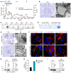
Figure 1. Anti-nephrin autoantibodies cause a podocytopathy with minimal change lesions.
(A) Disease course of a patient with anti-nephrin–associated MCD. Albuminuria given as urinary albumin-to-creatinine ratio (g/g). PLEX, plasma exchange; CsA, cyclopsporine A; Rtx, rituximab. (B) Representative periodic acid-Schiff (PAS) staining (left) and electron microscopy (EM, right) of the patient. Scale bars: 50 μm (PAS) and 1 μm (EM). (C) Western blot of immunoprecipitates (human (hu), mouse (ms), rabbit (rb), rat, guinea pig (gp), and swine (sw) glomerular extracts with serum from the patient (P) or from a healthy control (HC) individual). (D) Coomassie-blue stainings of urine samples from rabbits before (day –1) and after (days 3 and 5) the transfer of human IgG containing anti-nephrin autoantibodies or control IgG. (E) PAS stainings (left) and EM images (right) of rabbits 5 days after IgG transfer. Scale bars: 50 μm (PAS) and 500 μm (EM). (F) Representative immunofluorescence stainings for human IgG (hIgG) in colocalization with collagen IV (left), complement C3 in colocalization with wheat germ agglutinin (WGA, middle), and rabbit IgG (rbIgG) in colocalization with collagen IV (right) of kidneys from rabbits 5 days after IgG transfer. Panels are enlargements of the boxed areas in Supplemental Figure 2C. Scale bars: 5 μm.(G) Western blot of rabbit glomerular extracts (left) and of immunoprecipitates (rabbit glomerular extracts and anti-phosphotyrosine antibody,right). (H) Relative signal intensity given as the ratio of tyrosine-phosphorylated nephrin and input nephrin signal in (G). (I) Western blot of rabbit glomerular extracts (left) and of rabbit nephrin precipitated by human IgG present in glomerular extracts (right).

References

    1. Hengel FE, et al. Autoantibodies targeting nephrin in podocytopathies. N Engl J Med. 2024;391(5):422–433. doi: 10.1056/NEJMoa2314471. - DOI - PubMed
    1. Watts AJB, et al. Discovery of autoantibodies targeting nephrin in minimal change disease supports a novel autoimmune etiology. J Am Soc Nephrol. 2022;33(1):238–252. doi: 10.1681/ASN.2021060794. - DOI - PMC - PubMed
    1. Shirai Y, et al. A multi-institutional study found a possible role of anti-nephrin antibodies in post-transplant focal segmental glomerulosclerosis recurrence. Kidney Int. 2024;105(3):608–617. doi: 10.1016/j.kint.2023.11.022. - DOI - PubMed
    1. Seifert L, et al. An antigen-specific chimeric autoantibody receptor (CAAR) NK cell strategy for the elimination of anti-PLA2R1 and anti-THSD7A antibody-secreting cells. Kidney Int. 2024;105(4):886–889. doi: 10.1016/j.kint.2024.01.021. - DOI - PubMed