. 2025 Jan 16;20(1):e0313119.
doi: 10.1371/journal.pone.0313119. eCollection 2025.

Signalling pathways involved in urotensin II induced ventricular myocyte hypertrophy

Affiliations

Signalling pathways involved in urotensin II induced ventricular myocyte hypertrophy

Hadeel S Al Ali et al. PLoS One. .

Abstract

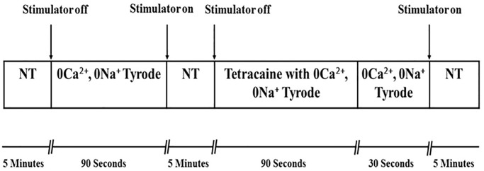
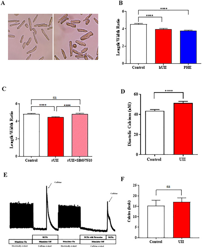
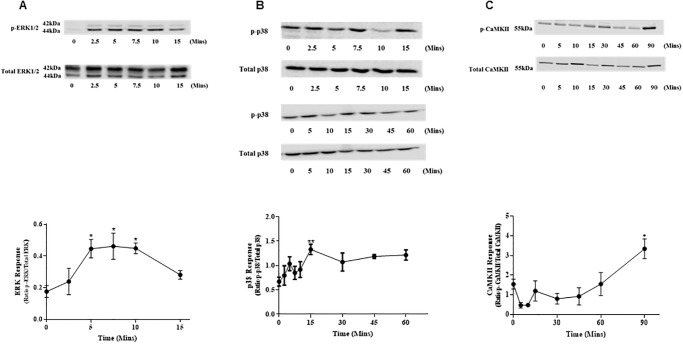
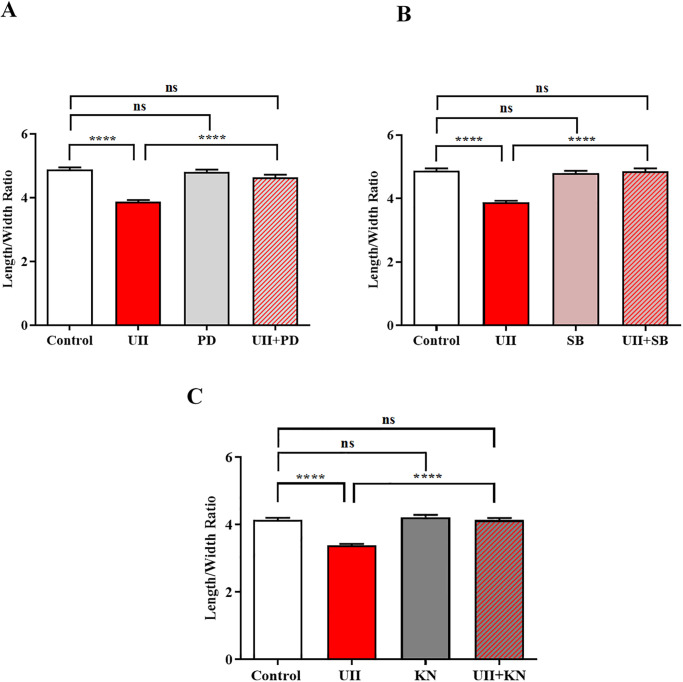
Sustained pathologic myocardial hypertrophy can result in heart failure(HF); a significant health issue affecting a large section of the population worldwide. In HF there is a marked elevation in circulating levels of the peptide urotensin II(UII) but it is unclear whether this is a result of hypertrophy or whether the high levels contribute to the development of hypertrophy. The aim of this study is to investigate a role of UII and its receptor UT in the development of cardiac hypertrophy and the signalling molecules involved. Ventricular myocytes isolated from adult rat hearts were treated with 200nM UII for 48hours and hypertrophy was quantified from measurements of length/width (L/W) ratio. UII resulted in a change in L/W ratio from 4.53±0.10 to 3.99±0.06; (p<0.0001) after 48hours. The response is reversed by the UT-antagonist SB657510 (1μM). UT receptor activation by UII resulted in the activation of ERK1/2, p38 and CaMKII signalling pathways measured by Western blotting; these are involved in the induction of hypertrophy. JNK was not involved. Moreover, ERK1/2, P38 and CaMKII inhibitors completely blocked UII-induced hypertrophy. Sarcoplasmic reticulum (SR) Ca2+-leak was investigated in isolated myocytes. There was no significant increase in SR Ca2+-leak. Our results suggest that activation of MAPK and CaMKII signalling pathways are involved in the hypertrophic response to UII. Collectively our data suggest that increased circulating UII may contribute to the development of left ventricular hypertrophy and pharmacological inhibition of the UII/UT receptor system may prove beneficial in reducing adverse remodeling and alleviating contractile dysfunction in heart disease.


Conflict of interest statement

The authors have declared that no competing interests exist.

Figures

Fig 1
Fig 1. Schematic diagram of the sarcoplasmic reticulum Ca2+-leak experiment.
Ventricular myocytes loaded with Fluo-3 were used to determine SR Ca2+-leak, using tetracaine (1mM) in EGTA solution to block Ca2+-leak through ryanodine receptors (RyR2).
Fig 2
Fig 2. Ventricular myocyte hypertrophy and SR Ca2+-leak.
(A) Isolated ventricular myocyte images in culture media (left). Ventricular myocytes treated with 200nM UII for 48 hours (right). (B) Length/width ratio in response to hUII and phenylephrine (PHE). Measurement of length/width ratio after 48 hours treatment with hUII (200nM) or phenylephrine (10μM). N = 6 hearts; 126, 209 & 141 cells, respectively. ****p<0.0001. One-way ANOVA followed by Sidak’s post hoc test. (C) Effect of UT receptor antagonist on rUII induced hypertrophy. Measurement of length/width ratio after rUII (200nM) or cells pretreated with SB657510 (1μM) before exposure to rUII for 24 hours. N = 6 hearts; 420, 519 & 441 cells, respectively. ****p<0.0001. One-way ANOVA followed by Sidak’s post hoc test. (D) Measurement of intracellular diastolic Ca2+-transient in cultured ARVMs. Diastolic [Ca2+]i recorded from ventricular myocytes after 24 hours tissue culture in the absence (control) and presence of 200nM UII. N = 8 hearts; 94 cells. ****p<0.0001. Paired t-test. (E) An example recording of showing the protocol and [Ca2+]i to determine SR Ca2+-leak. (F) Diastolic SR Ca2+-leak (nM) in cultured ventricular myocytes. Cells cultured for 24 hours in the absence (control) and presence of UII (200nM) for 24 hours. The difference in the diastolic [Ca2+]i was measured in the presence and absence of 1mM tetracaine for both control and treated cells. N = 6 hearts; 19 control cells & 25 treated cells. Unpaired t-test. All results are expressed as mean ± S.E.M.
Fig 3
Fig 3. Activation of MAPK and CaMKII in ventricular myocytes.
(A) Phosphorylation time course of ERK1/2 in ventricular myocytes by 200nM UII. Phosphorylation of ERK1/2 and total ERK was detected by Western blot analysis. N = 6 hearts. *p<0.05. (B) UII (200nM) induced p38 phosphorylation time course in ventricular myocytes. Ventricular myocyte p38 response was detected by Western blot analysis. N = 7 hearts. **p<0.01. (C) Time course for UII-induced phosphorylation of CaMKII in ventricular myocytes after treatment with 200nM UII. Phosphorylation of CaMKII and total CaMKII was detected by Western blot analysis. N = 5 hearts. *p<0.05. The results are expressed as mean ± S.E.M. One-way ANOVA followed by Dunn’s post hoc test. Uncropped blots are shown in the S1 File.
Fig 4
Fig 4. Role of MAPK and CaMKII in UII-induced myocyte hypertrophy.
(A) Effect of ERK1/2 inhibitor (PD184352; PD) on UII-induced hypertrophy in cultured ventricular myocytes. PD184352 blocked the UII-induced hypertrophy after 48 hours. N = 6 hearts; 358, 420, 253 & 195 cells, respectively. ****p<0.0001. (B) Effect of p38 inhibitor (SB202190; SB) on UII-induced hypertrophy in cultured ventricular myocytes. SB202190 blocked UII-induced hypertrophy after 48 hours. N = 6 hearts; 358, 420, 278 & 212 cells, respectively. ****p<0.0001. (C) Effect of CaMKII inhibitor (KN-93; KN) on UII-induced hypertrophy in cultured ventricular myocytes. KN-93 blocked UII-induced hypertrophy after 48 hours. N = 6 hearts; 320, 386, 282 & 223 cells, respectively. ****p<0.0001. Results are expressed as mean ± S.E.M. One-way ANOVA followed by Sidak’s post hoc test.

Similar articles

References

    1. Richey PA, Brown SP. Pathological versus physiological left ventricular hypertrophy: a review. Journal of sports sciences. 1998;16(2):129–41. doi: 10.1080/026404198366849 . - DOI - PubMed
    1. Molkentin JD, Dorn GW II. Cytoplasmic signaling pathways that regulate cardiac hypertrophy. Annual review of physiology. 2001;63(1):391–426. doi: 10.1146/annurev.physiol.63.1.391 . - DOI - PubMed
    1. Wang Y, Huang S, Sah VP, Ross J, Brown JH, Han J, et al.. Cardiac muscle cell hypertrophy and apoptosis induced by distinct members of the p38 mitogen-activated protein kinase family. Journal of Biological Chemistry. 1998;273(4):2161–8. doi: 10.1074/jbc.273.4.2161 . - DOI - PubMed
    1. Zhang W, Elimban V, Nijjar MS, Gupta SK, Dhalla NS. Role of mitogen-activated protein kinase in cardiac hypertrophy and heart failure. Experimental Clinical Cardiology. 2003;8(4):173. . - PMC - PubMed
    1. Wang Y. Mitogen-activated protein kinases in heart development and diseases. Circulation. 2007;116(12):1413–23. doi: 10.1161/CIRCULATIONAHA.106.679589 . - DOI - PMC - PubMed

MeSH terms