. 2025 Jan 16;16(1):22.
doi: 10.1038/s41419-025-07344-2.

Melanophilin-induced primary cilia promote pancreatic cancer metastasis

Affiliations

Melanophilin-induced primary cilia promote pancreatic cancer metastasis

Yu-Ying Chao et al. Cell Death Dis. .

Abstract

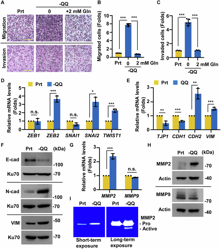
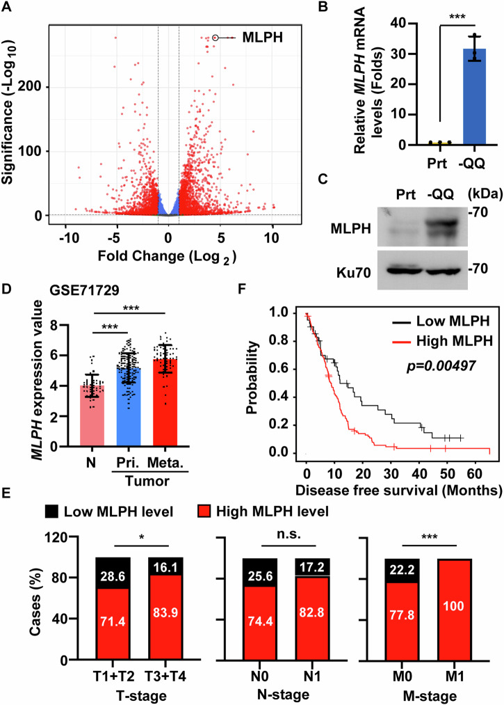
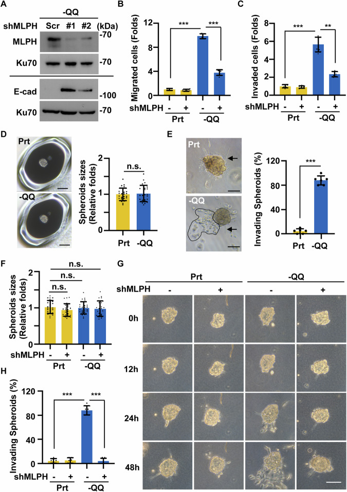
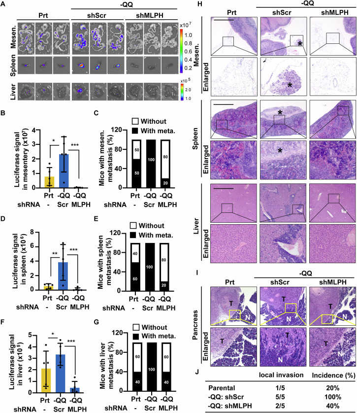
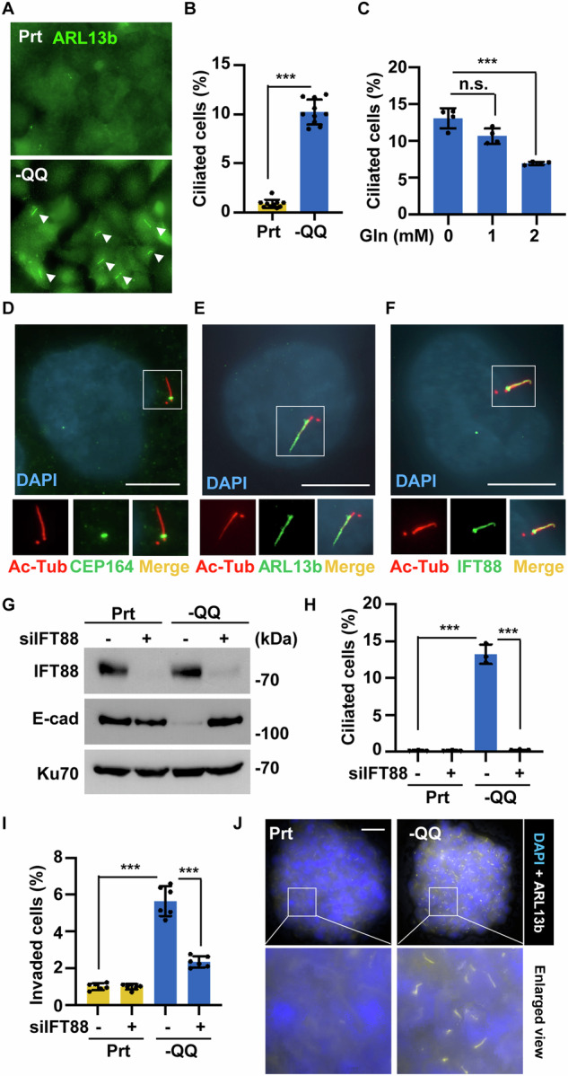
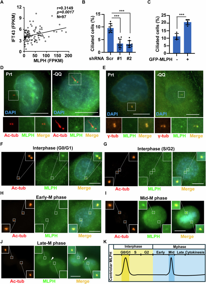
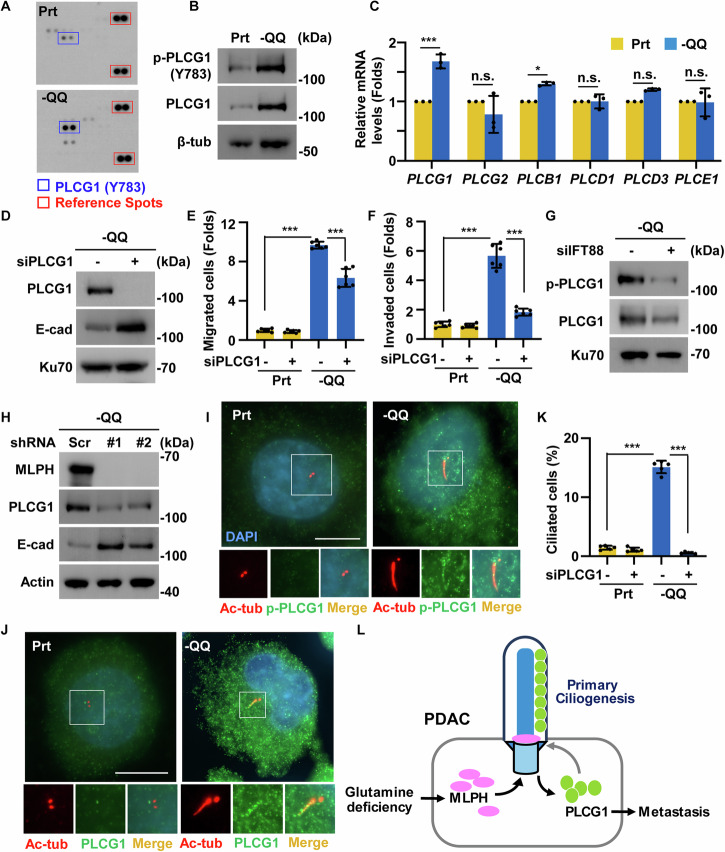
Pancreatic ductal adenocarcinoma (PDAC) is one of the most malignant tumors because of its high metastatic ability. The glutamine (Gln)-deficient microenvironment contributes to PDAC metastasis; however, the underlying molecular mechanisms remain unclear. Here, we demonstrated that melanophilin (MLPH) promotes PDAC metastasis by inducing the regrowth of primary cilia. Using RNA sequencing, we found that MLPH was upregulated in Gln-deficient conditions. MLPH facilitated PDAC metastasis in vitro and in vivo. Clinically, high MLPH expression is positively correlated with metastasis and poor PDAC prognosis. MLPH localized to the centrosome and facilitated the regrowth of primary cilia. The primary ciliogenesis upregulated phospholipase C γ-1 (PLCG1) to promote PDAC metastasis. Interestingly, PLCG1 was localized to the primary cilia, and depletion of PLCG1 alleviated primary ciliogenesis, suggesting a feedforward role for PLCG1 in mediating primary ciliogenesis. Thus, our study revealed a novel function of the MLPH-primary cilia-PLCG1 axis in facilitating PDAC metastasis under Gln deficiency both in vitro and in vivo.


Conflict of interest statement

Competing interests: The authors declare no competing interests. Ethics approval: All animal experiments were conducted in accordance with the guidelines approved by the animal ethics committee of National Cheng Kung University and complied with all relevant ethical guidelines (#113180). The retrospective clinical study was approved by the Institutional Review Board (IRB) of the NCKUH (IRB number: NCKUH B-ER-110-420) and was conducted following the ethical research guidelines.

Figures

Fig. 1
Fig. 1. Glutamine deprivation promotes EMT and invasion in PANC-1 cells.
AC Gln-deficient condition promoted PANC-1 migration and invasion, and supplementation of Gln restored these phenotypes. A The cell invasion and migration ability of parental (Prt) cells and -QQ PANC-1 cells supplemented with or without two mM Gln were evaluated using Trans-well analysis. Scale bar, 200 μm. B Quantitative results of the relative migrated cell numbers were measured in (A). C Quantitative results of the relative invaded cell numbers were measured in (A). DF Gln deficiency induced EMT in PANC-1 cells. D EMT-related transcription factors were upregulated in -QQ cells. Quantitative results of relative mRNA levels of ZEB1, ZEB2, SNAI1, SNAI2, and TWIST1 in Prt or -QQ PANC-1 cells. E Epithelial markers were downregulated, and mesenchymal markers were upregulated in -QQ cells. Quantitative results of relative mRNA levels of ZO-1, E-cadherin (E-cad, CDH1), N-cadherin (N-cad, CDH2), and vimentin (VIM) in Prt or -QQ PANC-1 cells. F E-cadherin (E-cad), N-cadherin (N-cad), and vimentin (VIM) were upregulated -QQ cells. Extracts of Prt or -QQ PANC-1 cells were analyzed by western blot assay with antibodies against E-cad, N-cad, VIM, and Ku70 (loading control). GI Upregulated MMP2 was observed in -QQ cells. G MMP2 but not MMP9 mRNA levels increased in -QQ PANC-1 cells. Quantitative results of relative mRNA levels of MMP2 and MMP9 in the Prt or -QQ PANC-1 cells. H MMP2 protein levels increased in -QQ cells. Extracts of Prt or -QQ PANC-1 cells were analyzed with western blot assay with antibodies against MMP2, MMP9, and actin. I MMP2 activity increased in -QQ cells. MMP activities were analyzed with gelatin zymography in Prt and -QQ PANC-1 cells. Pro: pro-MMP2, active: activated MMP2. Data are represented as the mean ± SD of three independent experiments. n.s. no significance, *P < 0.05, **P < 0.01, ***P < 0.001.
Fig. 2
Fig. 2. MLPH levels were positively correlated with PDAC tumorigenesis and metastasis.
AC MLPH was upregulated in -QQ PANC-1 cells. A Differentially expressed genes in parental (Prt) or -QQ PANC-1 cells were identified by the Bulk RNA sequence analysis shown in the Volcano Plot. Red dots represent genes with significant differences, and blue dots represent genes without significant differences. B MLPH mRNA levels increased in -QQ cells. Quantitative results of relative mRNA levels of MLPH in Prt or -QQ PANC-1 cells. C MLPH protein levels increased in -QQ cells. Extracts of Prt and -QQ PANC-1 cells were analyzed by western blot assay with antibodies against MLPH and Ku70 (loading control). D MLPH was upregulated in metastatic PDAC tissues compared to benign pancreas tissue in PDAC patients. The mRNA gene expression of MLPH in normal (N), primary tumor (Pri.), and metastatic tumor (Meta.) were obtained from the GEO database: GEO71729. E PDAC patients with higher MLPH expression showed poorer prognosis. Data analyzed from NCKUH cohort. F High MLPH expression positively correlated with poor disease-free survival. Disease-free survival was analyzed using the Kaplan–Meier method. Data were analyzed from The Cancer Genome Atlas (TCGA) database. P values were determined using the log-rank test. Data are represented as the mean ± SD of three independent experiments. n.s. no significance, *P < 0.05, ***P < 0.001.
Fig. 3
Fig. 3. MLPH upregulation promotes PDAC invasion in the 3D tumor spheroids.
A MLPH depletion restored E-cadherin expression. MLPH was depleted efficiently. Extracts of -QQ PANC-1 cells infected with lentivirus containing shRNA against scramble control depletion (shScr), or MLPH (#1 and #2) were analyzed by western blot assay with antibodies against MLPH, E-cadherin (E-cad), or Ku70 (loading control). MLPH promoted PDAC migration and invasion. Quantitative results of (B) migrated or (C) invaded parental (Prt) or -QQ cells in the absence or presence of shRNA against MLPH (shMLPH#1). D Tumor spheroid sizes in Prt or -QQ PANC-1 groups showed no difference. Scale bar, 100 µm. E Invaded cells were observed in the -QQ tumor spheroids. -QQ spheroids showed invaded cells that escaped from the spheroids (dashed circle). Scale bar, 50 µm. Multiple protrusions were observed in both Prt and -QQ PANC-1 spheroids (arrow). F Depletion of MLPH did not affect tumor spheroid sizes. Quantitative results of Prt or -QQ 3D tumor spheroids in the absence or presence of shRNA against MLPH (shMLPH#1). G, H Invading spheroids increased in a time-dependent manner. Observation of invaded cells in scramble control or MLPH-deficient Prt or -QQ spheroids. Scale bar, 50 µm. H Quantitative results of (G). Data are represented as the mean ± SD of three independent experiments. n.s. no significance, **P < 0.01, ***P < 0.001.
Fig. 4
Fig. 4. MLPH upregulation promotes PDAC metastasis under Gln deficiency in the orthotopic mouse model.
A Representative IVIS images were shown in the mesentery (Mesen.), spleen, and liver of Prt, scramble control depletion -QQ (-QQ-shScr), and MLPH depleted-QQ (-QQ-shMLPH) groups. Quantitative relative luciferase signals in (B) mesentery, (D) spleen, and (F) liver, and percentages of metastasis of (C) mesentery (Mesen.), (E) spleen, and (G) liver. H Representative H&E staining of the mesentery (Mesen.), spleen, and liver of Prt, -QQ-shScr, and -QQ-shMLPH groups. Metastatic colonies were labeled as asterisks. Scale bar, 100 μm. IJ MLPH promoted pancreatic local invasion. I Representative H&E staining of the pancreas of Prt, -QQ-shScr, and -QQ-shMLPH groups. T: tumor tissues, N: adjacent normal tissues. Scale bar, 100 μm. J Quantitative results of pancreatic local invasion were calculated in Prt, -QQ-shScr, and -QQ-shMLPH groups. Data are represented as the mean ± SD of three independent experiments. n.s. no significance, *P < 0.05, **P < 0.01, ***P < 0.001.
Fig. 5
Fig. 5. Primary cilia promote EMT and metastasis upon Gln deficiency.
A, B Primary cilia formation was observed in -QQ PANC-1 cells. (A) Primary cilia were detected by immunofluorescence staining with an antibody against ARL13b (green) in Prt or -QQ PANC-1 cells. DNA was stained with DAPI (blue). Scale bar, 20 µm. B Quantitative results of the frequency of ciliated cells in (A). C Gln supplementation inhibited primary ciliogenesis in -QQ cells. Quantitative results of the frequency of ciliated cells supplemented with different dosages of Gln in -QQ PANC-1 cells. Ciliary components were detected by immunofluorescence staining with antibodies against acetylated tubulin (Ac-tub, DF), CEP164 (D), ARL13b (E), and IFT88 (F). DNA was stained with DAPI. Scale bar, 10 µm. GI Primary cilia contributed to EMT and cell invasion. (G) IFT88 was depleted efficiently. Extracts of parental (Prt) or -QQ PANC-1 cells in the absence or presence of siRNA against IFT88 (siIFT88) were analyzed by western blot assay with antibodies against IFT88, E-cadherin (E-cad), or Ku70. (H) Quantitative results of the frequency of ciliated cells in Prt or -QQ PANC-1 cells in the absence or presence of siRNA against IFT88 (siIFT88). I Quantitative results of the relative invaded cells in Prt or -QQ PANC-1 cells in the presence or absence of siRNA against IFT88 (siIFT88). J The frequency and length of primary cilia were significantly increased in the -QQ PDAC spheroids. Primary cilia were detected by immunofluorescence staining with an antibody against ARL13b (white) in Prt or -QQ PANC-1 spheroids. DNA was stained with DAPI (blue). Scale bar, 10 µm. Data are represented as the mean ± SD of three independent experiments. n.s. no significance, ***P < 0.001.
Fig. 6
Fig. 6. MLPH promotes primary cilia formation upon Gln deficiency.
A IFT43 level positively correlated with MLPH expression. Scatter plot showed the relationship between genome-wide co-expression correlations for IFT43 and MLPH in pancreatic cancer tissues of NCKUH. (B-E) MLPH regulated primary ciliogenesis. B Depletion of MLPH decreased primary cilia formation in -QQ PANC-1 cells. Quantitative results of frequency of ciliated cells in -QQ PANC-1 cells infected with lentivirus containing shRNA against scramble control depletion -QQ (Scr) and MLPH (#1 and #2). C Overexpression of MLPH induced primary ciliogenesis. Quantitative results of frequency of ciliated cells in -QQ cells transfected with GFP or GFP-tagged MLPH. D MLPH localized to the base of primary cilia. Double staining of Prt and -QQ PANC-1 cells with antibodies against acetylated tubulin (Ac-tub, red) and MLPH (green). DNA was stained with DAPI (blue). Scale bar, 10 µm. E MLPH localized to centrosome. Double staining of Prt and -QQ PANC-1 cells with antibodies against gamma-tubulin (γ-tub, orange) and MLPH (green). DNA was stained with DAPI (blue). Scale bar, 10 µm. The localization of MLPH on centrosome from (FG) interphase to (HJ) mitosis. Double staining of -QQ PANC-1 cells with antibodies against MLPH (green) and acetylated tubulin (Ac-tub, red). DNA was stained with DAPI (blue). Scale bars, 10 μm. Insets are magnification of centrioles. K Schematic representation of the expression of centriolar MLPH from interphase to mitosis. Data are represented as the mean ± SD of three independent experiments. ***P < 0.001.
Fig. 7
Fig. 7. Primary cilia upregulate phospholipase C gamma 1(PLCG1) to facilitate PDAC metastasis.
A, B PLCG1 was activated in -QQ PANC-1 cells. A Phosphorylated PLCG1 increased as shown by human phospho-kinase array analysis in Prt and -QQ PANC-1 cells. The red line shows reference spots, whereas the blue line indicates the phosphorylation of Y783 of PLCG1. B PLCG1 was phosphorylated at tyrosine 783 in -QQ cells. Extracts of parental (Prt) or -QQ PANC-1 cells were analyzed by western blot assay with antibodies against phosphorylated PLCG1 at Y783, PLCG1, and tubulin. C The PLCG1 mRNA level increased in -QQ cells. Quantitative results of relative phospholipase C family mRNA levels were analyzed by qPCR in Prt or -QQ PANC-1 cells. DF PLCG1 promoted EMT, cell migration, and invasion. D Depletion of PLCG1 alleviated EMT in -QQ cells. Extracts of -QQ PANC-1 cells transfected with siRNA against PLCG1 (siPLCG1) were analyzed by western blot assay with antibodies against PLCG1, E-cadherin (E-cad), and Ku70. E, F Depletion of PLCG1 alleviated cell migration and invasion of -QQ cells. Quantitative results of the relative (E) migrated and (F) invaded cell numbers in Prt or -QQ PANC-1 cells in the presence or absence of siRNA against PLCG1 (siPLCG1). G Depletion of IFT88 decreased PLCG1 expression. Extracts of -QQ PANC-1 cells in the absence or presence of siRNA against IFT88 (siIFT88) were analyzed by western blot assay with antibodies against phosphorylated PLCG1 (Y783), PLCG1, and Ku70. H MLPH depletion decreased PLCG1 expression and restored EMT. Extracts of -QQ-shScr or -QQ-shMLPH#1 or #2 PANC-1 cells were analyzed by western blot assay with antibodies against MLPH, PLCG1, E-cadherin (E-cad), and actin. IJ PLCG1 and phosphorylated PLCG1 at Y783 (p-PLCG1) localized to the primary cilia. Immunofluorescence staining of Prt or -QQ PANC-1 cells with antibodies against acetylated tubulin (Ac-tub) and (I) p-PLCG1 and (J) PLCG1. DNA was stained with DAPI (blue). Scale bar, 10 µm. K Depletion of PLCG1 inhibited primary ciliogenesis. Quantitative results of the frequency of ciliated cells of Prt or -QQ PANC-1 cells were shown in the presence or absence of siRNA against PLCG1 (siPLCG1). L Graphic abstract. When PDAC cells suffered a Gln deprivation condition, MLPH was upregulated and recruited to the centrosome to facilitate primary ciliogenesis. The primary cilia induced and activated PLCG1 to promote EMT, invasion, and metastasis. Besides, PLCG1 was recruited to the primary cilia, thereby feedforward maintaining primary ciliogenesis. Data are represented as the mean ± SD of three independent experiments. n.s. no significance, *P < 0.05, ***P < 0.001.

Similar articles

Cited by

References

    1. Perera RM, Bardeesy N. Pancreatic cancer metabolism: breaking it down to build it back up. Cancer Discov. 2015;5:1247–61. - PMC - PubMed
    1. Hruban RH, Wilentz RE, Kern SE. Genetic progression in the pancreatic ducts. Am J Pathol. 2000;156:1821–5. - PMC - PubMed
    1. Chiang CL, Ma Y, Hou YC, Pan J, Chen SY, Chien MH, et al. Dual targeted extracellular vesicles regulate oncogenic genes in advanced pancreatic cancer. Nat Commun. 2023;14:6692. - PMC - PubMed
    1. Wang CA, Chang IH, Hou PC, Tai YJ, Li WN, Hsu PL, et al. DUSP2 regulates extracellular vesicle-VEGF-C secretion and pancreatic cancer early dissemination. J Extracell Vesicles. 2020;9:1746529. - PMC - PubMed
    1. Rhim AD, Mirek ET, Aiello NM, Maitra A, Bailey JM, McAllister F, et al. EMT and dissemination precede pancreatic tumor formation. Cell. 2012;148:349–61. - PMC - PubMed