. 2025 Jan 16;15(1):2132.
doi: 10.1038/s41598-025-85187-9.

Contribution of hypoxia-inducible factor 1alpha to pathogenesis of sarcomeric hypertrophic cardiomyopathy

Affiliations

Contribution of hypoxia-inducible factor 1alpha to pathogenesis of sarcomeric hypertrophic cardiomyopathy

Sarala Raj Murthi et al. Sci Rep. .

Abstract

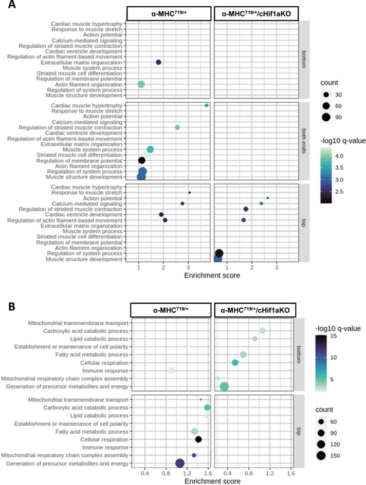
Hypertrophic cardiomyopathy (HCM) caused by autosomal-dominant mutations in genes coding for structural sarcomeric proteins, is the most common inherited heart disease. HCM is associated with myocardial hypertrophy, fibrosis and ventricular dysfunction. Hypoxia-inducible transcription factor-1α (Hif-1α) is the central master regulators of cellular hypoxia response and associated with HCM. Yet its exact role remains to be elucidated. Therefore, the effect of a cardiomyocyte-specific Hif-1a knockout (cHif1aKO) was studied in an established α-MHC719/+ HCM mouse model that exhibits the classical features of human HCM. The results show that Hif-1α protein and HIF targets were upregulated in left ventricular tissue of α-MHC719/+ mice. Cardiomyocyte-specific abolishment of Hif-1a blunted the disease phenotype, as evidenced by decreased left ventricular wall thickness, reduced myocardial fibrosis, disordered SRX/DRX state and ROS production. cHif1aKO induced normalization of pro-hypertrophic and pro-fibrotic left ventricular remodeling signaling evidenced on whole transcriptome and proteomics analysis in α-MHC719/+ mice. Proteomics of serum samples from patients with early onset HCM revealed significant modulation of HIF. These results demonstrate that HIF signaling is involved in mouse and human HCM pathogenesis. Cardiomyocyte-specific knockout of Hif-1a attenuates disease phenotype in the mouse model. Targeting Hif-1α might serve as a therapeutic option to mitigate HCM disease progression.

Keywords: HIF1A; Hypertrophic cardiomyopathy; Hypertrophy; Hypoxia; Myocardial fibrosis.


Conflict of interest statement

Declarations. Competing interests: Wolf CM: Honoraria: Novo Nordisk and Bristol-Myers Squibb; Dr. Wolf is a consultant for Day One Biopharmaceuticals, Inc., BioMarin Pharmaceuticals, Adrenomed AG, and Pliant Therapeutics. Dr. Wolf has ownership interest (Preventage Therapeutics). There are no other competing financial and non-financial interests

Figures

Fig. 1
Fig. 1
α-MHC719/+ mice develop left ventricular hypertrophy over time. (A,B) Exemplary transthoracic echocardiographic imaging (A) and echocardiographic myocardial wall thickness measurements (B) of 12 weeks old wildtype (12 weeks old WT + CSA), α-MHC719/+ mice (12 weeks old α-MHC719/+  + CSA) treated with Cyclosporine, 30 weeks old wildtype mice (30 weeks old WT) and 30 weeks old α-MHC719/+ mice (30 weeks old α-MHC719/+) without Cyclosporine treatment (n = 6 each group). 12 weeks old α-MHC719/+  + CSA and 30 weeks old α-MHC719/+ mice show increase of the end-diastolic left ventricular septal (*) and posterior wall thickness (#), and reduced end-diastolic LV diameter (§) when compared to the 12 weeks old WT + CSA and 30 weeks old WT mice on parasternal short axis view. The phenotype arises in the 30 weeks old α-MHC719/+ mice naturally and Cyclosporine (CSA) treatment enhances the phenotype in the young 12 weeks old α-MHC719/+. Data are presented as mean ± SEM with significances indicated by p values. No significant difference between 12 weeks WT mice treated with CSA and 30 weeks old WT mice without CSA treatment was detected.
Fig. 2
Fig. 2
Hif-1α is upregulated in left ventricular tissue of α-MHC719/+ mice displaying the pathognomic features of hypertrophic cardiomyopathy. (A,B) Proteins were isolated from LV tissue from both wild type (WT) and α-MHC719/+ mice. Western blot (A) and quantification (B) of proteins extracted from LV tissue performed for Hif-1α, Myh7, Desmin (Des) and Tpm4 with Actin as loading control. Compared to WT increased relative fold change (FC) of Hif-1α, Myh7, Des and Tpm4 in α-MHC719/+ mice (n = 3 each group). Data are presented as mean ± SEM with significances indicated by p values. (C,D) Differentially expressed genes on LV transcriptome (C) or proteome (D) analysis of α-MHC719/+ vs. WT. Names indicate top ten regulated genes or proteins. Original western blot in (A) presented in (Supplemental Fig. 8).
Fig. 3
Fig. 3
Cardiomyocyte specific Hif-1α knockout decreases HCM related protein expression. (A) Hif1a gene expression on RT-PCR was decreased in α-MHC719/+/cHif1aKO mice compared to α-MHC719/+. (B) Western blot from protein extracted of freshly isolated adult murine cardiomyocytes from WT, α-MHC719/+, WT/cHif1aKO, and α-MHC719/+/cHif1aKO (n = 3 in each group). Increased expression of Hif-1α on Western blot from protein extracted of freshly isolated adult murine cardiomyocytes after incubation under hypoxia conditions (1% oxygen) for 16 h in WT and α-MHC719/+ mice, but not in WT/cHif1aKO and α-MHC719/+/cHif1aKO mice (n = 3 in each group). Ponceau S (PoncS) served as loading control. (C,D) FFPE heart sections were stained with an antibody against CAIX. Nuclei were visualized with DAPI. (C) Representative staining is shown. (D) Average number of CAIX positive signal/ cells was measured in four high power fields per heart section of WT (n = 3), α-MHC719/+ (n = 5), WT/cHif1aKO (n = 4) and α-MHC719/+/cHif1aKO (n = 6) mice. Scatter plots with mean ± SEM with significances indicated by p values for all plots. Scale bar = 50 µm. (E,F) Western blot (E) and quantification (F) of classical HCM proteins Myh7. Desmin (Des) and Tpm4 from freshly isolated adult murine cardiomyocytes of WT, α-MHC719/+, WT/cHif1aKO and α-MHC719/+/cHif1aKO mice (n = 3 in each group). Ponceau S (PoncS) served as loading control. fc fold change. Data are presented as mean ± SEM with significances indicated by p values. Original western blot in (B) and (E) presented in (Supplemental Figs. 9, 10).
Fig. 4
Fig. 4
Cardiomyocyte-specific Hif-1α knockout in the α-MHC719/+ HCM mouse model attenuates disease phenotype. (A,B) Exemplary transthoracic echocardiographic imaging (A) and echocardiographic myocardial wall thickness measurements (B) of WT (n = 6), α-MHC719/+ (n = 13), WT/cHif1aKO (n = 5), and α-MHC719/+/cHif1aKO (n = 8) mice. Marked increase of the end-diastolic left ventricular septal (*) and posterior wall (#) thickness, and reduced end-diastolic LV diameter (§) in α-MHC719/+ compared to the WT mice on parasternal long axis view, which is ameliorated in α-MHC719/+/cHif1aKO mice. (C) Initial rapid decay amplitude corresponding to disordered relaxed state (DRX) heads conformations in LV myocardial tissue is abnormal in α-MHC719/+ (n = 5) but not in α-MHC719/+/cHif1aKO (n = 6) compared to WT (n = 6) and WT/cHif1aKO (n = 4) mice. (D) Cardiomyocyte-specific Hif1α knockout in the α-MHC719/+ HCM mouse model attenuates disease phenotype: Increased heart weight–body weight ratios in α-MHC719/+ (n = 5) but not in α-MHC719/+/cHif1aKO (n = 6) compared to WT (n = 6) and WT/cHif1aKO (n = 4) mice. (E,F) Myocardial fibrosis: Exemplary Masson trichrome staining of the myocardium in WT, α-MHC719/+, WT/cHif1aKO, and α-MHC719/+/cHif1aKO mice is shown. The magnification bar represents 100 μm (E). The area of fibrosis, stained in blue, was measured morphometrically in WT (n = 6), α-MHC719/+ (n = 5), WT/cHif1aKO (n = 4), and α-MHC719/+/cHif1aKO (n = 6) mice as a percentage of the entire myocardium (F). Myocardial fibrosis (blue), as indicated by arrows in E, markedly increased in α-MHC719/+ mice compared to WT. When compared to α-MHC719/+ mice, the myocardial fibrosis in α-MHC719/+/cHif1aKO mice were found to be less pronounced. fc fold change. Scatter plots show mean ± SEM, with significances indicated by p-values for all plots.
Fig. 5
Fig. 5
Normalization of proteins and signaling pathways involved in pathological myocardial remodeling on proteomic analysis of α-MHC719/+ mice lacking Hif-1α in cardiomyocytes. (A) Shown is a subset of genes known to be involved in pathologic myocardial remodeling and significantly dysregulated in α-MHC719/+ mice that show counter regulation or normalization towards WT levels in α-MHC719/+/cHif1aKO mice, represented as the log2 fold change relative to WT. # denotes q-value < 0.05; ns denotes non-significantly regulated. (B) Known pro-hypertrophic, pro-fibrotic and pro-inflammatory proteins dysregulated in α-MHC719/+ compared to WT mice are unchanged in α-MHC719/+/cHif1aKO compared to WT mice.
Fig. 6
Fig. 6
Normalization of potential mechanistic pathways involved in pathological myocardial remodeling on transcriptome analysis of α-MHC719/+ mice lacking Hif-1α in cardiomyocytes. (AC) Enrichment bubble graph for hypoxia/ROS (A), stress (B) or inflammation (C) related pathways dysregulated in α-MHC719/+ mice. (D) Gene expression pattern of key genes involved in hypoxia (Hx), immune and stress related pathways dysregulated in in α-MHC719/+ (green) compared to WT mice are unchanged in α-MHC719/+/cHif1aKO (red) compared to WT mice.
Fig. 7
Fig. 7
Pathways differential affected in the proteome of α-MHC719/+ mice and α-MHC719/+ mice lacking Hif-1α in cardiomyocytes. Enrichment bubble plot for cardiac related pathways (A) or potential mechanistic related pathways (B) in the hearts of α-MHC719/+ mice or α-MHC719/+/cHif1aKO mice.
Fig. 8
Fig. 8
α-MHC719/+ mice showed oxidative stress, inflammation and dysregulated angiogenesis reversed in α-MHC719/+ mice lacking Hif-1α in cardiomyocytes. (A) Superoxide generation rate was measured by electron paramagnetic resonance using 1-Hydroxy-3-methoxycarbonyl-2, 2, 5, 5-tetramethylpyrrolidine in macerated LV tissue from α-MHC719/+ (n = 5), α-MHC719/+/cHif1aKO (n = 6), WT (n = 3) and WT/cHif1aKO mice (n = 4). (B,C) FFPE heart sections were stained with an antibody against 8-Hydroxydeoxyguanosine (8OHdG). Nuclei were visualized with DAPI. (B) Representative staining is shown. (C) Fluorescence intensity of 60 nuclei was measured in four high power fields per heart section (n = 3–6). (D) Il1b gene expression on RT-PCR is decreased in α-MHC719/+/cHif1aKO mice compared to α-MHC719/+. (E,F) Representative cardiac sections of HCM and cardiomyocyte-specific Hif-1α knock out mice line exhibiting cardiomyocytes in green, CD31 – stained vessels visualized in red by antibody staining. (a,e) WT, (b,f) α-MHC719/+, (c,g) WT/cHif1aKO and (d,h) α-MHC719/+/cHif1aKO mice line (n = 3–4 each group). Scale bar: 100 µm (ad). Zoom in on CD31 stained Endothelial cells 50 µm (eh). Scatter plots show mean ± SEM, with significances indicated by p-values for all plots.

References

    1. Maron, B. J. et al. Prevalence of hypertrophic cardiomyopathy in a general population of young adults. Echocardiographic analysis of 4111 subjects in the CARDIA study. Coronary artery risk development in (young) adults. Circulation92, 785–789. 10.1161/01.cir.92.4.785 (1995). - PubMed
    1. Maron, B. J. Contemporary insights and strategies for risk stratification and prevention of sudden death in hypertrophic cardiomyopathy. Circulation121, 445–456. 10.1161/CIRCULATIONAHA.109.878579 (2010). - PubMed
    1. Watkins, H. Sudden death in hypertrophic cardiomyopathy. N. Engl. J. Med.342, 422–424. 10.1056/NEJM200002103420609 (2000). - PubMed
    1. Seidman, C. E. & Seidman, J. G. Identifying sarcomere gene mutations in hypertrophic cardiomyopathy: a personal history. Circ. Res.108, 743–750. 10.1161/CIRCRESAHA.110.223834 (2011). - PMC - PubMed
    1. Geisterfer-Lowrance, A. A. et al. A molecular basis for familial hypertrophic cardiomyopathy: a beta cardiac myosin heavy chain gene missense mutation. Cell62, 999–1006. 10.1016/0092-8674(90)90274-i (1990). - PubMed

Publication types

Substances