

Quantification of the Anterior-Centripetal Movement of the Ciliary Muscle During Accommodation Using Dynamic OCT Imaging
- PMID: 39820463
- PMCID: PMC11745204
- DOI: 10.1167/tvst.14.1.17
Quantification of the Anterior-Centripetal Movement of the Ciliary Muscle During Accommodation Using Dynamic OCT Imaging
Abstract
Purpose: Although the lens undoubtedly plays a major role in presbyopia, altered lens function could be in part secondary to age-related changes of the ciliary muscle. Ciliary muscle changes with accommodation have been quantified using optical coherence tomography, but so far these studies have been limited to quantifying changes in ciliary muscle thickness, mostly at static accommodative states. Quantifying ciliary muscle thickness changes does not effectively capture the dynamic anterior-centripetal movement of the ciliary muscle during accommodation. To address this issue, we present a method to quantify the movement of the ciliary muscle during accommodation using trans-scleral optical coherence tomography images obtained dynamically.
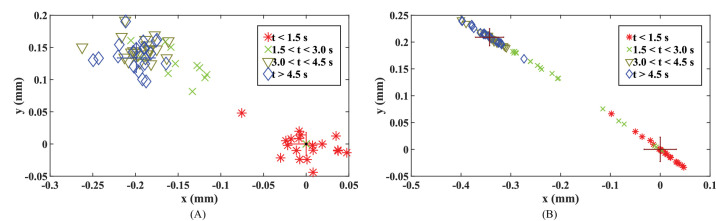
Methods: An image processing framework including distortion correction, geometric transformation, and Procrustes analysis, was used to quantify the anterior-centripetal movement of the ciliary muscle apex and centroid during accommodation. The method was applied in a preliminary study to quantify ciliary muscle displacement and its relation to lens thickness change with accommodation on two young adults and two prepresbyopes.
Results: The magnitude and the direction relative to the pupil plane of the apex/centroid displacement in response to a two diopters (2D) stimulus were 0.16/0.20 mm at 11.3°/30.5° and 0.26/0.34 mm at 6.6°/33.2° for the young adults and 0.20/0.20 mm at 29.7°/40.6° and 0.24/0.40 mm at 33.0°/31.7° for the prepresbyopes, respectively.
Conclusions: This study demonstrates the feasibility of quantifying dynamic anterior-centripetal movement of the ciliary muscle during accommodation using optical coherence tomography. The method better captures the functional response of the muscle than the quantification of thickness changes.
Translational relevance: We provide a method that holds potential to better understand the age-related changes of the ciliary muscle on presbyopia.
Conflict of interest statement
Disclosure:
Figures


References
-
- Cabeza-Gil I, Grasa J, Calvo B. A validated finite element model to reproduce Helmholtz's theory of accommodation: a powerful tool to investigate presbyopia. Ophthalm Physiol Opt. 2021; 41(6): 1241–1253. - PubMed
-
- Croft MA, Glasser A, Kaufman PL. Accommodation and presbyopia. Int Ophthalmol Clin. 2001; 41(2): 33–46. - PubMed
MeSH terms
Grants and funding
LinkOut - more resources
Full Text Sources
Miscellaneous
