

Physiopathological effects of entrance versus distal spread-out Bragg peak on mouse spinal cord neurons
- PMID: 39820768
- PMCID: PMC11739576
- DOI: 10.1038/s41598-024-84902-2
Physiopathological effects of entrance versus distal spread-out Bragg peak on mouse spinal cord neurons
Abstract
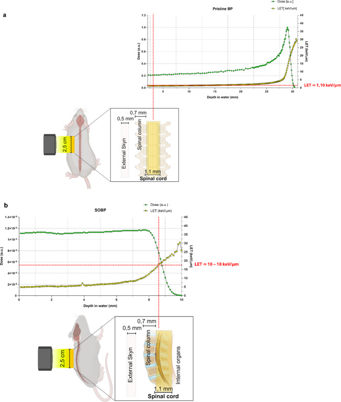
Recent investigations into radiation-induced side effects have focused on understanding the physiopathological consequences of irradiation on late-responding tissues like the spinal cord, which can lead to chronic progressive myelopathy. Proton therapy, an advanced radiation treatment, aims to minimize damage to healthy tissues through precise dose deposition. However, challenges remain, particularly regarding the variation in dose distribution, characterized by maximum deposition at the end of the proton range, known as the distal fall-off of a spread-out Bragg peak. This variation is critical for nearby organs at risk. In this preliminary study, we evaluated the effects of proton irradiation on the neuronal cell population in mouse spinal cord by comparing two positions of the particle range dose deposition profile. We irradiated the spinal cords at the entrance and the distal edge of the spread-out Bragg peak with a single proton dose. Results showed changes in the expression of synaptophysin, a presynaptic protein crucial for synaptic plasticity. Our findings suggest that examining early radiation-induced physiopathological effects on late-responding tissues can provide valuable insights into neuronal plasticity, informing clinical treatment planning for proton beam positioning.
Keywords: Bragg peak; Proton therapy; RBE; Radiation-induced off-target effects; Synaptic plasticity.
© 2025. The Author(s).
Conflict of interest statement
Declarations. Competing interests: The authors declare no competing interests. Ethical approval: Approval was granted by the Ethics Committee of University of Catania, Catania (Italy), approval Code: 248/2018-PR of 30/03/2018, according to Ministry of Health. All data generated or analysed during this study are included in this published article [and its supplementary information files].
Figures

Similar articles
-
Characterizing Proton-Induced Biological Effects in a Mouse Spinal Cord Model: A Comparison of Bragg Peak and Entrance Beam Response in Single and Fractionated Exposures.Int J Radiat Oncol Biol Phys. 2024 Jul 1;119(3):924-935. doi: 10.1016/j.ijrobp.2023.12.031. Epub 2024 Feb 2. Int J Radiat Oncol Biol Phys. 2024. PMID: 38310485
-
Dosimetric Assessment of a High Precision System for Mouse Proton Irradiation to Assess Spinal Cord Toxicity.Radiat Res. 2021 Jun 1;195(6):541-548. doi: 10.1667/RADE-20-00153.1. Radiat Res. 2021. PMID: 33826742
-
Determination of the proton RBE in the rat spinal cord: Is there an increase towards the end of the spread-out Bragg peak?Radiother Oncol. 2018 Jul;128(1):115-120. doi: 10.1016/j.radonc.2018.03.002. Epub 2018 Mar 21. Radiother Oncol. 2018. PMID: 29573823
-
New insights in the relative radiobiological effectiveness of proton irradiation.Radiat Oncol. 2018 Jan 16;13(1):6. doi: 10.1186/s13014-018-0954-9. Radiat Oncol. 2018. PMID: 29338744 Free PMC article. Review.
-
Normal tissue complications from low-dose proton therapy.Health Phys. 2012 Nov;103(5):586-9. doi: 10.1097/HP.0b013e3182611114. Health Phys. 2012. PMID: 23032888 Review.
References
Publication types
MeSH terms
Substances
Grants and funding
LinkOut - more resources
Full Text Sources
