. 2025 Jan 16;15(1):2180.
doi: 10.1038/s41598-025-86486-x.

Iron metabolism in a mouse model of hepatocellular carcinoma

Affiliations

Iron metabolism in a mouse model of hepatocellular carcinoma

Dilay Yilmaz et al. Sci Rep. .

Abstract

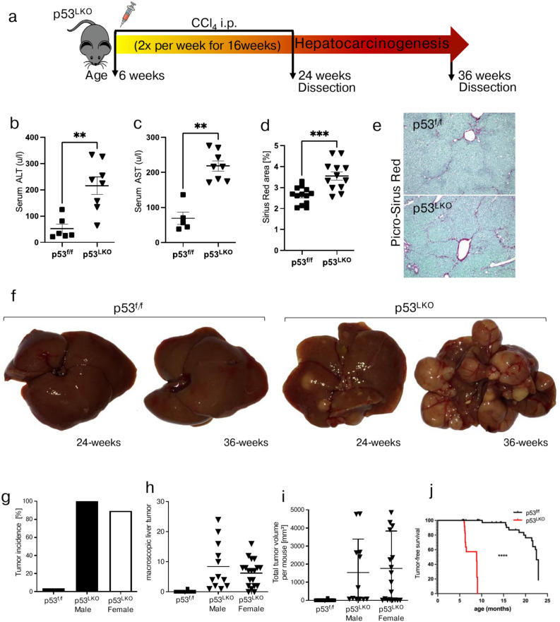
Hepatocellular carcinoma (HCC) remains the most prevalent type of primary liver cancer worldwide. p53 is one of the most frequently mutated tumor-suppressor genes in HCC and its deficiency in hepatocytes triggers tumor formation in mice. To investigate iron metabolism during liver carcinogenesis, we employed a model of chronic carbon tetrachloride injections in liver-specific p53-deficient mice to induce liver fibrosis, cirrhosis and subsequent carcinogenesis. A transcriptome analysis of liver carcinoma was employed to identify p53-dependent gene expression signatures with subsequent in-depth analysis of iron metabolic parameters being conducted locally within liver cancers and at systemic levels. We show that all mutant mice developed liver cancer by 36-weeks of age in contrast to 3.4% tumors identified in control mice. All liver cancers with a p53-deficient background exhibited a local iron-poor phenotype with a "high transferrin receptor 1 (Tfr1) and low hepcidin (Hamp)" signature. At systemic levels, iron deficiency was restricted to female mice. Additionally, liver tumorigenesis correlated with selective deficits of selenium, zinc and manganese. Our data show that iron deficiency is a prevalent phenomenon in p53-deficient liver cancers, which is associated with alterations in Hamp and Tfr1 and a poor prognosis in mice and patients.

Keywords: Hepcidin; Iron metabolism; Liver cancer; Trace elements; Transferrin receptor; p53.


Conflict of interest statement

Declarations. Competing interests: The authors declare no competing interests.

Figures

Fig. 1
Fig. 1
Liver carcinoma formation in p53LKO mice is highly accelerated following chronic CCl4 treatment. (a) Study design. Mice with liver-specific Trp53 deletion (short: p53LKO) and p53f/f control mice, both males and females, were injected intraperitoneally (i.p.) with carbon tetrachloride (CCl4; 0.5 ml/kg body weight) at 6 weeks of age, twice per week for total 16 weeks. Mice were sacrificed at 24 and 36 weeks of age. (b, c) Serum ALT (alanine transaminase) and AST (aspartate transaminase), indicative of liver damage in CCl4 treated mice (n = 6;8). (d) Bar graphs depict liver fibrosis analysed by Desmet stage (n = 17;11). (e) Representative photographs of Picro-Sirius-red staining of liver from p53f/f mice (top) and of liver from p53LKO mice (bottom). (f) Representative photographs of macroscopic liver carcinoma formation from p53f/f and p53LKO at the age of 24 and 36 weeks. (g) Bar chart represents tumor incidence in p53f/f (n = 1/29) and p53LKO male (n = 12/12) and female mice (n = 17/19). (h, i) Dot plots depict macroscopic tumor formation and total tumor volume per mouse in p53f/f and p53LKO male and female mice. (j) Tumor-free survival curve of p53f/f and p53LKO mice treated with chronic CCl4 injections.
Fig. 2
Fig. 2
Transcriptome analysis of liver carcinoma reveals p53-dependent gene expression signatures associated with iron metabolism. Transcriptome analysis of liver carcinoma from p53LKO and p53f./f male mice. (a) Volcano plot representation of genes that differ significantly in HCC in p53LKO in comparison to HCC from p53f./f mice (n = 5;5). Plotted is significance (adjusted p-value) versus log2 fold change (FC) on the y and x-axis, respectively. Depicted in blue are genes significantly different with adjusted p < 0.05 and the FC > 2. (b) Pie charts illustrate the spectrum of upregulated and downregulated genes and the number of differentially regulated genes between HCC of p53LKO and p53f./f mice. (c) GSEA pathways associated with iron metabolism are plotted in red. (d, e) Pre-ranked GSEA enrichment plots of representative signaling pathways with corresponding heat maps displaying genes grouped according to the expression intensities. Euclidean distance was used for hierarchical clustering. NES, normalized enrichment score; GSEA, gene set enrichment analysis. (f, g) Heat map of genes differentially regulated between HCC of p53LKO and p53f./f mice in respective signaling pathways (d, e).
Fig. 3
Fig. 3
Liver carcinoma show a reduced trace element content and dysregulated iron metabolism. (a, b) Total trace element content (iron, selenium, zinc and manganese) in NTL and HCC of p53LKO male and female mice, respectively. (c, d) mRNA expression levels of Tfr1, Hamp (hepcidin), Hfe, Smad7, Bmp6, and Fpn1 in NTL and HCC of p53LKO male and female mice, respectively. (e, f) Correlation analysis between Tfr1, Hamp and iron levels in HCC tissues from p53LKO male and female mice, respectively. (g, h) Representative immunoblot analysis and quantification blots of TFR1 relative to β-actin levels (expressed in arbitrary units) in NTL and HCC of p53LKO female mice.
Fig. 4
Fig. 4
High transferrin receptor 1 and low hepcidin expression in liver carcinoma is associated with a poor prognosis. (a-c) TFRC expression levels in non-tumor (NT) and HCC (T) of human patients with (d-f) plots comparing probability of survival of TFRChigh and TFRClow expressing HCC, in both genders, in male, and female patients, respectively. (g-i) HAMP expression levels in NT and T of human patients with (j-l) plot comparing probability of survival of HAMPhigh and HAMPlow expressing HCC , in both genders, in male, and female patients, respectively.
Fig. 5
Fig. 5
Hepatocellular carcinoma is characterized by a profound systemic iron deficiency in p53LKO female mice. (a) Serum iron and (b) hepcidin levels in p53f./f and p53LKO male mice. (c) Relative Hamp mRNA expression level in p53f./f and p53LKO male mice. (d) Systemic iron levels in the liver , spleen, kidney, lung and pancreas of p53f./f and p53LKO male mice. (e-g) Serum iron, erythropoietin (EPO) and hepcidin levels in p53f./f and p53LKO female mice. (h) Systemic iron levels in the liver, spleen, kidney, lung, and pancreas of p53f./f and p53LKO female mice. (i) Relative Hamp, TfR1 and Fpn1 mRNA expression level in the livers of p53f./f and p53LKO female mice. (j,k) Representative immunoblot analysis and quantification blots of TFR1, FtH and Fpn relative to β-actin levels (expressed in arbitrary units) in the spleen of p53f./f and p53LKO female mice. M: Molecular weight marker (kDa), + : positive control for Fpn expression

References

    1. Sung, H. et al. Global cancer statistics 2020: GLOBOCAN estimates of incidence and mortality worldwide for 36 cancers in 185 countries. CA. Cancer J. Clin.10.3322/caac.21660 (2021). - PubMed
    1. Llovet, J. M. et al. Hepatocellular carcinoma. Nat. Rev. Dis. Prim.7(1), 6. 10.1038/s41572-020-00240-3 (2021). - PubMed
    1. Balogh, J. et al. Hepatocellular carcinoma: A review. J. Hepatocell. Carcinoma3, 41–53. 10.2147/JHC.S61146 (2016). - PMC - PubMed
    1. Kan, Z. et al. Whole-genome sequencing identifies recurrent mutations in hepatocellular carcinoma. Genome. Res.23(9), 1422–1433. 10.1101/gr.154492.113 (2013). - PMC - PubMed
    1. Li, S. & Mao, M. Next generation sequencing reveals genetic landscape of hepatocellular carcinomas. Cancer Lett.340(2), 247–253. 10.1016/j.canlet.2012.09.027 (2013). - PubMed

Publication types

MeSH terms