. 2025 Mar;55(3):47.
doi: 10.3892/ijmm.2025.5488. Epub 2025 Jan 17.

Protective role of triiodothyronine in sepsis‑induced cardiomyopathy through phospholamban downregulation

Affiliations

Protective role of triiodothyronine in sepsis‑induced cardiomyopathy through phospholamban downregulation

Qiumin Xie et al. Int J Mol Med. 2025 Mar.

Abstract

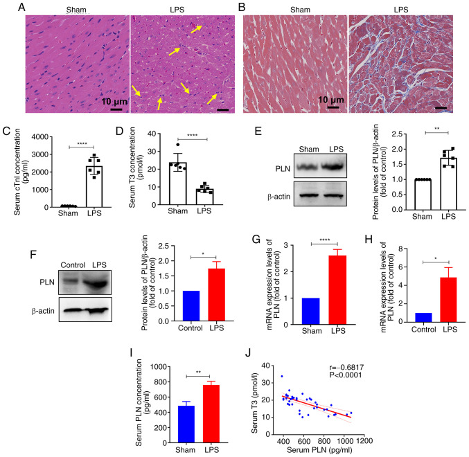
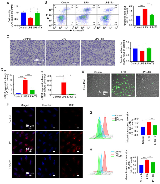
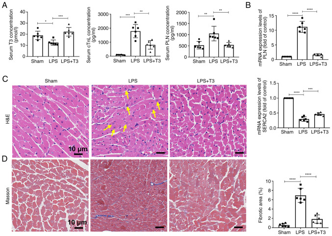
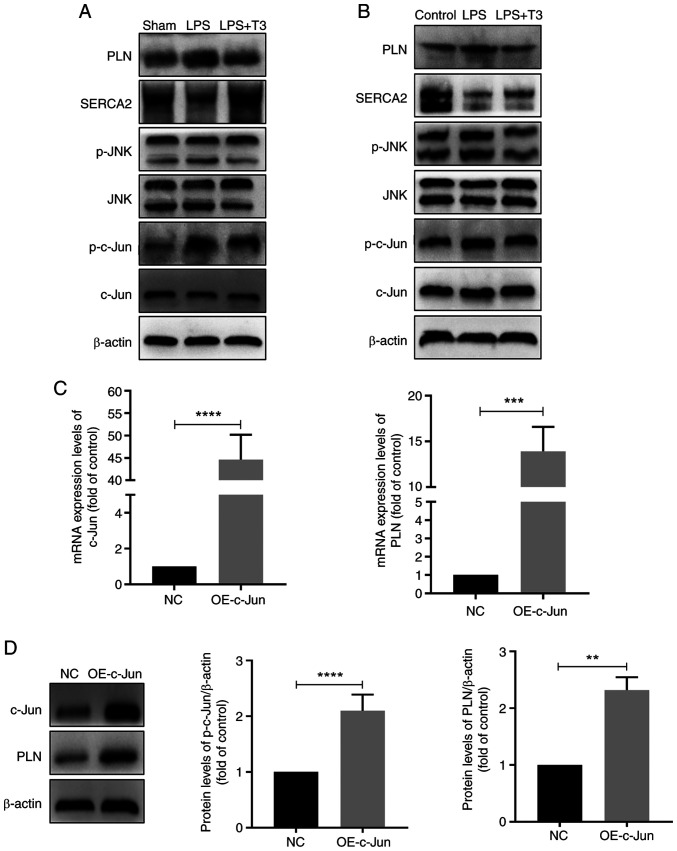
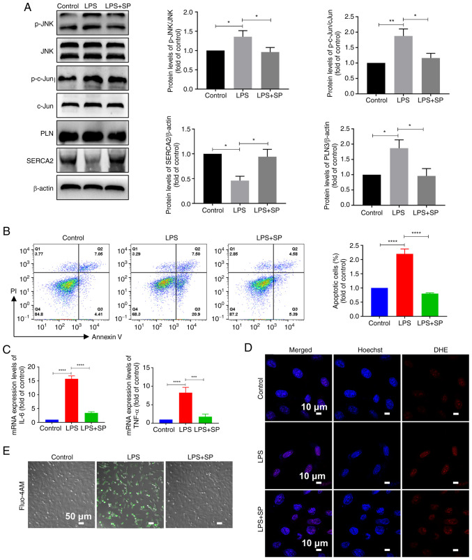
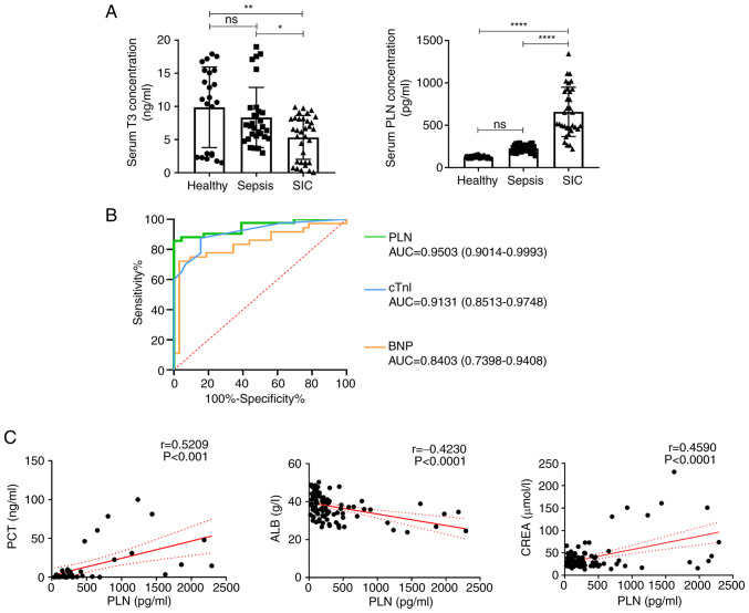
Sepsis is often a cause of mortality in patients admitted to the intensive care unit. Notably, the heart is the organ most susceptible to the impact of sepsis and this condition is referred to as sepsis‑induced cardiomyopathy (SIC). Low triiodothyronine (T3) syndrome frequently occurs in patients with sepsis, and the heart is one of the most important target organs for the action of T3. Phospholamban (PLN) is a key protein associated with Ca2+‑pump‑mediated cardiac diastolic function in the myocardium of mice with SIC, and PLN is negatively regulated by T3. The present study aimed to explore whether T3 can protect cardiac function during sepsis and to investigate the specific molecular mechanism underlying the regulation of PLN by T3. C57BL/6J mice and H9C2 cells were used to establish in vivo and in vitro models, respectively. Myocardial damage was detected via pathological tissue sections, a Cell Counting Kit-8 assay, an apoptosis assay and crystal violet staining. Intracellular calcium levels and reactive oxygen species were detected by Fluo‑4AM and DHE fluorescence. The protein and mRNA expression levels of JNK and c‑Jun were measured by western blotting and reverse transcription‑quantitative PCR to investigate the molecular mechanisms involved. Subsequently, 100 clinical patients were recruited to verify the clinical application value of PLN in SIC. The results revealed a significant negative correlation between PLN and T3 in the animal disease model. Furthermore, the expression levels of genes and proteins in the JNK/c‑Jun signaling pathway and PLN expression levels were decreased, whereas the expression levels of sarcoplasmic reticulum calcium ATPase were increased after T3 treatment. These results indicated that T3 alleviated myocardial injury in SIC by inhibiting PLN expression and its phosphorylation, which may be related to the JNK/c‑Jun signaling pathway. Accordingly, PLN may have clinical diagnostic value in patients with SIC.

Keywords: biomarker; calcium homeostasis; myocardial injury; reactive oxygen species.


Conflict of interest statement

The authors declare that they have no competing interests.

Figures

Figure 1
Figure 1
Serum T3 levels are significantly negatively correlated with myocardial PLN levels in sepsis. (A) Hematoxylin and eosin staining of mouse myocardial tissues. Yellow arrows indicate inflammatory cells. Scale bar, 10 µm; n=6. (B) Masson staining was applied to mouse myocardial tissues. Scale bar, 10 µm; n=6. ELISA of the serum levels of (C) cTnI and (D) T3 (n=6). WB of PLN protein levels and semi-quantitative analysis in the (E) myocardial tissues of mice (n=6) and in (F) H9C2 cells (n=3). Quantitative PCR analysis of the mRNA expression levels of PLN in the (G) myocardial tissues of mice (n=6) and in (H) H9C2 cells (n=3). (I) ELISA measured the serum levels of PLN in the serum of mice (n=6). (J) Spearman correlation analysis of T3 and PLN concentrations in mouse serum. All experiments were repeated three times. Data are presented as the mean ± standard deviation. *P<0.05, **P<0.01, ****P<0.0001. cTnI, cardiac troponin I; LPS, lipopolysaccharide; PLN, phospholamban. qPCR, quantitative PCR; T3, triiodothyronine.
Figure 2
Figure 2
T3 treatment alleviates cardiomyocyte damage in vitro. (A) Cell Counting Kit-8 assay assessed H9C2 cell viability (n=3). (B) Apoptosis detection and statistical analysis of H9C2 cells (n=3). (C) Crystal violet staining and statistical analysis of H9C2 cells (n=3). (D) Quantitative PCR detected the mRNA expression levels of IL-6 and TNF-α in H9C2 cells (n=3). (E) Fluo-4AM fluorescence images of H9C2 intracellular calcium levels. Scale bar, 50 µm; n=3. (F) DHE fluorescence images of H9C2 intracellular reactive oxygen species levels. Scale bar, 10 µm; n=3. (G) Flow cytometric detection of (G) Fluo-4 AM and (H) DHE mean fluorescence intensity in H9C2 cells (n=3). All experiments were repeated three times. Data are presented as the mean ± standard deviation. *P<0.05, **P<0.01, ***P<0.001, ****P<0.0001. IL-6, interleukin 6; LPS, lipopolysaccharide; OD, optical density; PI, propidium iodide; PLN, phospholamban; T3, triiodothyronine; TNF-α, tumor necrosis factor α.
Figure 3
Figure 3
T3 treatment alleviates cardiomyocyte damage in vivo. (A) Serum ELISA results for mice (n=6). (B) Quantitative PCR detection of mRNA expression levels in mice (n=6). (C) H&E staining was applied to mouse myocardial tissue (n=6). Yellow arrows indicate inflammatory cells. Scale bar, 10 µm. (D) Masson staining and quantitative analysis were applied to mouse myocardial tissue (n=6). Scale bar, 10 µm. All experiments were conducted in triplicate using independent samples. The data are presented as the mean ± standard deviation. *P<0.05, **P<0.01, ***P<0.001, ****P<0.0001. cTnI, cardiac troponin I; H&E, hematoxylin and eosin; LPS, lipopolysaccharide; PLN, phospholamban; SERCA2, sarcoplasmic reticulum calcium ATPase; T3, triiodothyronine.
Figure 4
Figure 4
T3 reduces PLN expression through JNK/c-Jun signaling pathway inhibition. WB detection of protein levels in the (A) myocardial tissues of mice (n=6) and in (B) H9C2 cells (n=3). (C) Quantitative PCR detection of mRNA expression levels in H9C2 cells (n=3). (D) WB detection of protein levels in H9C2 cells (n=3). All in vitro experiments were performed using triplicate samples and were repeated three times. Data are presented as the mean ± standard deviation. **P<0.01, ***P<0.001, ****P<0.0001. LPS, lipopolysaccharide; NC, negative control; OE, overexpression; p-, phosphorylated; PLN, phospholamban; SERCA2, sarcoplasmic reticulum calcium ATPase; T3, triiodothyronine; WB, western blotting.
Figure 5
Figure 5
Inhibition of the JNK/c-Jun signaling pathway improves cardiomyocyte damage. (A) Western blotting detection of protein levels and semi-quantitative analysis in H9C2 cells (n=3). (B) Apoptosis assay and statistical analysis in H9C2 cells (n=3). (C) Quantitative PCR detection of mRNA expression levels in H9C2 cells (n=3). (D) Fluorescence images of H9C2 intracellular DHE reactive oxygen species levels. Scale bar, 10 µm; n=3. (E) Fluo-4 AM fluorescence images of H9C2 intracellular calcium levels. Scale bar, 50 µm; n=3. All experiments were repeated three times. Data are presented as the mean ± standard deviation. *P<0.05, **P<0.01, ***P<0.001, ****P<0.0001. IL-6, interleukin 6; LPS, lipopolysaccharide; p-, phosphorylated; PI, propidium iodide; PLN, phospholamban; SP, SP600125; TNF-α, tumor necrosis factor α.
Figure 6
Figure 6
Clinical value of elevated PLN in patients with SIC. (A) Levels of T3 and PLN in each group of patients were measured by ELISA. (B) Receiver operating characteristic curves were constructed according to the serum levels of PLN, cTnI and BNP. (C) Correlation curves of serum PLN with PCT, ALB and CREA. Data are presented as the mean ± standard deviation. ns, no significance; *P<0.05, **P<0.01, ****P<0.0001. ALB, albumin; AUC, area under the curve; BNP, brain natriuretic peptide; CREA, creatinine; cTnI, cardiac troponin I; PCT, procalcitonin; PLN, phospholamban; SIC, sepsis-induced cardiomyopathy; T3, triiodothyronine

Similar articles

References

    1. Singh S, Mohan S, Singhal R. Definitions for sepsis and septic shock. JAMA. 2016;316:458. doi: 10.1001/jama.2016.6368. - DOI - PubMed
    1. Gohil SK, Cao C, Phelan M, Tjoa T, Rhee C, Platt R, Huang SS. Impact of policies on the rise in sepsis incidence, 2000-2010. Clin Infect Dis. 2016;62:695–703. doi: 10.1093/cid/civ1019. - DOI - PMC - PubMed
    1. Mahla RS, Vincent JL, Sakr Y, ICON and SOAP Investigators Sepsis is a global burden to human health: Incidences are underrepresented: Discussion on 'comparison of European ICU patients in 2012 (ICON) versus 2002 (SOAP)'. Intensive Care Med. 2018;44:1197–1198. doi: 10.1007/s00134-018-5239-0. - DOI - PubMed
    1. Suzuki T, Suzuki Y, Okuda J, Kurazumi T, Suhara T, Ueda T, Nagata H, Morisaki H. Sepsis-induced cardiac dysfunction and β-adrenergic blockade therapy for sepsis. J Intensive Care. 2017;5:22. doi: 10.1186/s40560-017-0215-2. - DOI - PMC - PubMed
    1. Allison SJ. Sepsis: NET-induced coagulation induces organ damage in sepsis. Nat Rev Nephrol. 2017;13:133. doi: 10.1038/nrneph.2017.7. - DOI - PubMed

MeSH terms