. 2025 Jan 3;9(1):pkaf001.
doi: 10.1093/jncics/pkaf001.

Head and neck paraganglioma in Pacak-Zhuang syndrome

Affiliations

Head and neck paraganglioma in Pacak-Zhuang syndrome

Jared S Rosenblum et al. JNCI Cancer Spectr. .

Abstract

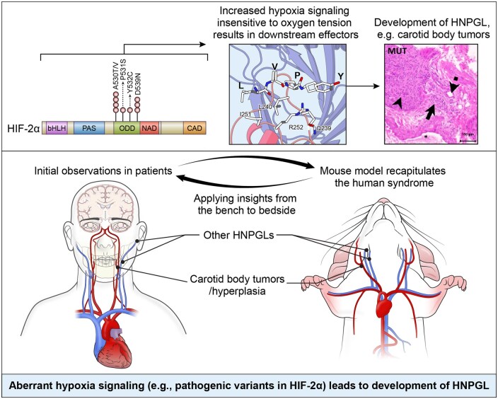
Background: Head and neck paragangliomas (HNPGLs) are typically slow-growing, hormonally inactive tumors of parasympathetic paraganglia. Inactivation of prolyl-hydroxylase domain-containing 2 protein causing indirect gain-of-function of hypoxia-inducible factor-2α (HIF-2α), encoded by EPAS1, was recently shown to cause carotid body hyperplasia. We previously described a syndrome with multiple sympathetic paragangliomas caused by direct gain-of-function variants in EPAS1 (Pacak-Zhuang syndrome, PZS) and developed a corresponding mouse model.

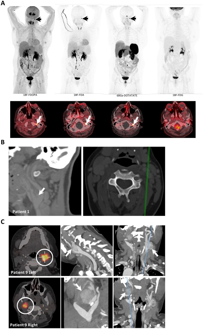
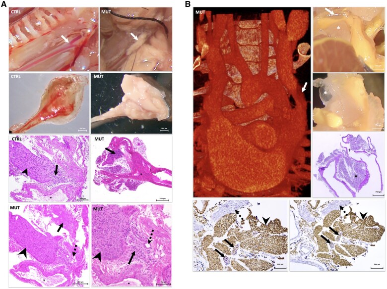
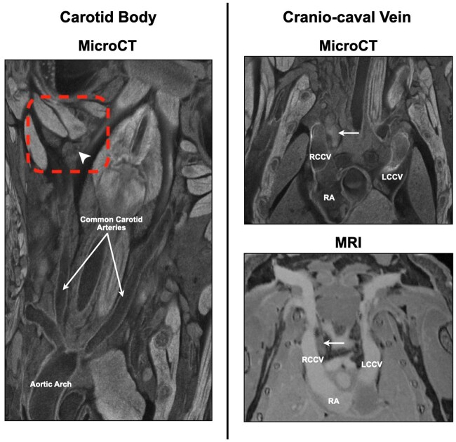
Methods: We evaluated a cohort of patients with PZS (n = 9) for HNPGL by positron emission tomography, magnetic resonance imaging, and computed tomography and measured carotid body size compared to literature reference values. Resected tumors were evaluated by histologic sectioning and staining. We evaluated the corresponding mouse model at multiple developmental stages (P8 and adult) for lesions of the head and neck by high resolution ex vivo imaging and performed immunohistochemical staining on histologic sections of the identified lesions.

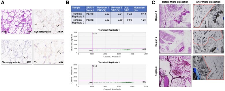
Results: hree patients had imaging consistent with HNPGL, one of which warranted resection and was confirmed on histology. Three additional patients had carotid body enlargement (Z-score > 2.0), and 3 had carotid artery malformations. We found that 9 of 10 adult variant mice had carotid body tumors and 6 of 8 had a paraganglioma on the cranio-caval vein, the murine homologue of the superior vena cava; these were also found in 4 of 5 variant mice at post-natal day 8. These tumors and the one resected from a patient were positive for tyrosine hydroxylase, synaptophysin, and chromogranin A. Brown fat in a resected patient tumor carried the EPAS1 pathogenic variant.

Conclusions: These findings (1) suggest HNPGL as a feature of PZS and (2) show that these pathogenic variants are sufficient to cause the development of these tumors, which we believe represents a continuous spectrum of disease starting from hyperplasia.


Conflict of interest statement

The authors have no conflicts of interest to declare.

Figures

Graphical Abstract
Graphical Abstract
The sequence of HIF-2a, which is encoded by EPAS1, is shown with locations of variants seen in the individuals with Pacak-Zhuang syndrome (PZS) marked in the sequence. These variants lead to increased hypoxia signaling that is insensitive to oxygen tension and the development of head and neck paraganglioma (HNPGL), as found in the individuals with PZS and in the corresponding mouse model.
Figure 1.
Figure 1.
Carotid body hyperplasia and tumors of the head and neck in patients with PZS. Panel A: In patient 5 (currently 55-year-old), a female patient with PZS, 46-year-old at the time of the scan, a lesion of the left skull base (arrows) is appreciated on the maximum intensity projection and fused axial images of positron emission tomography/computed tomography (PET/CT) scans using 18F-fluorodihydroxyphenylalanine (18F-FDOPA), 18F-flurodopamine (18F-FDA), and 68Ga-DOTA(0)-Tyr(3)-octreotate (68Ga-DOTATATE), consistent with glomus jugulare; 18F-fluorodeoxyglucose (18F-FDG) does not demonstrate the lesion. Multiple additional foci consistent with hepatic and para-lumbar paraganglioma are also appreciated best on 18F-FDOPA and 18F-FDA. The pituitary is positive in the 68Ga-DOTATATE scan. Panel B: In patient 1, a 42-year-old female patient with PZS, sagittal CT of the neck shows an anomalous trifurcation of the left carotid artery; the carotid body is seen (arrow). This section corresponds to the line on the axial section. Panel C: Images of lesions in patient 9, a 39-year-old female patient with PZS. Left—Axial slices of PET image following provision of 68Ga-DOTATATE shows increased uptake bilaterally (circles). Middle—Corresponding sagittal section on the simultaneously acquired low-dose CT for attenuation correction; paraganglioma marked by an arrow. Right—Corresponding coronal section showing location of the lesions (arrow) in the neck. Resection of this tumor confirmed paraganglioma on the basis of cytologic appearance of nested paraganglia cells and positive immunohistochemical staining (synaptophysin). Abbreviations: PET/CT = positron emission tomography/computed tomography; 18F-FDOPA = 18F-fluorodihydroxyphenylalanine; 18F-FDA = 18F-flurodopamine; 68Ga-DOTATATE = 68Ga-DOTA(0)-Tyr(3)-octreotate; 18F-FDG, 18F-fluorodeoxyglucose.
Figure 2.
Figure 2.
Paragangliomas in the syndrome mouse model. Panel A: Representative images shown. Gross dissection of the left carotid artery in the variant (MUT) mouse following fixation in 4% paraformaldehyde revealed a mass (arrow) at the carotid bifurcation; this was not present in the control mouse which was unfixed (CTRL). The carotid artery, including the bifurcation, was isolated from a variant mouse and visualized at 4× magnification, demonstrating the mass, which has a gross appearance consistent with a carotid body paraganglioma; the isolated carotid artery and bifurcation from the control mouse sample is also shown. Histologic sectioning, staining with hematoxylin and eosin (H&E), and visualization of the control sample at 10× magnification demonstrated the carotid body paraganglia (arrow) adjacent to the ganglion (arrowhead); the carotid artery is in view (asterisk). Visualization of the H&E-stained section from the variant sample at 4× magnification demonstrated a mass (arrow) at the carotid bifurcation (asterisk). The same section 20× magnification demonstrates the ganglion (arrowhead), abnormal vasculature (dashed arrow) within the paraganglia of the carotid body which is continuous with the mass (arrow) along the carotid (asterisk). The same region of abnormal vasculature is shown at 40×. Panel B: Coronal anteroposterior view of 3D reconstruction of micro-CT of the chest in a Microfil® polymer-perfused variant mouse shows angiographic blush (arrow) around a narrowed left cranio-caval vein, homologous to the human jugular vein, suggesting a glomus jugulare or glomus vagale tumor. Gross dissection of the same specimen confirmed a mass (asterisk and arrow) surrounding the polymer-filled left cranio-caval vein (yellow); the mass (arrow) was removed en bloc with the segment of the cranio-caval vein and bronchus (asterisk). Histologic sectioning and H&E staining at 4× magnification demonstrated the mass located between the vein and airway. Immunohistochemistry (IHC) with anti-synaptophysin antibody (lower left) on adjacent sections demonstrated positive signal in both adipocytes (arrowheads) and clusters of paraganglia (arrows) scattered throughout; nerve (dashed arrows) in field did not stain positively. IHC with chromogranin A antibody (lower right) showed the same pattern of signal positivity.
Figure 3.
Figure 3.
Carotid body and cranio-caval lesions in PZS. Left panel: High-resolution ex vivo micro-CT of the neck in PZS mouse model at post-natal day 8 revealed lesions of the carotid bifurcation (dashed box) likely consistent with either hyperplasia or a tumor of the carotid body (arrowhead). Right panel: High-resolution ex vivo micro-CT and MRI of the chest in the PZS model at the same stage similarly revealed lesions of the cranio-caval vein (CCV), which is homologous to the human superior vena cava, likely consistent with a tumor such as a paraganglioma (arrow). Right atrium, RA.
Figure 4.
Figure 4.
Variant allele frequency in brown fat in paraganglioma tissue resected from patient 2. Representative images are shown. Panel A: Hematoxylin and eosin (H&E) staining is shown. Tyrosine hydroxylase (TH), chromogranin A, and synaptophysin positivity are shown in a resected patient tumor; positivity in brown fat is marked with arrows while positivity in autonomic nerves innervating brown fat is indicated by arrowheads. Panel B: This middle panel shows tabulated results of the EPAS1 variant allele frequency in brown fat tissue within the resected region. The bottom of the panel shows representative digital droplet polymerase chain reaction measurement results from these tissues. Panel C: Micrographs of the regions of tissue measured before and after isolation of the brown fat for measurement of variant allele frequency. Images before and after dissection were collected at 4× and 2× magnification, respectively.

Similar articles

Cited by

References

    1. Därr R, Nambuba J, Del Rivero J, et al. Novel insights into the polycythemia-paraganglioma-somatostatinoma syndrome. Endocr Relat Cancer. 2016;23:899-908. - PMC - PubMed
    1. Pacak K, Jochmanova I, Prodanov T, et al. New syndrome of paraganglioma and somatostatinoma associated with polycythemia. J Clin Oncol. 2013;31:1690-1698. - PMC - PubMed
    1. Semenza GL. Hypoxia-inducible factors in physiology and medicine. Cell. 2012;148:399-408. - PMC - PubMed
    1. Zhuang Z, Yang C, Lorenzo F, et al. Somatic HIF2A gain-of-function mutations in paraganglioma with polycythemia. N Engl J Med. 2012;367:922-930. - PMC - PubMed
    1. Chowdhury R, Leung IKH, Tian Y-M, et al. Structural basis for oxygen degradation domain selectivity of the HIF prolyl hydroxylases. Nat Commun. 2016;7:12673. - PMC - PubMed

MeSH terms

Substances

LinkOut - more resources