. 2025 Jan 17;29(1):39.
doi: 10.1007/s10006-025-01331-9.

Exosomes derived from bone marrow mesenchymal stem cells ameliorate chemotherapeutically induced damage in rats' parotid salivary gland

Affiliations

Exosomes derived from bone marrow mesenchymal stem cells ameliorate chemotherapeutically induced damage in rats' parotid salivary gland

Ahmed Zakaria et al. Oral Maxillofac Surg. .

Abstract

Objective: A nanometer-sized vesicles originating from bone marrow mesenchymal stem cells (BMMSCs), called exosomes, have been extensively recognized. This study defines the impact of BMMSCs and their derived exosomes on proliferation, apoptosis and oxidative stress (OS) levels of CP-induced parotid salivary gland damage.

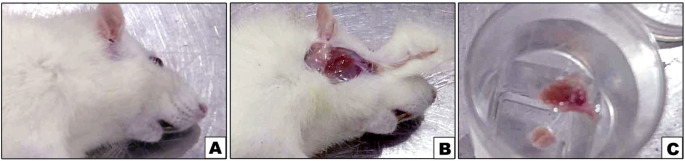
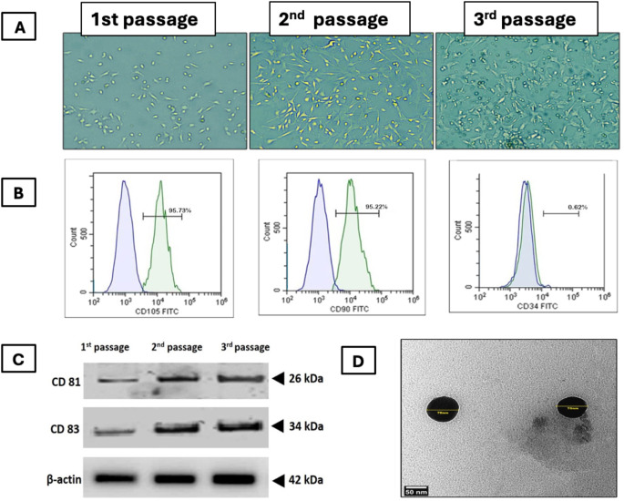
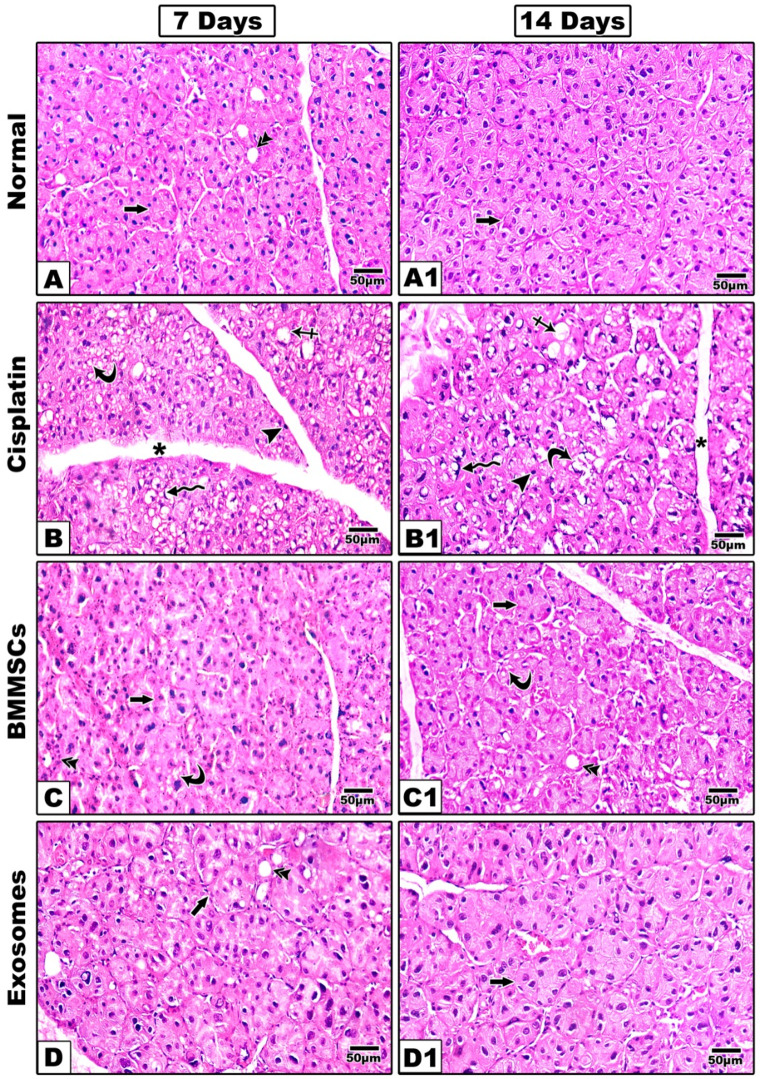
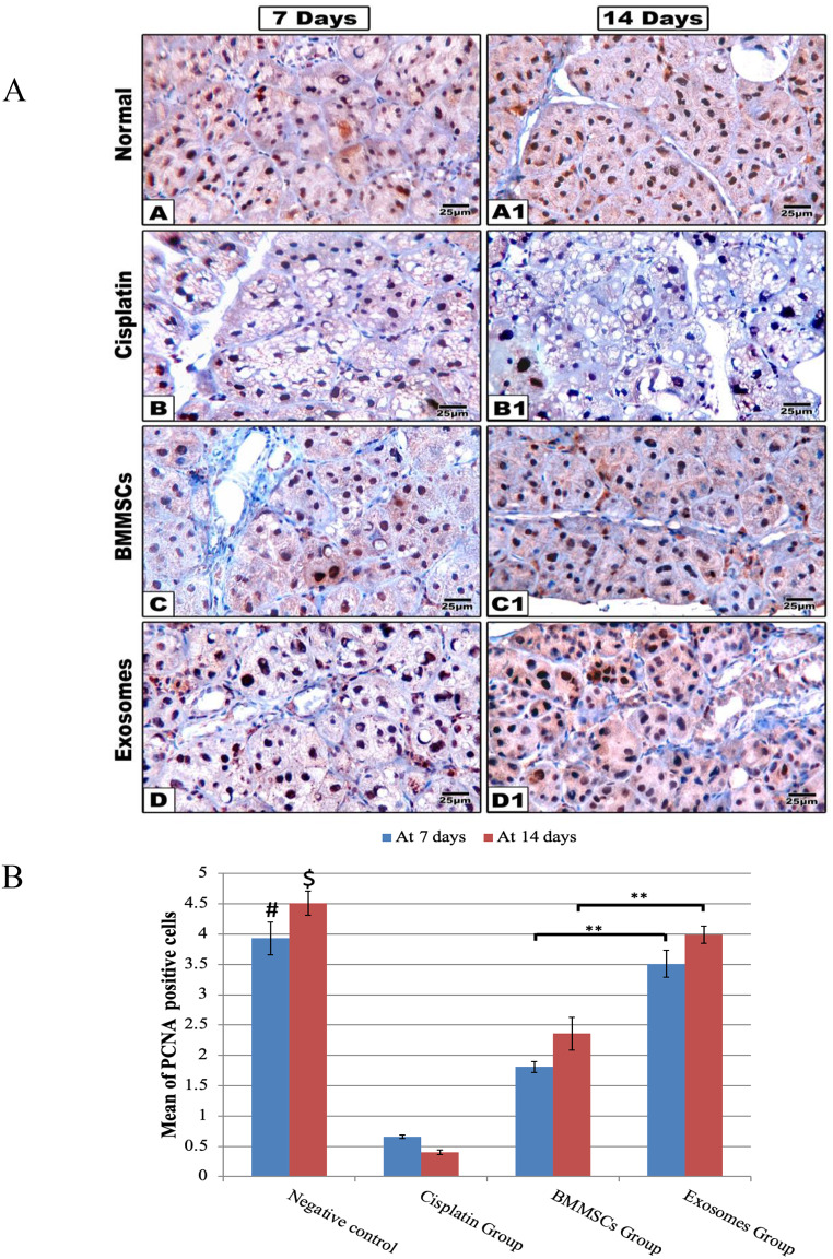
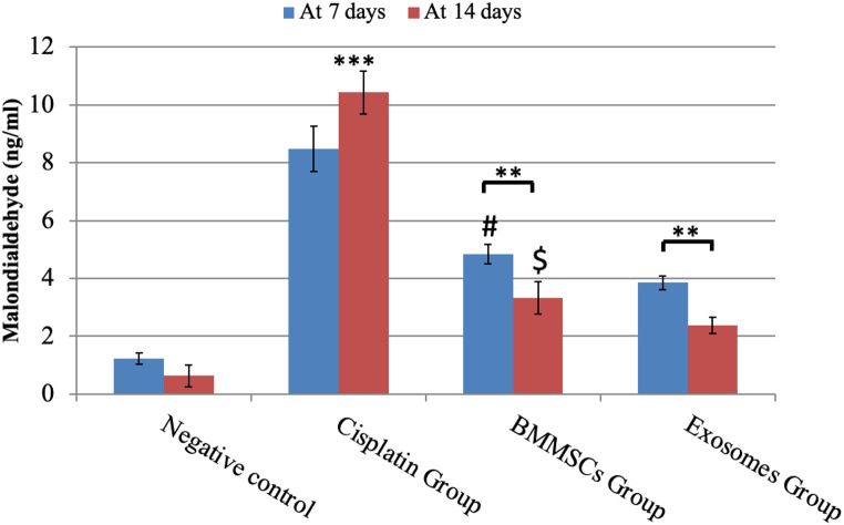
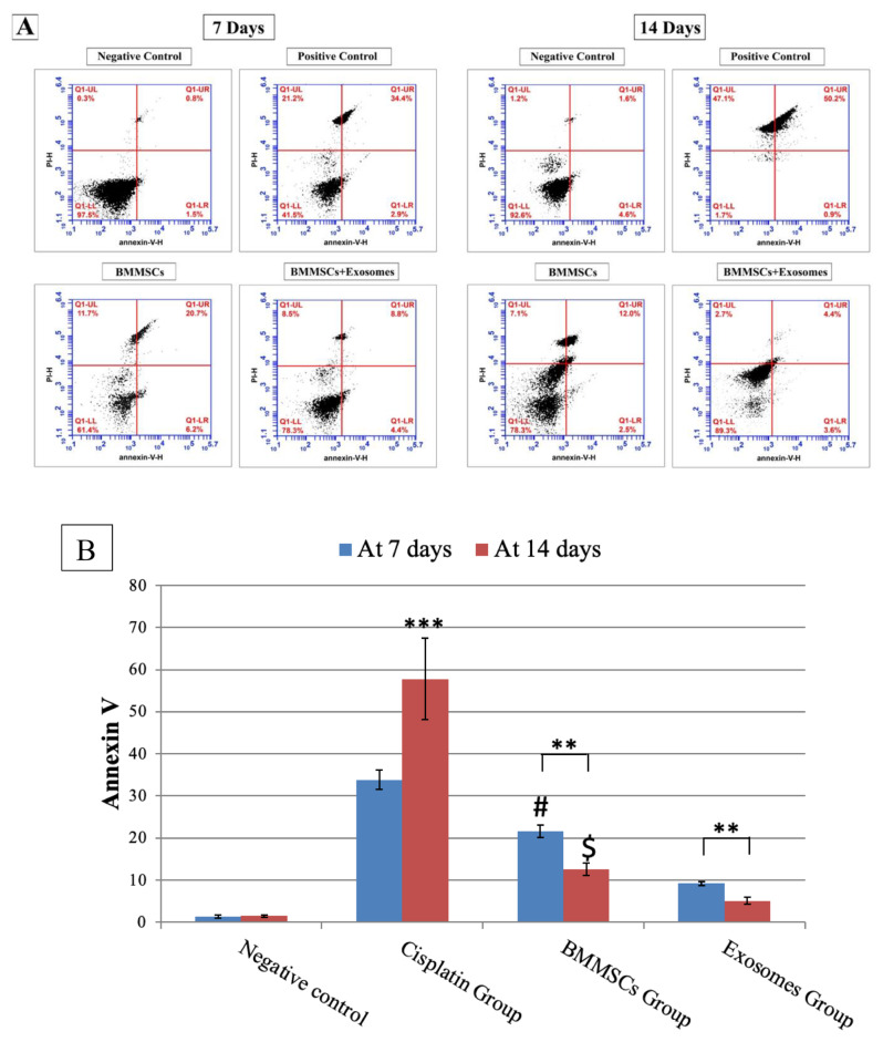
Methods: BMMSCs were isolated from the tibia of four white albino rats and further characterized by flowcytometric analysis. BMMSCs-derived exosomes were harvested and underwent characterization using transmission electron microscopy (TEM), western blot analysis and BCA assay. Fifty-six healthy white albino male rats weighting from 200 to 250 g were allocated into 4 groups (n = 14); Group I, rats received phosphate buffered saline (PBS), group II, rats were intraperitoneally injected with CP, group III& IV received CP and after 3 days they were intravenously injected with either BMMSCs (group III) or BMMSCs-exosomes (group IV). Histological, and immunohistochemical studies using proliferating cell nuclear antigen (PCNA) were done after 7 and 14 days. The OS was measured using malondialdehyde (MDA) and apoptosis was measured by annexin V-FITC/PI.

Results: BMMSCs and exosomes treated groups showed better histological features approximating the normal architecture of the control group. The percentage of PCNA positively stained cells were significantly higher in the exosomes treated group in comparison to all other groups. MDA assay test revealed that the exosomes were able to reduce the OS when compared to the cell-based therapy using BMMSCs. Annexin V revealed that BMMSCs-exosomes significantly reduced the percentage of apoptotic cells compared to other treated groups.

Conclusions: BMMSCs-exosomes could improve the CP-induced cytotoxicity in rats' parotid salivary gland.

Keywords: Bone marrow; Chemotherapy; Exosomes; Malondialdehyde; Parotid gland; Stem cells.


Conflict of interest statement

Declarations. Ethics approval and consent to participate: Experimental protocols of the current study were carried out in adherence to the appropriate criteria and were approved by the ethical committee of the Faculty of Dentistry at Mansoura University. Competing interests: The authors declare no competing interests.

Figures

Fig. 1
Fig. 1
Photographs A, B and C showing the surgical procedure of parotid gland isolation where B showing the parotid gland exposure and C showing the isolated glands
Fig. 2
Fig. 2
Characterization of BMMSCs and their derived exosomes. (A) phase contrast microscopic images showing BMMSCs at different passages, (B) Flowcytometric characterization of BMMSCs. (C) Western blotting of BMMSCs-exosomes for common markers CD83 and CD81 where, 1st passage unfractionated material and 2nd passage corresponds to the first ultracentrifugation and 3rd passage corresponds to the second ultracentrifugation. (D) TEM image showing nanosized vesicles
Fig. 3
Fig. 3
Representative histological images of (H&E) are shown at 7- and 14-days post-treatment. The group received CP (B, B1) shows remarkable structural alterations in the acini and ducts with multiple intracytoplasmic vacuoles (curved arrow) and ballooning of the acini (crossed arrow). The nuclei were irregular and different in shape and size and some nuclei showed also a signet ring appearance (zigzag arrow) other showing hyperchromatic nuclei (arrow head). With noticeable widening of connective tissue septa (asterisk). In BMMSCs treated group (C, C1); The acinar outline was still irregular (arrow) and acinar cells showed mitotic figures, but intracytoplasmic vacuoles were still detected at 7 days (curved arrow) however at 14 days sections showed improved arrangement of acinar and ductal cells with marked decrease in intracytoplasmic vacuoles. Groups treated with BMMSCs-exosomes (D, D1) revealed that the acinar (arrow), and ductal outline (double arrow) began to regain their normal architecture, the acinar cells showed mitotic figures noteworthy that intracytoplasmic vacuoles were still present although they were decreased in number sections showed well-arranged acini and ducts with almost normal appearance and marked reduction in the intracytoplasmic vacuoles. Scale 100x
Fig. 4
Fig. 4
(A) Representative histological images of PCNA immune proliferation marker are shown at 7- and 14-days post-treatment. The group received CP (B, B1) shows little expression of PCNA immune marker. While PCNA expression markedly increased in BMMSCs (C, C1) and the exosomes (D, D1) treated groups, scale 100x. (B) The Bar chart represents the histomorphometric analysis of PCNA positive expressions at 7- and 14-days post-treatment. Data were presented in means ± SEM. ** p < 0.01, # and $ means significant in relation to CP and BMMSCs groups after 7 and 14 days respectively, *** p < 0.001
Fig. 5
Fig. 5
MDA analysis in parotid glands exposed to different treatments for 7 and 14 days. Data were expressed in means ± SDs; n = 14. ** P < 0.01, *** P < 0.001.# Means significant to exosomes group after 7 days P < 0.05. $ means significant to exosomes group after 14 days P < 0.05
Fig. 6
Fig. 6
A: Showing flow plots for Annexin V (early marker of apoptosis) in parotid salivary gland after 7 and 14 days of treatment analysed by flow cytometry. Apoptosis was measured by Annexin V-FITC/PI staining of live cells. B: Demonstrating the percentage of apoptotic cells at 7- and 14-days post-treatment. Data are presented in means ± SDs. ***P < 0.001, **P < 0.01. # Means significant to exosomes group after 7 days P < 0.05. $ means significant to exosomes group after 14 days P < 0.05

Similar articles

References

    1. Patel VN, Hoffman MP (2013) Salivary gland development: a template for regeneration. Semin Cell Dev Biol 25:52–60. 10.1016/j.semcdb.2013.12.001 - PMC - PubMed
    1. Lombaert IM, Knox SM, Hoffman MP (2011) Salivary gland progenitor cell biology provides a rationale for therapeutic salivary gland regeneration. Oral Dis 17:445–449 - PMC - PubMed
    1. Bakhtiari S, Sehatpour M, Mortazavi H, Bakhshi M (2018) Orofacial manifestations of adverse drug reactions: a review study. Clujul Med 91:27–36. 10.15386/cjmed-748 - PMC - PubMed
    1. Maciejczyk M, Szulimowska J, Taranta-Janusz K, Wasilewska A, Zalewska A (2020) Salivary gland dysfunction, protein glycooxidation and nitrosative stress in children with chronic kidney disease. J Clin Med 9:1285 - PMC - PubMed
    1. Aljerf L, Alhaffar I (2017) Salivary distinctiveness and modifications in males with diabetes and Behçet’s Disease. Biochem Res Int 2017:1–12 - PMC - PubMed

LinkOut - more resources