. 2025 Mar;21(3):e14550.
doi: 10.1002/alz.14550. Epub 2025 Jan 17.

Plasma N-terminal tau fragment is an amyloid-dependent biomarker in Alzheimer's disease

Affiliations

Plasma N-terminal tau fragment is an amyloid-dependent biomarker in Alzheimer's disease

Guoyu Lan et al. Alzheimers Dement. 2025 Mar.

Abstract

Introduction: Novel fluid biomarkers for tracking neurodegeneration specific to Alzheimer's disease (AD) are greatly needed.

Methods: Using two independent well-characterized cohorts (n = 881 in total), we investigated the group differences in plasma N-terminal tau (NT1-tau) fragments across different AD stages and their association with cross-sectional and longitudinal amyloid beta (Aβ) plaques, tau tangles, brain atrophy, and cognitive decline.

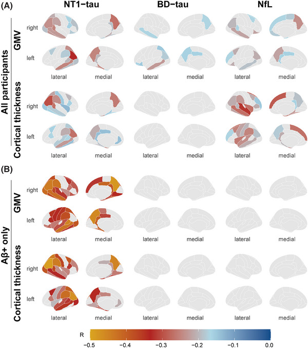
Results: Plasma NT1-tau significantly increased in symptomatic AD and displayed positive associations with Aβ PET (positron emission tomography) and tau PET. Higher baseline NT1-tau levels predicted greater tau PET, with 2- to 10-year intervals and faster longitudinal Aβ PET increases, AD-typical neurodegeneration, and cognitive decline. Plasma NT1-tau showed negative correlations with baseline regional brain volume and thickness, superior to plasma brain-derived tau (BD-tau) and neurofilament light (NfL) in Aβ-positive participants.

Discussion: This study suggests that plasma NT1-tau is an Aβ-dependent biomarker and outperforms BD-tau and NfL in detecting cross-sectional neurodegeneration in the AD continuum.

Highlights: Plasma N-terminal tau (NT1-tau) was specifically increased in the A+/T+ stage. Plasma NT1-tau was positively associated with greater amyloid beta (Aβ) and tau PET (positron emission tomography) accumulations. Higher plasma NT1-tau predicted greater tau burden and faster Aβ increases. Plasma NT1-tau was more related to neurodegeneration than plasma brain-derived tau (BD-tau) and neurofilament light (NfL).

Keywords: Alzheimer's disease; BD‐tau; NT1‐tau; NfL; cognitive decline; neurodegeneration.


Conflict of interest statement

The authors report no competing interests. Author disclosures are available in the Supporting Information.

Figures

FIGURE 1
FIGURE 1
Comparison of plasma NT1‐tau across the clinical diagnosis and biological stages. Plasma NT1‐tau levels in the GHABS cohort by Aβ status and clinical diagnosis (A) and A/T stages (B). Plasma NT1‐tau levels in the ADNI cohort by Aβ status and clinical diagnosis (C) and A/T stages (D). The boxplots depict the median (horizontal bar), IQR (hinges), and 1.5 × IQR (whiskers). Each point represents an individual, and green dashed lines represent the median values of the Aβ– CU or A–/T– group. Multiple comparisons were corrected using the Benjamini–Hochberg method (FDR <0.05). The adjusted p‐values of the comparisons are shown at the top of the bar, controlling for age and sex. Aβ, amyloid beta; ADNI, Alzheimer's Disease Neuroimaging Initiative; CI, cognitively impaired; CU, cognitively unimpaired; FDR, false discovery rate; GHABS, Greater‐Bay‐Area Healthy Aging Brain Study; IQR, interquartile range; MCI, mild cognitive impairment.
FIGURE 2
FIGURE 2
Association of plasma NT1‐tau with Aβ PET and tau PET. Association of plasma NT1‐tau with baseline FSP Aβ PET (n = 278) (A) and FTP tau PET (n = 141) (B) in the GHABS cohort. Association of plasma NT1‐tau with baseline FBP Aβ PET (n = 532) (C) and future FTP tau PET (n = 154) (D) in the ADNI cohort. The points (blue, Aβ–; red, Aβ+) and solid lines represent the participants and regression lines (95% CI). βstd and p were computed using GLM, controlling for age and sex. Spearman correlation coefficient (rho) was also calculated. ***p < 0.001; **p < 0.01; *p < 0.05. βstd, Standardized β; Aβ, amyloid beta; ADNI, Alzheimer's Disease Neuroimaging Initiative; CI, confidence interval; FBP, [18F]‐florbetapir; FSP, [18F]‐D3FSP; FTP, [18F]‐flortaucipir; GHABS, Greater‐Bay‐Area Healthy Aging Brain Study; GLM, Generalized linear models; PET, positron emission tomography; SUVR, standard uptake value ratio.
FIGURE 3
FIGURE 3
Association of plasma NT1‐tau with longitudinal Aβ PET. Association of plasma NT1‐tau with longitudinal changes in composite FBP Aβ PET (n = 381) in the ADNI cohort (A). LME models were used with composite FBP Aβ PET SUVR as the outcome and the time × NT1‐tau interaction as the predictor with random intercepts and time slopes. Age and sex were used as covariates. For illustration purposes only, the regression lines of longitudinal changes are shown for participants with plasma NT1‐tau levels below versus above the median. The trajectory of longitudinal changes in composite FBP Aβ PET, stratified by NT1‐tau status (B). Brain‐wise ROI analyses for the associations between plasma NT1‐tau and longitudinal FBP Aβ PET across 68 brain regions (C). The threshold was set at p < 0.05 after Benjamini–Hochberg corrections. ***p < 0.001. Aβ, amyloid beta; ADNI, Alzheimer's Disease Neuroimaging Initiative; FBP, [18F]‐florbetapir; LME, linear mixed‐effects; PET, positron emission tomography; ROI, region of interest; SUVR, standardized uptake value ratio.
FIGURE 4
FIGURE 4
Cross‐sectional association of plasma NT1‐tau, BD‐tau, and NfL with regional GMV and cortical thickness. Correlation coefficients of plasma NT1‐tau, BD‐tau, and NfL with baseline regional GMV and cortical thickness across 68 brain regions in the whole cohort (A) and Aβ+ participants (B). The threshold was set at p < 0.05 after Benjamini–Hochberg corrections. Aβ, amyloid beta; BD‐tau, brain‐derived tau; GMV, gray matter volume; NfL, neurofilament light.
FIGURE 5
FIGURE 5
Association of plasma NT1‐tau, BD‐tau, and NfL with longitudinal cortical thinning and hippocampal atrophy. Association of plasma NT1‐tau, BD‐tau, and NfL with longitudinal changes in temporal‐MetaROI cortical thickness (A) and residual HCV (B) in the GHABS cohort (n = 106). LME models were used with temporal‐MetaROI cortical thickness, residual HCV as outcome, and the time × plasma biomarkers interaction as predictor with random intercepts and time slopes. Age and sex were used as covariates. For illustration purposes only, the regression lines of longitudinal changes are shown for participants with plasma biomarker levels below versus above the median. ***p < 0.001; **p < 0.01; *p < 0.05. BD‐tau, brain‐derived tau; HCV, hippocampal volume; LME, linear mixed‐effects; NfL, neurofilament light.

Similar articles

References

    1. Guo T, Dukart J, Brendel M, Rominger A, Grimmer T, Yakushev I. Rate of β‐amyloid accumulation varies with baseline amyloid burden: implications for anti‐amyloid drug trials. Alzheimer's Dement. 2018;14:1387‐1396. doi:10.1016/j.jalz.2018.05.013 - DOI - PubMed
    1. Guo T, Shaw LM, Trojanowski JQ, Jagust WJ, Landau SM. Association of CSF Aβ, amyloid PET, and cognition in cognitively unimpaired elderly adults. Neurology. 2020;95:e2075‐e2085. doi:10.1212/WNL.0000000000010596 - DOI - PMC - PubMed
    1. Cai Y, Du J, Li A, et al. Initial levels of β‐amyloid and tau deposition have distinct effects on longitudinal tau accumulation in Alzheimer's disease. Alzheimers Res Ther. 2023;15:1‐14. doi:10.1186/s13195-023-01178-w - DOI - PMC - PubMed
    1. Guo T, Korman D, La Joie R, et al. Normalization of CSF pTau measurement by Aβ40 improves its performance as a biomarker of Alzheimer's disease. Alzheimers Res Ther. 2020;12:97. doi:10.1186/s13195-020-00665-8 - DOI - PMC - PubMed
    1. Jack CR Jr, Andrews JS, Beach TG, et al. Revised criteria for diagnosis and staging of Alzheimer's disease: Alzheimer's Association Workgroup. Alzheimers Dement. 2024:20:5143‐5169. doi:10.1002/alz.13859 - DOI - PMC - PubMed

MeSH terms