Case Reports
. 2025 Aug;54(8):1753-1759.
doi: 10.1007/s00256-025-04872-y. Epub 2025 Jan 17.

Multifocal osteochondromatous proliferation and paraneoplastic hematologic dyscrasia in the context of latent Epstein-Barr virus reactivation: a case of oncologic and infectious pathophysiology

Affiliations
Case Reports

Multifocal osteochondromatous proliferation and paraneoplastic hematologic dyscrasia in the context of latent Epstein-Barr virus reactivation: a case of oncologic and infectious pathophysiology

Zeyad Hossam Atta Khalil et al. Skeletal Radiol. 2025 Aug.

Abstract

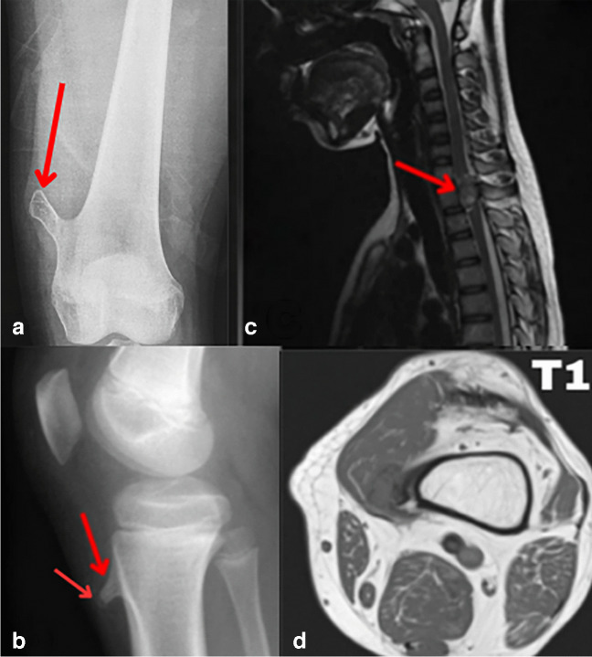
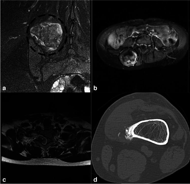
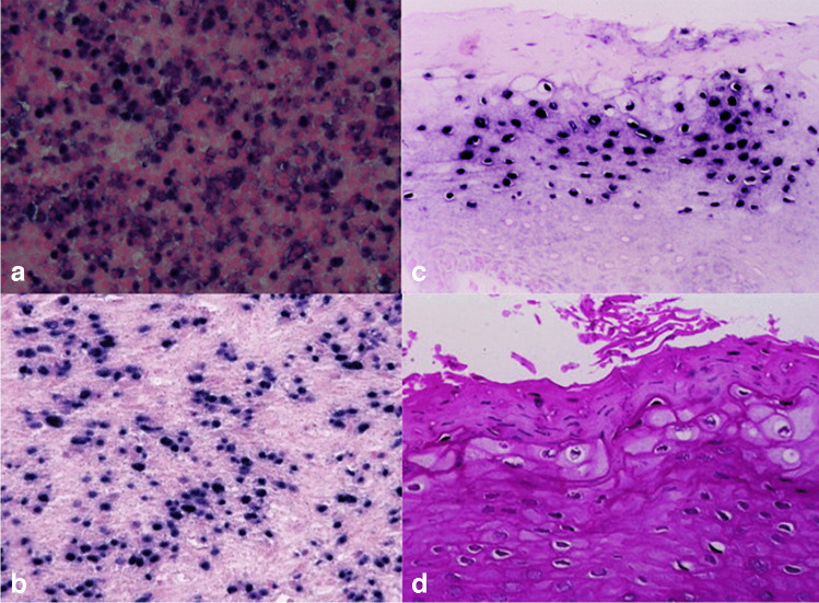
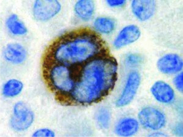
This case report describes a 15-year-old male with multifocal osteochondromatous proliferation and paraneoplastic hematologic dyscrasia, linked to latent Epstein-Barr virus reactivation. Radiographic and advanced imaging revealed widespread skeletal lesions consistent with osteochondromatosis. Hematologic evaluation indicated pancytopenia with dysplastic megakaryocytes and marrow infiltration. Immunohistochemical staining confirmed latent Epstein-Barr virus infection, suggesting its role in the pathogenesis of both the osteochondromatous and hematologic abnormalities. This case highlights the correlation between Epstein-Barr virus reactivation, bone proliferation, and paraneoplastic hematologic processes, which we believe has not yet been reported in the literature, emphasizing the need for a comprehensive diagnostic approach.

Keywords: Bone marrow dysplasia; Epstein-Barr virus (EBV); Multifocal osteochondromatous proliferation; Paraneoplastic hematologic dyscrasia.


Conflict of interest statement

Declarations. Competing of interest: The authors declare no competing interests. Informed consent: Written informed consent was obtained from the patient for the publication of this case report and the accompanying images. The patient has been informed that the information and images will be anonymized, ensuring that no identifying details will be disclosed. This consent process was conducted by ethical guidelines established by the corresponding institution where the study has been reported, ensuring the patient’s autonomy, confidentiality, and rights were fully protected.

Figures

Fig. 1
Fig. 1
The AP radiograph image of the knee (a) shows a well-defined lesion in the medial aspect of the femoral metaphyses directed away from the joint. The knee joint is normal in appearance. The lesion was hard in nature and found as a lump in the left thigh with the thought of potential continuity, which was disapproved, due to the absence of clear continuity on advanced imaging. The lateral radiograph image (b) reveals a sessile ossific tibial metaphyseal lesion and a small bony protrusion from the anterior metaphysis, separated from the growth plate, with chondroid calcification and no continuity with the adjacent bone marrow, which was initially thought as well to be continuous but appeared on advanced imaging otherwise. A second arrow points to the chondroid calcification found at the anterior margin of the lesion. A STIR MR of the cervical spine in image (c) demonstrates a well-defined lesion extending into the spinal canal, causing mass effect and early myelopathic changes in the dorsal spinal cord; STIR MRI was performed specifically in this case due to its capability to suppress fat signals, which was essential for visualizing the osteochondromatous lesion without interference from overlapping fat signals. Posterior scalloping of the T2 vertebral body is also evident, highlighting the lesion’s pressure impact and remodeling effects. The T1-weighted MRI shown here in (d) demonstrates the lack of continuity of the lesion in the distal femur with the medulla. The lesion’s low intensity on the T1 image is also evident
Fig. 2
Fig. 2
The coronal and axial T2 FS post-contrast. Images (a) and (b) show a large, multiloculated osteochondroma arising from the posterior right iliac crest, extending slightly into the superior sacroiliac joint and laterally toward the right paraspinal muscles and is non-continuous. Post-contrast images show patchy enhancement in the posterior osseous component, though its significance is unclear. No fractures or bursa formations are identified, and there is no associated soft tissue mass. Degenerative changes in the right sacroiliac joint include small subchondral cysts. The pelvis appears to be clear of masses. Image (c) shows an axial T2 MRI detailing the exact location of the T2 lesion relative to the vertebral body. A pedunculated bony outgrowth originates from the right superior articular process of the T2 vertebra. This lesion presents with a T2 hypointense stalk and a T2 hyperintense signal in its cap. Post-contrast imaging reveals heterogeneous enhancement of the lesion, which extends into the spinal canal, resulting in myelopathic changes in the adjacent dorsal spinal cord. Image (d) illustrates a CT scan displaying a widespread ossified mass adjacent to the medial cortex of the distal femur, with no continuity observed between the mass and the medullary cavity
Fig. 3
Fig. 3
In situ hybridization for Epstein-Barr virus (EBV) from bone marrow biopsy of femoral metaphyseal lesion. a Bone marrow smear showing cellularity. b Positive EBER staining in bone marrow cells, indicated by dark nuclear signals, confirming EBV presence. c Higher magnification highlighting nuclear EBER positivity in hematopoietic cells. d ISH staining of the osteochondromatous lesion showing nuclear EBER signals in stromal and chondrocytic cells, indicating EBV’s involvement in both the hematologic and skeletal abnormalities
Fig. 4
Fig. 4
Immunohistochemical staining of the excised osteochondromatous lesion, showing strong nuclear and cytoplasmic positivity for Epstein-Barr virus (EBV) latent membrane protein 1 within a chondrocytic cell. The presence of latent membrane protein 1, marked by the brown staining, indicates active EBV involvement within the lesion’s cellular matrix. Surrounding cells exhibit typical morphology without latent membrane protein 1 expression, highlighting the localized EBV infection in the osteochondromatous tissue

Similar articles

References

    1. Bajwa SN, Reddy R, Wagh YS, Agarwal M, Katariya A. Bizarre parosteal osteochondromatous proliferation- a case series of typical and atypical presentations. J Orthop Case Rep. 2019;10(1):45–50. 10.13107/jocr.2019.v10.i01.1630. - PMC - PubMed
    1. Alvarez C, Tredwell S, De Vera M, Hayden M. The genotype-phenotype correlation of hereditary multiple exostoses. Clin Genet. 2006;70(2):122–30. 10.1111/j.1399-0004.2006.00653.x. - PubMed
    1. R K, K P, B G, Naseer B S, Ds K. Multiple heriditary exostoses in a family for three generation of Indian origin with review of literature. J Clin Diagn Res. 2014;8(10):LD01–3. 10.7860/JCDR/2014/8228.4942 - PMC - PubMed
    1. Tepelenis K, Papathanakos G, Kitsouli A, et al. Osteochondromas: an updated review of epidemiology, pathogenesis, clinical presentation, radiological features and treatment options. In Vivo. 2021;35(2):681–91. 10.21873/invivo.12308. - PMC - PubMed
    1. Bukowska-Olech E, Trzebiatowska W, Czech W, et al. Hereditary multiple exostoses-a review of the molecular background, diagnostics, and potential therapeutic strategies. Front Genet. 2021;10(12):759129. 10.3389/fgene.2021.759129. - PMC - PubMed

Publication types

MeSH terms

LinkOut - more resources