. 2025 May;62(5):6503-6520.
doi: 10.1007/s12035-025-04686-y. Epub 2025 Jan 16.

Dose-Dependent Effect of a New Biotin Compound in Hippocampal Remyelination in Rats

Affiliations

Dose-Dependent Effect of a New Biotin Compound in Hippocampal Remyelination in Rats

Burak Yulug et al. Mol Neurobiol. 2025 May.

Abstract

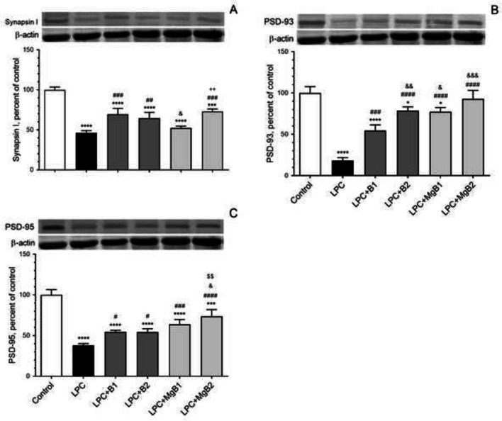
Demyelination is commonly observed in neurodegenerative disorders, including multiple sclerosis (MS). Biotin supplementation is known to stabilize MS progression. To reduce the effective dose of biotin, we synthesized a new and superior form of biotin, a complex of magnesium ionically bound to biotin (MgB) and compared its dose-dependent effect with biotin alone after inducing demyelination using lysolecithin (LPC) in rats. Myelination was assessed using luxol fast blue staining and immunostaining against MBP protein, revealing that the most significant remyelination occurred in the MgB groups. Additionally, both biotin and MgB-treated animals showed dose-dependent improvements in spatial memory. Moreover, we detected a decrease in inflammatory proteins in both treatment groups, which was more prominent in high-dose MgB-treated animals and correlated with decreased expression of NF-κB p65, OP, and MMP-9 proteins. Further analysis of biotin-related proteins demonstrated that both biotin and, notably, MgB reversed the demyelination-dependent reduction of these proteins. Furthermore, biotin, particularly MgB, improved neuronal transmission proteins, Synapsin-1, PSD-93, and PSD-95. Additionally, both treatment groups exhibited increased BDNF, GAP43, and ICAM levels, with significant increments observed in high-dose MgB-treated animals. Increased GFAP, indicative of reactive gliosis, was observed in LPC-treated animals, and this effect was notably reversed by high-dose MgB treatment. The current data emphasize the dose-dependent beneficial effect on the remyelination process. Furthermore, the combination of biotin with Mg resulted in a more potent effect compared to biotin by itself. The strong influence of MgB encourages proof-of-concept studies using MgB in patients with MS.

Keywords: Biotin; Demyelination; Magnesium-biotin; Multiple sclerosis; Remyelination.


Conflict of interest statement

Declarations. Ethics Approval: This study complied with ethical concerns and was approved by the Firat University Animal Research Ethics Committee, Approved number: 257169. Consent to Participate: Not applicable. Consent for Publication: Not applicable. Conflict of Interest: James Komorowski, Sara Perez Ojalvo, and Sarah Sylla are employed by JDS Therapeutics, LLC (Purchase, NY, USA). The other authors declare no conflict of interest.

Figures

Fig. 1
Fig. 1
Effects of biotin and magnesium biotinate (MgB) supplementation on entries to target quadrant (A), probe trial (B), and latency (C) to find the hidden platform from the first day to the fifth day in the Morris water maze task in hippocampal demyelinated rats (n = 7). Different symbols (*, #, &, $, and +) indicate difference compared to the Control, LPC, LPC + B1, LPC + B2, LPC + MgB1, and LPC + MgB2 groups, respectively) Statistical significance between groups was shown as compared to Control *p < 0.05, **p < 0.01, ***p < 0.001, ****p < 0.0001; compared to LPC #p < 0.05, ####p < 0.0001; compared to LPC + B1; &&&p < 0.001, &&&&p < 0.0001; compared to LPC + MgB1; ++++p < 0.0001, respectively, using ANOVA and Tukey’s post-hoc test. B1: Biotin 0.9 mg/rat/day; B2: Biotin 9 mg/rat/day; MgB1: magnesium biotinate (Biotin 0.9 mg/rat/day); MgB2: magnesium biotinate (Biotin 9 mg/rat/day)
Fig. 2
Fig. 2
Representative images of histopathological changes stained by H&E (A) and Luxol fast blue (B) in response to Biotin B1, B2, MgB1, and MgB2 treatments in demyelinated rat hippocampus. (n = 7). B1: Biotin 0.9 mg/rat/day; B2: Biotin 9 mg/rat/day; MgB1: magnesium biotinate (Biotin 0.9 mg/rat/day); MgB2: magnesium biotinate (Biotin 9 mg/rat/day)
Fig. 3
Fig. 3
Representative images of gliosis in response to Biotin B1, B2, MgB1, and MgB2 treatments in demyelinated rat hippocampus. The left panel shows immunohistochemical staining against myelin basic protein (MBP), labeling myelin (red), and the right panel shows immunohistochemical staining against the glial fibrillary basic protein (GFAP), labeling astrocytes (orange). B.i LPC causes extensive demyelination in the rat hippocampus, compared to the saline-treated group in (A.i). Both doses of biotin accelerate remyelination; however, complete remyelination is not observed (C.i and D.i). An increase in the astrocyte population is observed in the LPC-treated group (B.ii) compared to saline-treated control group (A.ii). The reactive state of astrocytes continues at a low dose of biotin (C.ii) but decreases when a high dose is applied (D.ii). MgB1 and MgB2 also accelerate remyelination (E.i and F.i) and attenuate reactive states (E.ii and F.ii). Almost complete remyelination is observed in these groups (E and F). Images were taken from the CA1 region of the hippocampus with 40 × (i) and 20 × (ii) objectives. Scale bars: i, 50 µm; ii, 100 µm
Fig. 4
Fig. 4
Effects of biotin and magnesium biotinate (MgB) supplementation on hippocampal interleukin 6 (IL-6, A), interleukin 17A (IL-17A, B), tumor necrosis factor-alpha (TNF-α, C), nuclear factor kappa light chain enhancer of activated B cells (NF-κB p65, D), chemokine (C–C motif) ligand 3 (CCL-3, E), chemokine (C–C motif) ligand 5 (CCL-5, F) and chemokine (C-X-C motif) ligand 16 (CXCL-16, G), osteoprotegerin (OPG, H) and matrix metallopeptidase 9 (MMP-9, I) protein levels in hippocampal demyelinated rats (n = 7). Data are expressed as a percent of the control value. Each bar represents the mean and standard error of the mean. Blots were repeated at least three times. Western blot analysis was performed with actin included, ensuring equal protein loading. The data are percentages of the control. Different symbols (*, #, &, $, and + indicates difference compared to the Control, LPC, LPC + B1, LPC + B2, LPC + MgB1, and LPC + MgB2 groups, respectively). Statistical significance between groups was shown as compared to Control *p < 0.05, **p < 0.01, ***p < 0.001, ****p < 0.0001; compared to LPC #p < 0.05, ##p < 0.01, ###p < 0.001, ####p < 0.0001; compared to LPC + B1; &p < 0.05, &&p < 0.01, &&&p < 0.001, &&&&p < 0.0001; compared to LPC + B2; $p < 0.05, $$p < 0.01, $$$p < 0.001, $$$$p < 0.0001; compared to LPC + MgB1 +p < 0.05, +++p < 0.001, ++++p < 0.0001, using ANOVA and Tukey’s post-hoc test. B1, Biotin 0.9 mg/rat/day; B2, Biotin 9 mg/rat/day; MgB1, magnesium biotinate (Biotin 0.9 mg/rat/day); MgB2, magnesium biotinate (Biotin 9 mg/rat/day)
Fig. 5
Fig. 5
Effects of biotin and magnesium biotinate (MgB) supplementation on hippocampal acetyl-CoA carboxylase 1 (ACC1, A), acetyl-CoA carboxylase 2 (ACC2, B), pyruvate carboxylase (PC, C), propionyl-CoA carboxylase (PCC, D), and 3-methylcrotonyl-CoA carboxylase (MCC, E) protein levels in hippocampal demyelinated rats (n = 7). Data are expressed as a percent of the control value. Each bar represents the mean and standard error of the mean. Blots were repeated at least three times. Western blot analysis was performed with actin included, ensuring equal protein loading. The data are percentages of the control. Different symbols (*, #, &, $, and +) indicates difference compared to the Control, LPC, LPC + B1, LPC + B2, LPC + MgB1, and LPC + MgB2 groups, respectively). Statistical significance between groups were shown by as compared to Control *p < 0.05, **p < 0.01, ***p < 0.001, ****p < 0.0001; compared to LPC #p < 0.05, ##p < 0.01, ###p < 0.001, ####p < 0.0001; compared to LPC + B1; &&p < 0.01, &&&p < 0.001, &&&&p < 0.0001; compared to LPC + B2; $p < 0.05, $$$$p < 0.0001; compared to LPC + MgB1.++++p < 0.0001, using ANOVA and Tukey’s post-hoc test. B1, Biotin 0.9 mg/rat/day; B2, Biotin 9 mg/rat/day; MgB1, magnesium biotinate (Biotin 0.9 mg/rat/day); MgB2, magnesium biotinate (Biotin 9 mg/rat/day)
Fig. 6
Fig. 6
Effects of biotin and magnesium biotinate (MgB) supplementation on hippocampal brain-derived neurotrophic factor (BDNF, A), growth-associated protein (GAP43, B), glial fibrillary acidic protein (GFAP, C), and intercellular adhesion molecule 1 (ICAM-1, D) protein levels in hippocampal demyelinated rats (n = 7). Data are expressed as a percent of the control value. Each bar represents the mean and standard error of the mean. Blots were repeated at least three times. Western blot analysis was performed with actin included, ensuring equal protein loading. The data are percentages of the control. Different symbols (*, #, &, $, and +) indicates difference compared to the Control, LPC, LPC + B1, LPC + B2, LPC + MgB1, and LPC + MgB2 groups, respectively). Statistical significance between groups were shown as compared to Control *p < 0.05, **p < 0.01, ***p < 0.001, ****p < 0.0001; compared to LPC #p < 0.05, ##p < 0.01, ###p < 0.001, ####p < 0.0001; compared to LPC + B1; &p < 0.05, &&p < 0.01, &&&p < 0.001, &&&&p < 0.0001; compared to LPC + B2; $p < 0.05, $$p < 0.01; compared to LPC + MgB1.+p < 0.05, using ANOVA and Tukey’s post-hoc test. B1, Biotin 0.9 mg/rat/day; B2, Biotin 9 mg/rat/day; MgB1, magnesium biotinate (Biotin 0.9 mg/rat/day); MgB2, magnesium biotinate (Biotin 9 mg/rat/day)
Fig. 7
Fig. 7
Effects of biotin and magnesium biotinate (MgB) supplementation on hippocampal Synapsin-I (A), postsynaptic density protein 93 (PSD-93, B) and postsynaptic density protein 95 (PSD-95, C) protein levels in hippocampal demyelinated rats (n = 7). Data are expressed as a percent of the control value. Each bar represents the mean and standard error of the mean. Blots were repeated at least three times. Western blot analysis was performed with actin included, ensuring equal protein loading. The data are percentages of the control. Different symbols (*, #, &, $, and +) indicates difference compared to the Control, LPC, LPC + B1, LPC + B2, LPC + MgB1, and LPC + MgB2 groups, respectively). Statistical significance between groups were shown as compared to Control *p < 0.05, ***p < 0.001, ****p < 0.0001; compared to LPC #p < 0.05, ##p < 0.01, ###p < 0.001, ####p < 0.0001; compared to LPC + B1; &p < 0.05, &&p < 0.01, &&&p < 0.001; compared to LPC + B2; $$p < 0.01; compared to LPC + MgB1.++p < 0.01, using ANOVA and Tukey’s post-hoc test. B1, Biotin 0.9 mg/rat/day; B2, Biotin 9 mg/rat/day; MgB1, magnesium biotinate (Biotin 0.9 mg/rat/day); MgB2, magnesium biotinate (Biotin 9 mg/rat/day)

References

    1. Sedel F, Bernard D, Mock DM, Tourbah A (2016) Targeting demyelination and virtual hypoxia with high-dose biotin as a treatment for progressive multiple sclerosis. Neuropharmacology 110:644–653. 10.1016/j.neuropharm.2015.08.028 - PubMed
    1. Sedel F, Papeix C, Bellanger A, Touitou V, Lebrun-Frenay C, Galanaud D, Gout O, Lyon-Caen O et al (2015) High doses of biotin in chronic progressive multiple sclerosis: a pilot study. Mult Scler Relat Disord 4:159–169. 10.1016/j.msard.2015.01.005 - PubMed
    1. Ludwin SK, Rao VT, Moore CS, Antel JP (2016) Astrocytes in multiple sclerosis. Mult Scler J 22:1114–1124. 10.1177/1352458516643396 - PubMed
    1. Stadelmann C, Wegner C, Brück W (2011) Inflammation, demyelination, and degeneration - recent insights from MS pathology. Biochim Biophys Acta-Mol Basis Dis 1812:275–282. 10.1016/j.bbadis.2010.07.007 - PubMed
    1. Ayers MM, Hazelwood LJ, Catmull DV, Wang D, McKormack Q, Bernard CC, Orian JM (2004) Early glial responses in murine models of multiple sclerosis. Neurochem Int 45:409–419. 10.1016/j.neuint.2003.08.018 - PubMed

LinkOut - more resources