

Pubic Arch Angle Postural Mobility Evaluation in Different Patient's Positions for Clinical Research
- PMID: 39822419
- PMCID: PMC11737889
- DOI: 10.7759/cureus.75898
Pubic Arch Angle Postural Mobility Evaluation in Different Patient's Positions for Clinical Research
Abstract
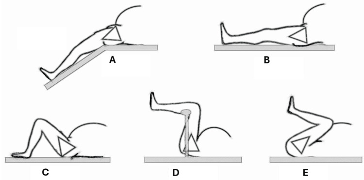
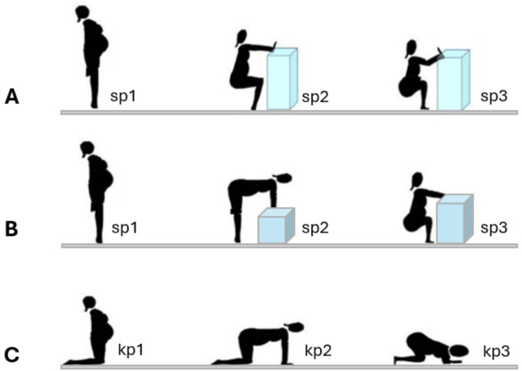
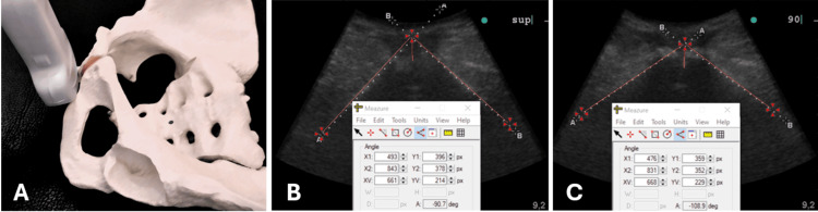
Childbirth is a dynamic process involving mutual adaptation between the maternal pelvis and the presenting fetal part. The ability of the pelvis to maintain optimal mobility during labor plays a crucial role in achieving favorable obstetric outcomes. The pubic arch angle (PAA) increases amplitude during pregnancy, showing pelvic tissue adjustment. The PAA evaluated with ultrasound in a single position predicts the risk of dystocia in labor and, consequently, anal sphincter trauma and incontinence after delivery. The hip flexion degree was found to reduce lumbar lordosis, shift the sacral promontory, affect the pubic arch angle, and increase pelvic diameter, creating more space for the fetus to descend during labor. Studies with magnetic resonance have demonstrated the modification of pelvic diameters and the PAA with maternal position change in the degree of hip joint flexion. The present technical report intends to describe the technique for evaluating the PAA amplitude change in supine, kneeling, and standing patients' different leg positions. The procedure is designed for clinical research in labor biomechanics. The supine leg positions for pubic angle measurement can vary from hyperextension, as in Walcher's position, to neutral supine position, mild hip flexion, and hyperflexion, which is the position of the McRoberts maneuver. The kneeling and standing positions mimic labor and delivery in the flexible sacrum maternal positions. The 2D ultrasound technique can assess the PAA in the clinical research setting during the obstetrical examination. The transducer transversely positioned on the perineum shows the pubic symphysis and the two symmetrical ischiopubic branches, as described in the literature. Evidence from ultrasound, magnetic resonance imaging, and computational modeling highlights the adaptability of pelvic structures influenced by hip flexion and soft tissue elasticity. Preliminary studies confirm significant positional differences in pubic arch angle and pelvic measurements, supporting the clinical relevance of assessing pelvic mobility. The proposed ultrasound-based approach for evaluating PAA measurements in various maternal positions offers a practical tool for research in labor management and predicting vaginal birth outcomes. Ongoing research aims to elucidate further the relationship between pelvic dimensions in different maternal positions, fetal progression, and obstetric outcomes, contributing to safer, more effective childbirth practices.
Keywords: childbirth; mobility limitation; obstetrics; pelvimetry; posture; pubic arch angle; ultrasound.
Copyright © 2024, Siccardi et al.
Conflict of interest statement
Human subjects: Consent for treatment and open access publication was obtained or waived by all participants in this study. Animal subjects: All authors have confirmed that this study did not involve animal subjects or tissue. Conflicts of interest: In compliance with the ICMJE uniform disclosure form, all authors declare the following: Payment/services info: All authors have declared that no financial support was received from any organization for the submitted work. Financial relationships: All authors have declared that they have no financial relationships at present or within the previous three years with any organizations that might have an interest in the submitted work. Intellectual property info: Marco Siccardi admits to owning the intellectual property of the "Digital Distance Indicator" mentioned in the article's discussion session. Other relationships: All authors have declared that there are no other relationships or activities that could appear to have influenced the submitted work.
Figures
References
-
- Pubic arch angle in prolonged second stage of labor: clinical significance. Gilboa Y, Kivilevitch Z, Spira M, Kedem A, Katorza E, Moran O, Achiron R. Ultrasound Obstet Gynecol. 2013;41:442–446. - PubMed
-
- Correlation between bituberous diameter and mode of delivery in a cohort of low-risk nulliparous women. Neri S, Di Pasquo E, Corrado NA, et al. Eur J Obstet Gynecol Reprod Biol. 2023;287:75–79. - PubMed
-
- Longitudinal changes of subpubic arch angle throughout pregnancy. Martelli F, Youssef A, Capogna MV, et al. Gynecol Obstet Invest. 2020;85:100–106. - PubMed
LinkOut - more resources
Full Text Sources