Value of CT diagnostic techniques based on imaging post-processing systems in the early diagnosis and treatment of lung cancer
- PMID: 39822500
- PMCID: PMC11733320
- DOI: 10.62347/VJSR2965
Value of CT diagnostic techniques based on imaging post-processing systems in the early diagnosis and treatment of lung cancer
Abstract
Objective: To evaluate the application value of CT diagnostic technology based on the Shukun Imaging Post-Processing System for early screening and diagnosis of lung cancer.
Methods: A total of 35 patients diagnosed with lung cancer postoperatively and 53 patients with benign nodules were included in this retrospective study, all of whom were treated in the Department of Thoracic and Cardiovascular Surgery of the Second Affiliated Hospital of Fujian Medical University from January 2020 to December 2023. All patients underwent chest spiral CT examinations. Original thin-slice axial CT images were processed using Shukun software for three-dimensional reconstruction of the lesions, surrounding lung tissue, and trachea. The diagnoses and malignant risk indicators of pulmonary nodules were established based on the final imaging results.
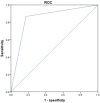
Results: Statistical analysis showed that the sensitivity of Shukun processing technology in diagnosing early-stage lung cancer was 82.86%, with a specificity of 88.46%, when compared to postoperative pathological analysis. Univariate logistic regression analysis indicated that features such as burr sign, lobulation sign, pleural traction sign, vascular convergence sign, vacuole sign, and nodule size derived from Shukun processing had significant predictive value for malignant nodules (P<0.05).
Conclusion: Shukun processing technology can effectively reconstruct ordinary CT tomographic images into three-dimensional representations, enhancing the visualization of spatial relationships between the tumor and adjacent anatomical structures, including trachea, pleura, bronchi, and blood vessels. It has high clinical diagnostic value for the early diagnosis of malignant pulmonary nodules.
Keywords: Pulmonary nodule; Shukun imaging post-processing system; chest CT; diagnostic efficacy; lung cancer.
AJTR Copyright © 2024.
Conflict of interest statement
None.
Figures


Similar articles
-
Application of CT Postprocessing Reconstruction Technique in Differential Diagnosis of Benign and Malignant Solitary Pulmonary Nodules and Analysis of Risk Factors.Comput Math Methods Med. 2022 Aug 9;2022:9739047. doi: 10.1155/2022/9739047. eCollection 2022. Comput Math Methods Med. 2022. Retraction in: Comput Math Methods Med. 2023 Jun 28;2023:9892068. doi: 10.1155/2023/9892068. PMID: 35983523 Free PMC article. Retracted.
-
Predicting malignant potential of solitary pulmonary nodules in patients with COVID-19 infection: a comprehensive analysis of CT imaging and tumor markers.BMC Infect Dis. 2024 Sep 27;24(1):1050. doi: 10.1186/s12879-024-09952-3. BMC Infect Dis. 2024. PMID: 39333962 Free PMC article.
-
Artificial Intelligence Algorithm-Based Feature Extraction of Computed Tomography Images and Analysis of Benign and Malignant Pulmonary Nodules.Comput Intell Neurosci. 2022 Sep 14;2022:5762623. doi: 10.1155/2022/5762623. eCollection 2022. Comput Intell Neurosci. 2022. PMID: 36156972 Free PMC article.
-
LUCIS: lung cancer imaging studies.Dan Med J. 2012 Nov;59(11):B4542. Dan Med J. 2012. PMID: 23171752 Review.
-
EarlyCDT Lung blood test for risk classification of solid pulmonary nodules: systematic review and economic evaluation.Health Technol Assess. 2022 Dec;26(49):1-184. doi: 10.3310/IJFM4802. Health Technol Assess. 2022. PMID: 36534989 Free PMC article.
Cited by
-
Predictive model of malignancy probability in pulmonary nodules based on multicenter data.Front Oncol. 2025 May 28;15:1588147. doi: 10.3389/fonc.2025.1588147. eCollection 2025. Front Oncol. 2025. PMID: 40502634 Free PMC article.
References
-
- Mazzone PJ, Lam L. Evaluating the patient with a pulmonary nodule: a review. JAMA. 2022;327:264–273. - PubMed
-
- Khodayari Moez E, Warkentin MT, Brhane Y, Lam S, Field JK, Liu G, Zulueta JJ, Valencia K, Mesa-Guzman M, Nialet AP, Atkar-Khattra S, Davies MPA, Grant B, Murison K, Montuenga LM, Amos CI, Robbins HA, Johansson M, Hung RJ. Circulating proteome for pulmonary nodule malignancy. J Natl Cancer Inst. 2023;115:1060–1070. - PMC - PubMed
-
- Astaraki M, Zakko Y, Toma Dasu I, Smedby Ö, Wang C. Benign-malignant pulmonary nodule classification in low-dose CT with convolutional features. Phys Med. 2021;83:146–153. - PubMed
-
- Sears CR, Mazzone PJ. Biomarkers in lung cancer. Clin Chest Med. 2020;41:115–127. - PubMed
LinkOut - more resources
Full Text Sources
