Review
. 2024 Oct 10:4:103928.
doi: 10.1016/j.bas.2024.103928. eCollection 2024.

Enlightening the invisible: Applications, limits and perspectives of intraoperative fluorescence in neurosurgery

Affiliations
Review

Enlightening the invisible: Applications, limits and perspectives of intraoperative fluorescence in neurosurgery

Giulia Cossu et al. Brain Spine. .

Abstract

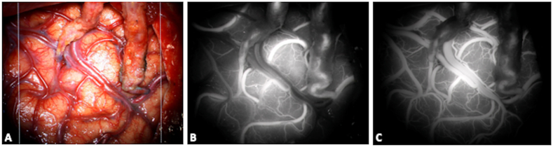
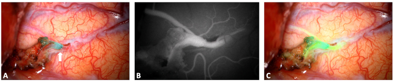
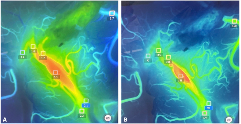
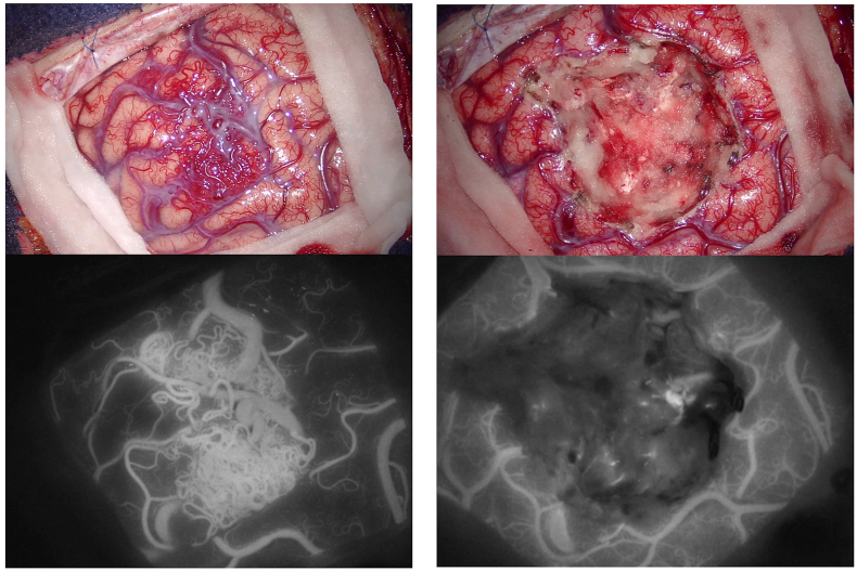
Introduction: The introduction of intraoperative fluorophores represented a significant advancement in neurosurgical practice. Nowadays they found different applications: in oncology to improve the visualization of tumoral tissue and optimize resection rates and in vascular neurosurgery to assess the exclusion of vascular malformations or the permeability of bypasses, with real-time intraoperative evaluations.

Research question: A comprehensive knowledge of how fluorophores work is crucial to maximize their benefits and to incorporate them into daily neurosurgical practice. We would like to revise here their applications and clinical relevance.

Material and methods: A focused literature review of relevant articles dealing with the versatile applications of fluorophores in neurosurgery was performed.

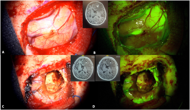
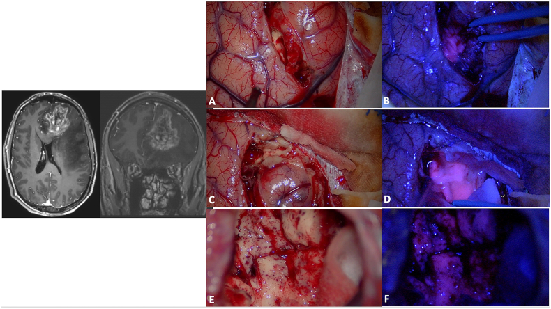
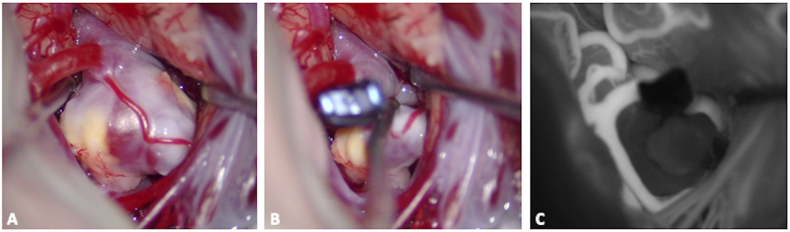
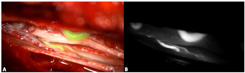
Results: The fundamental mechanisms of action of intraoperative fluorophores are enlightened, examining their interactions with target tissues and the principles driving fluorescence-guided surgery. The clinical applications of the principal fluorophores, namely fluorescein sodium, 5-ALA and indocyanine green, are detailed, in regards to the management of vascular malformations, brain tumors and pathologies treated through endoscopic endonasal approaches.

Discussion and conclusion: Future perspective dealing with the development of new technologies or of new molecules are discussed. By critically assessing the efficacy and applications of the different fluorophores, as well as charting their potential future uses, this paper seeks to guide clinicians in their practice and provide insights for driving innovation and progress in fluorescence-based surgery and research.

Keywords: 5-ALA; Fluorescein sodium; Fluorescence; Fluorophores; Indocyanine green; Microneurosurgery.


Conflict of interest statement

None.

Figures

Fig. 1
Fig. 1
A 74 years old patient was diagnosed for a cerebral metastasis on the left frontal lobe. Intraoperative anatomical views are provided in picture A and C. A bolus of intravenous fluorescein sodium was administered at anesthesia induction (3 mg/kg) and a YELLOW 560 nm filter was used to visualize the tumor (Picture B and D). The tumor was well-defined and fluorescein sodium helped in achieving a complete resection (panel D). A preoperative is also provided (small upper panel) and compared to a postoperative MRI with and without gadolinium administration (lower right and left panel respectively). (For interpretation of the references to color in this figure legend, the reader is referred to the Web version of this article.)
Fig. 2
Fig. 2
Surgical resection of a high-grade glioma in the left frontal lobe (preoperative MRI on the left; A, C and E: intraoperative anatomical views). The high-grade portion presented a superficial part at the level of the superior frontal gyrus and then extended in the depth till the frontal horn of the lateral ventricle. During the initial phase of surgical resection, the use of a dedicated BLUE 400 filter may help in identifying the tumor that shows a strong pink color, while the surrounding brain remains blue (B and D). Towards the end of the resection, the identification of normal tissue under white light may be difficult (E) and 5-ALA fluorescence may guide the surgeons in identifying residual tumor (F: residual tumor is still present at the bottom of the surgical cavity and is easily recognized by its pink color). A careful hemostasis should be performed to allow a proper visualization of the fluorophore as the blood could mask fluorescent tissue. Furthermore, it should be kept in mind that the peripheral portion of the tumor may be less fluorescent and that low grade portions may be false negative. Furthermore, 5-ALA resection should be accompanied by a careful anatomical study of preoperative images, and coupled with the use of intraoperative imaging and/or electrophysiological evaluations in eloquent areas. (For interpretation of the references to color in this figure legend, the reader is referred to the Web version of this article.)
Fig. 3
Fig. 3
Surgical clipping of a middle cerebral artery aneurysm on the right side. After the dissection of the sylvian fissure, the aneurysm was identified, along with M1 and the superior and inferior divisions (M2), Panel A. Once the definitive clip was positioned at the aneurysm neck (Panel B), an intraoperative bolus of ICG helped in establishing the complete exclusion of the aneurysm (black) along with the adequate filling of M1 and of the two M2 (white; Panel C). This procedure, coupled with intraoperative doppler evaluations, can limit the incidence of parent arteries' and bifurcation's stenosis and of residual filling of the aneurysm.
Fig. 4
Fig. 4
Double barrel bypass: the frontal and the parietal branch of the STA were anastomosed with two M4 arteries belonging to the superior and inferior MCA division respectively (A, anatomical view). An intraoperative bolus of ICG was administered showing an optimal permeability of the two bypasses (B: early phase of the injection; C: late phase, with an associated venous enhancement).
Fig. 5
Fig. 5
Extracranial-intracranial anastomosis on the left side for a patient with a Moya disease: the permeability of the STA-M4 bypass is checked with an intraoperative administration of indocyanin green bolus. A: Anatomical visualization of the different structures. The parietal branch of the left STA is used as donor artery, while the receiving artery is a temporal M4 branch. The white arrow indicated the anastomosis. B: Microscope-integrated ICG-videoangiography shows the classical black and white image and allows evaluation of bypass permeability. C: Augmented-reality systems integrated into the microscope may allow a real-time evaluation of blood flow while superimposing colored ICG image with the anatomical 3D view. (For interpretation of the references to color in this figure legend, the reader is referred to the Web version of this article.)
Fig. 6
Fig. 6
Through the use of a flow 800 software, the delay map (A, in seconds) and speed map (B, in mm/second) of ICG videoangiography can also be recorded and analyzed. In this case, they were used to evaluate bypass functionality along with flow direction and cortical perfusion. They can also be applied to the surgical resection of arterio-venous malformations or during the treatment of arterio-venous fistulae (not shown).
Fig. 7
Fig. 7
Intraoperative view of a superficial parietal arteriovenous malformation (superior parietal lobule). Intravenous ICG videoangiography may help in identifying arterial feeders, nidus and venous drainage (left panels), while helping in differentiating them from normal veins, that should be preserved. At the end of the resection a repeated ICG injection may help in establishing the completeness of the resection of the nidus (right panels).
Fig. 8
Fig. 8
Intraoperative image of a spinal dural arterio-venous fistula. Intraoperative ICG images help in identifying the arterial feeder(s) and the draining vein(s), to guide the surgical exclusion. While black and white images show high contrast but a limited anatomical resolution (B), the superposition of color images allow real time manipulation and dissection (A). (For interpretation of the references to color in this figure legend, the reader is referred to the Web version of this article.)
Fig. 9
Fig. 9
Preoperative T1-weighted MRI of a patient with a Cushing disease, after gadolinium administration (A and B). The ACTH-secreting micro pituitary neuro-endocrine tumor (PitNET) was visualized on the right portion of the pituitary gland, as assessed in the sagittal (A) and coronal plane (B) of the MRI (white arrow). The tumor showed less contrast enhancement than the normal pituitary gland and it appears as hypointense on this sequence. An intraoperative picture is provided, showing the dura mater of the sella turcica after bone removal (C). The aspiration in the clival recess. A bolus of ICG is then administered and the contrast enhancement is visualized through the dura as it strongly accumulates in the normal pituitary gland and less in the tumor (D). This confirms that the PitNET, marked with a white star, is localized on the right side of the sella and a selective approach can be performed, guided by fluorescence.

References

    1. Acerbi F., Broggi M., Schebesch K.M., Hohne J., Cavallo C., De Laurentis C., Eoli M., Anghileri E., Servida M., Boffano C., et al. Fluorescein-guided surgery for resection of high-grade gliomas: a multicentric prospective phase II study (FLUOGLIO) Clin. Cancer Res. 2018;24(1):52–61. - PubMed
    1. Ahrens L.C., Krabbenhoft M.G., Hansen R.W., Mikic N., Pedersen C.B., Poulsen F.R., Korshoej A.R. Effect of 5-aminolevulinic acid and sodium fluorescein on the extent of resection in high-grade gliomas and brain metastasis. Cancers. 2022;14(3) - PMC - PubMed
    1. Akcakaya M.O., Goker B., Kasimcan M.O., Hamamcioglu M.K., Kiris T. Use of sodium fluorescein in meningioma surgery performed under the YELLOW-560 nm surgical microscope filter: feasibility and preliminary results. World Neurosurg. 2017;107:966–973. - PubMed
    1. Alander J.T., Kaartinen I., Laakso A., Patila T., Spillmann T., Tuchin V.V., Venermo M., Valisuo P. A review of indocyanine green fluorescent imaging in surgery. Int. J. Biomed. Imag. 2012;2012 - PMC - PubMed
    1. Albert F.K., Forsting M., Sartor K., Adams H.P., Kunze S. Early postoperative magnetic resonance imaging after resection of malignant glioma: objective evaluation of residual tumor and its influence on regrowth and prognosis. Neurosurgery. 1994;34(1):45–60. - PubMed

LinkOut - more resources