. 2025 Jun 10;80(7):glae303.
doi: 10.1093/gerona/glae303.

Nicotinamide Prevents the Plasticity Impairments and the Cognitive Dysfunction Caused by Bone Fracture in Older Mice

Affiliations

Nicotinamide Prevents the Plasticity Impairments and the Cognitive Dysfunction Caused by Bone Fracture in Older Mice

Tatiana Guncay et al. J Gerontol A Biol Sci Med Sci. .

Abstract

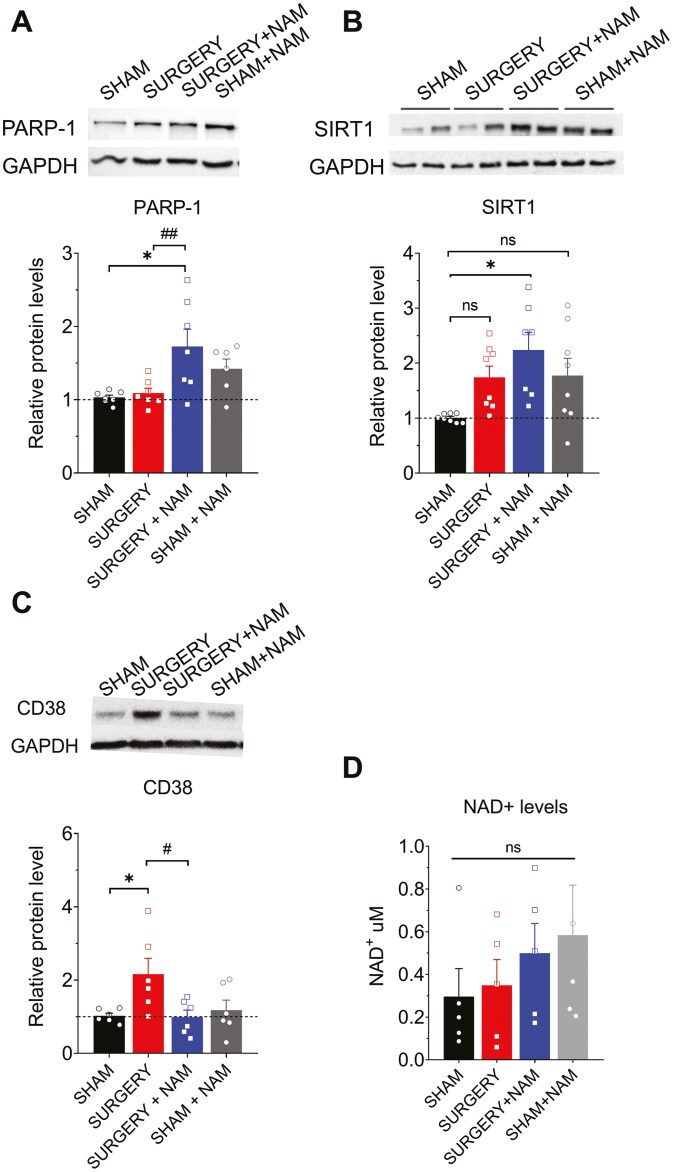
Postoperative delirium (POD), an acute cognitive dysfunction linked to morbidity and mortality, is characterized by memory impairments and disturbances in consciousness, particularly in patients aged 65 and older. Neuroinflammation and NAD+ imbalance are key mechanisms behind POD, leading to synaptic and cognitive deterioration. However, how surgery contributes to POD and neuroinflammation remains unclear, and effective treatments are lacking. Here we used a rodent model of bone fracture to examine the impact of surgery on synaptic plasticity, inflammation, and cognition. Additionally, we explored whether treatment with nicotinamide (NAM), a NAD+ precursor, reduced the neuroinflammation and metabolic imbalance caused by surgery. Female C57BL/6J mice aged 20-22 months underwent tibial fracture surgery and received pre- and post-surgery NAM treatment. Neuroinflammation, synaptic plasticity, and cognition were assessed 72 hours post-surgery via long-term potentiation (LTP) assays, dendritic spine counting, and behavioral tests (open field maze and Y-maze). Tibial fracture surgery decreased LTP, dendritic spine density, and hippocampal-dependent memory function, and increased hippocampal inflammatory markers (IL-1β mRNA, CD38, and SIRT1 protein content); NAM pretreatment prevented these changes. Given surgery's adverse effects on LTP and dendritic spine density, we assessed cellular oxidative state and brain-derived neurotrophic factor (BDNF) protein levels. We found that surgery increased the oxidation of ryanodine receptor calcium channels (cellular redox sensors), and decreased BDNF protein levels; NAM supplementation mitigated both effects and prevented the cognitive decline and synaptic plasticity deficits while reducing inflammation post-surgery by lowering IL-1β and CD38 protein levels. We propose that the CD38 signaling pathway mediates these NAM protective effects.

Keywords: Aging; Animal model; Delirium; Memory; Synaptic plasticity.


Conflict of interest statement

The authors declare that there were no commercial or financial relationships present during the research that could be perceived as potential conflicts of interest.

Figures

Figure 1.
Figure 1.
Nicotinamide (NAM) prevents surgery-induced synaptic plasticity deficits in excitatory hippocampal synapses. (A) Representative fEPSP traces from hippocampal slices in the following groups: Sham, Sham + NAM, Surgery, and Surgery + NAM. (B) fEPSP slope versus stimulus intensity. (C) Fiber volley amplitude versus stimulus intensity. (D) Paired-pulse facilitation responses. (E) Representative fEPSP traces at 50 ms interstimulus intervals. Data are mean ± SEM; (8, 4), with the first number indicating hippocampal slices and the second animals used. Two-way ANOVA, p > .05. (F) Representative fEPSP traces 1–5 minutes before (trace 1) and 60 minutes after TBS (trace 2) from the same groups. (G) Time course of fEPSP slopes (Schaffer collateral–CA1) before and after TBS. Group sizes: Sham (11, 6), Sham + NAM (13, 7), Surgery (10, 6), Surgery + NAM (13, 7). (H) Average fEPSP slopes during the last 10 minutes after TBS. Surgery group had lower fEPSP slopes (*p = .044) compared to Sham. Surgery + NAM group had higher fEPSP slopes (#p = .036) compared to Surgery. Data are mean ± SEM. Two-way ANOVA followed by Holm–Sidak’s post hoc test. ANOVA = analysis of variance; fEPSP = field excitatory postsynaptic potential; TBS = theta-burst stimulation.
Figure 2.
Figure 2.
Effects of nicotinamide (NAM) treatment on hippocampal synaptic plasticity markers and dendritic spine density following surgery. (A) Representative blots of RyR2 expression (left) and RyR2 S-glutathionylation levels (right) in CA3–CA1 hippocampal homogenates from mice, obtained 72 hours post-surgery from the Sham, Sham + NAM, Surgery, and Surgery + NAM groups. Below, Western blot analysis levels normalized to total GAPDH levels. Enhanced RyR2 S-glutathionylation was observed in slices from the Surgery group (***p < .001) compared to slices from the Sham group. However, slices from the Surgery + NAM and the Sham + NAM groups showed partial attenuation of this effect; n = 5 animals per group. (B) Representative blots of BDNF expression in CA3–CA1 hippocampal homogenates from the 4 mentioned groups are depicted. Below, Western blot analysis levels normalized to total GAPDH levels. NAM administration significantly elevated BDNF levels after surgery compared to mice exposed to surgery without NAM treatment (#p = .036); n = 7 animals per group. (C) Representative images of neurites from the CA1 region of mouse hippocampal slices stained with FD rapid Golgi Stain are depicted for the 4 mentioned groups (left). The scale bar represents 10 µm. Right, spine density analysis was performed by calculating the ratio between the number of spines and the dendritic length (in 10 μm). The density of dendritic spines showed a significant decrease in the total number of spines following surgery (**p < .01). This decrease was prevented by NAM treatment (##p < .01). (D) Graphical representation of the percentage of spines as mushroom-like, thin, and stubby-shaped per total spines of apical dendrites in the CA1 subarea. The surgery group displayed a significant increase in thin-shaped spines (*p < .019), accompanied by a decrease in mushroom-like spines (**p < .01). This latter change was prevented by NAM treatment (##p < .01). A total of N = 60 dendrites from 4 animals per group were analyzed. Values represent mean ± SEM. Statistical significance was assessed with 2-way ANOVA followed by Holm–Sidak’s post hoc test. ANOVA = analysis of variance; BDNF = brain-derived neurotrophic factor.
Figure 3.
Figure 3.
After surgery, mice displayed impaired natural (open field test) and learned (Y-maze test) behaviors, measured 72 hours post-surgery; NAM administration prevented these changes. (A) Open field test. At 72 hours post-surgery, there were no significant differences in locomotor activity between Surgery and Sham groups. All groups traveled a similar distance during the exploration time. (B) When assessing the time spent exploring the center of the maze, the Surgery group (n = 9) exhibited a significant decrease compared to the Sham condition (n = 10; **p = .006). This deficit was not observed in the Surgery group that received preventive treatment with NAM (n = 11). (C) Illustrations of the Y-maze of the alternations based on the sequence of arm visits. (D) Y-maze test: The surgery did not significantly affect the total number of arm visits made by the mice compared to the sham condition. However, it significantly altered the number of visits to novel arms (E) without changing the percentage of time spent exploring the new arm (F). (G) When assessing the spontaneous alternation behavior, the Surgery group (n = 9) exhibited a significant decrease compared to the Sham condition (**p = .002). All these effects were prevented by NAM pretreatment (n = 11; #p = .01). Values represent mean ± SEM. Sham condition (n = 10), Surgery group (n = 9), Surgery + NAM group (n = 11), and Sham + NAM group (n = 9). Statistical significance was assessed with 2-way ANOVA followed by Holm–Sidak’s post hoc test. ANOVA = analysis of variance; NAM = nicotinamide.
Figure 4.
Figure 4.
Nicotinamide (NAM) administration prevents surgery-induced inflammation in the mouse hippocampus. (A) RT-PCR quantification in the Sham, Sham + NAM, Surgery, and Surgery + NAM groups. At 72 hours after surgery, hippocampal IL-1β levels were significantly increased in the Surgery group compared to the Sham group (**p < .01). However, the Surgery group treated with NAM showed a significant decrease in IL-1β mRNA levels compared to the Surgery group (###p < .001). (B, C) No significant differences were observed in IL-6 (B) or TNF-α (C) levels among the 4 groups. (D) Mean fluorescence quantification for the 4 groups across the 3 hippocampal sections. (E) Immunofluorescence staining for the Iba1 marker in the CA1, CA3, and dentate gyrus (DG) regions of hippocampal sections from the 4 groups. (F, G) Morphological analysis of microglia segmentation. Values represent mean ± SEM; (A–C) n = 5–6; (D–G) n = 3–4 animals per group. Statistical significance was assessed using 2-way ANOVA followed by Holm–Sidak’s post hoc test (A–E) and Kruskal–Wallis nonparametric test (F, G). *p < .05; **p <.001; ***p < .001. ANOVA = analysis of variance; RT-PCR = reverse transcription-polymerase chain reaction.
Figure 5.
Figure 5.
Effects of NAM on hippocampal NAD+-consuming enzymes and NAD+ cofactor availability following surgery. Representative blots of PARP-1 (A), SIRT1 (B), and CD38 (C) protein contents in CA3–CA1 hippocampal homogenates from mice, measured 72 hours post-surgery in the Sham, Sham + NAM, Surgery, and Surgery + NAM groups. Western blot analysis levels were normalized to total GAPDH levels. (A) Representative blots of PARP-1 expression. NAM administration significantly elevated PARP-1 levels in the samples from the Surgery + NAM group compared to the Sham group (*p = .01) and the Surgery group (##p = .007); n = 6–7 animals per group. (B) Representative blots of SIRT1 protein content in hippocampal samples from the 4 groups. NAM administration significantly elevated SIRT1 levels in the Surgery group compared to the Sham group (*p = .01); n = 7–8 animals per group. (C) Markedly increased CD38 expression was observed in slices from the Surgery group (*p < .017) compared to slices from the Sham group. Slices from the Surgery + NAM group showed an attenuation of this effect (#p = .014), while Sham + NAM group did not have changes; n = 5–6 animals per group. (D) Quantification of hippocampal NAD+ levels measured by spectrophotometry revealed no significant decline in the Surgery group compared to the Sham group. Preventive NAM treatment promoted slight increases in NAD+ levels. Values represent mean ± SEM. Statistical significance was assessed with 2-way ANOVA followed by Holm–Sidak’s post hoc test. ANOVA = analysis of variance; NAM = nicotinamide; PARP = poly-ADP-ribose polymerase.

References

    1. Wilson JE, Mart MF, Cunningham C, et al. Delirium. Nat Rev Dis Primers. 2020;6(1):1–26. https://doi.org/ 10.1038/s41572-020-00223-4 - DOI - PMC - PubMed
    1. Bravo M, Bustos S, Acuña E, et al. Epidemiology of delirium in hospitalized patients in Latin America: a systematic review. Acta Psychiatr Scand. 2023;147(5):420–429. https://doi.org/ 10.1111/acps.13468 - DOI - PubMed
    1. Aung Thein MZ, Pereira JV, Nitchingham A, Caplan GA.. A call to action for delirium research: meta-analysis and regression of delirium associated mortality. BMC Geriatr. 2020;20(1):1–12. https://doi.org/ 10.1186/s12877-020-01723-4 - DOI - PMC - PubMed
    1. Davis D, Muniz Terrera G, Keage H, et al. Delirium is a strong risk factor for dementia in the oldest-old: a population-based cohort study. Brain. 2012;135(Pt 9):2809–2816. https://doi.org/ 10.1093/brain/aws190 - DOI - PMC - PubMed
    1. González M, Uslar W, Villarroel L, Calderón J, Palma C, Carrasco M.. Hospital costs associated with delirium in older medical patients. Rev Esp Geriatr Gerontol. 2012;47(1):23– 26. https://doi.org/ 10.1016/j.regg.2011.03.005 - DOI - PubMed

MeSH terms