. 2025 Mar;12(2):e200359.
doi: 10.1212/NXI.0000000000200359. Epub 2025 Jan 17.

Conformational Antibodies to Proteolipid Protein-1 and Its Peripheral Isoform DM20 in Patients With CNS Autoimmune Demyelinating Disorders

Collaborators, Affiliations

Conformational Antibodies to Proteolipid Protein-1 and Its Peripheral Isoform DM20 in Patients With CNS Autoimmune Demyelinating Disorders

Stefano Masciocchi et al. Neurol Neuroimmunol Neuroinflamm. 2025 Mar.

Erratum in

  • Conformational Antibodies to Proteolipid Protein-1 and Its Peripheral Isoform DM20 in Patients With CNS Autoimmune Demyelinating Disorders.
    Masciocchi S, Businaro P, Greco G, Scaranzin S, Malvaso A, Morandi C, Zardini E, Risi M, Vegezzi E, Diamanti L, Bini P, Siquilini S, Giannoccaro MP, Morelli L, Liguori R, Patti F, De Giuli V, Portaccio E, Zanetta C, Bergamoni S, Simone AM, Lanzillo R, Bruno G, Gallo A, Bisecco A, Di Filippo M, Pauri F, Toriello A, Barone P, Tazza F, Bucello S, Banfi P, Fabris M, Volonghi I, Raciti L, Vigliani MC, Bocci T, Paoletti M, Colombo E, Filippi M, Pichiecchio A, Marchioni E, Franciotta D, Gastaldi M. Masciocchi S, et al. Neurol Neuroimmunol Neuroinflamm. 2025 May;12(3):e200389. doi: 10.1212/NXI.0000000000200389. Epub 2025 Mar 21. Neurol Neuroimmunol Neuroinflamm. 2025. PMID: 40117521 Free PMC article. No abstract available.

Abstract

Background and objectives: Antibodies to proteolipid protein-1 (PLP1-IgG), a major central myelin protein also expressed in the peripheral nervous system (PNS) as the isoform DM20, have been previously identified mostly in patients with multiple sclerosis (MS), with unclear clinical implications. However, most studies relied on nonconformational immunoassays and included few patients with non-MS CNS autoimmune demyelinating disorders (ADDs). We aimed to investigate conformational PLP1-IgG in the whole ADD spectrum.

Methods: We devised a new live cell-based assay (CBA) for PLP1-IgG and used it to test 2 cohorts (retrospective exploratory, n = 284; prospective validation, n = 824) of patients with ADDs and controls (n = 177). Patients were classified as MS, neuromyelitis optica spectrum disorders (NMOSDs), myelin oligodendrocyte glycoprotein antibody-associated disease (MOGAD), and other ADDs. PLP1-IgG-positive samples were tested for IgG subclasses, DM20-IgG, and on rat brain tissue-based assay (TBA). Complement-dependent cytotoxicity (CDC) was assessed on a live CBA and antigen specificity and conformational binding through immunoadsorption/colocalization/fixation experiments.

Results: PLP1-IgG were found in 0 of 177 controls and 42 of 1104 patients with ADDs mainly diagnosed as other ADDs (19/42) with frequent myelitis/encephalomyelitis (14/19) and coexisting PNS involvement (13/19). Four of 19 patients with other ADDs fulfilled the seronegative NMOSD criteria. PLP1-IgG were also found in patients with MOGAD (11/42), more frequently with PNS involvement (p = 0.01), and in patients with MS (12/42), more frequently with atypical features (p < 0.001). PLP1-IgG-positive MOGAD had higher EDSS scores (p < 0.001) and PLP1-IgG-positive MS had higher severity scores (MSSS, p < 0.001) compared with those PLP1-IgG-negative. Overall, PLP1-IgG were found in 24.1% of patients with CNS+PNS-ADD, 21.2% with atypical MS, 8.3% with MOGAD, 12.0% with seronegative NMOSD, and 1.4% with typical MS. Their frequency within each diagnostic subgroup was consistent between the exploratory and validation cohorts. PLP1-IgG a) colocalized with their target on CBA-TBA, where their binding was abolished after immunoadsorption and fixation-induced conformational epitope alteration; b) mostly pertained to the IgG1/IgG3 subclass (68.3%) and were able to induce CDC; and c) coreacted with DM20 in all 12 patients with PNS involvement tested.

Discussion: Conformational PLP1-IgG predominantly identify patients with non-MS ADDs. They should be tested mainly in those with CNS + PNS ADD, coherently with DM20-IgG coreactivity. PLP1-IgG could also be investigated as disease modifiers and prognostic markers in MS and MOGAD. Preliminary evidence supports their pathogenic potential.


Conflict of interest statement

M. Gastaldi received compensation for speaking activities and/or consulting services, from Alexion, UCB, and Roche; he is the MG is the co-beneficiary of a patent on the PLP1 CBA currently under evaluation from the UIBIM (IT n. 102023000005046). Prof. M. Filippi is Editor-in-Chief of the Journal of Neurology; Associate Editor of Human Brain Mapping, Neurological Sciences, and Radiology; he received compensation for consulting services from Alexion, Almirall, Biogen, Horizon, Merck, Novartis, Roche, Sanofi; speaking activities from Bayer, Biogen, Celgene, Chiesi Italia SpA, Eli Lilly, Genzyme, Horizon, Janssen, Merck-Serono, Neopharmed Gentili, Novartis, Novo Nordisk, Roche, Sanofi, Takeda, and TEVA; participation in Advisory Boards for Alexion, Biogen, Bristol-Myers Squibb, Horizon, Merck, Novartis, Roche, Sanofi, Sanofi-Aventis, Sanofi-Genzyme, Takeda; scientific direction of educational events for Biogen, Merck, Roche, Celgene, Bristol-Myers Squibb, Lilly, Novartis, Sanofi-Genzyme; he receives research support from Biogen Idec, Merck-Serono, Novartis, Roche, Italian Ministry of Health, Fondazione Italiana Sclerosi Multipla, and ARiSLA (Fondazione Italiana di Ricerca per la SLA). C. Zanetta received compensation for speaking activities, and/or consulting services, from Alexion, Astrazeneca, Biogen, Bristol Myers Squibb, Janssen, Merck, Novartis, Roche, Sandoz, and Sanofi. R. Lanzillo received personal compensations for speaking or consultancy from Biogen, Alexion, Sanofi, Merck, Bristol Myer Squibb, Jansenn, Novartis, Amgen and Roche. The remaining authors report no disclosures. Go to Neurology.org/NN for full disclosures.

Figures

Figure 1
Figure 1. Conformation-Dependent PLP1 Epitopes Are the Target Antigen of Myelin Antibodies
(A) Monoclonal antibodies against different epitopes of PLP1 and serum from a patient with ADD label the surface of live HEK293T cells transfected with PLP1-EGFP-tag (green). Merged images show colocalization of the staining (orange). No binding is observed with the MOG8-18C5 monoclonal antibody or with serum from a healthy control. (B) Structure of PLP1. Colored aminoacidic sequences identify the different epitopes recognized by the monoclonal antibodies (yellow = PLP150-69; green = PLP1178-191, and blue = PLP1200-219) or their intracellular portion, which is absent in the DM20 protein isoform (red). (C) Serum of a PLP1-IgG positive patient (green) binds to the myelinated areas (cerebellum, striatum and hippocampus) of lightly fixed rat brain sagittal slices (indirect immunofluorescence). PLP1 monoclonal antibody (red) binds to the same regions, colocalizing with patients' serum (yellow). DAPI stains cell nuclei. Scale bar: 50 µm. (D) Serum from the same patient stains both PLP1-CBA and TBA on lightly fixed rat brain (left column); unmodified staining after preadsorption on MOG-transfected cells (central column); abolition of the binding after preadsorption on PLP1-transfected cells on both CBA and TBA (right column). Created in BioRender. Gastaldi, M. (2024) BioRender.com/v24o621. ADD = autoimmune demyelinating disorder; CBA = cell-based assay; MOG = myelin oligodendrocyte glycoprotein; PLP1 = proteolipid protein-1; TBA = tissue-based assay.
Figure 2
Figure 2. Algorithm of the Study and PLP1-IgG Distribution in Disease Subgroups
Patients from (A) a retrospective exploratory cohort and (B) a prospective validation cohort were tested. (C) Proportions of PLP1-IgG–positive patients within the diagnostic subgroups of both the exploratory and validation cohorts. *Seventy-three of 824 patients without sufficient clinical information were excluded from the figure, all negative for MOG/AQP4/PLP1-IgG. **The SN-NMOSD group includes 3 PLP1-IgG–positive patients (2 from the exploratory and 1 from the validation cohort) also classified as CNS+PNS-ADD. ADD = autoimmune demyelinating disorder; AQP4 = aquaporin 4; CBA = cell-based assay; CNS+PNS ADD = ADD with peripheral and CNS involvement; MOG = myelin oligodendrocyte glycoprotein; MOGAD = myelin oligodendrocyte glycoprotein antibody–associated disease; MS = multiple sclerosis; n = number; NMOSD = neuromyelitis optica spectrum disorder; ON = optic neuritis; PLP1 = proteolipid protein-1; SN = seronegative; TM = transverse myelitis.
Figure 3
Figure 3. Diagnostic Groups and Clinical Features of PLP1-IgG–Positive Patients With Autoimmune Demyelinating Disorders
(A) Clinical phenotypes of patients with ADDs split into 3 groups: PLP1-IgG–positive other ADDs, PLP1-IgG–positive MOGAD, and PLP1-IgG–positive MS; (B) EDSS-measured disability over the disease course; (C) median EDSS scores at the last follow-up (or MSSS for patients with MS) in PLP1-IgG–positive patients with MOGAD or with MS, compared with those measured, over the disease course, in the corresponding PLP1-IgG–negative groups. ADD = autoimmune demyelinating disorder; b = bilateral; BsE = brainstem encephalitis; CerS = cerebellar syndrome; CNS+PNS ADD = ADD with peripheral and CNS involvement; EDSS = Expanded Disability Status Scale; FU = follow-up; EMyR = encephalomyeloradiculitis; LETM = longitudinally extensive transverse myelitis; My = myelitis; MyR = myeloradiculitis; MOGAD = myelin oligodendrocyte glycoprotein antibody–associated disease; MS = multiple sclerosis; MSSS = MS severity score; ON = optic neuritis; PLP1 = proteolipid protein-1; TDL = tumefactive demyelinating lesions.
Figure 4
Figure 4. PLP1 Antibodies Can Bind to the Isoform DM20
(A) Serum of a patient with PLP1-IgG binds to DM20-transfected CBA (green) and colocalizes with a PLP1 mAb (red) directed against an extracellular loop of the protein (top row). No binding is observed with serum from a HC (bottom row). (B) Binding to both PLP1- and DM20-transfected cells (green) is observed with both PLP1mAb (first row) and serum from a patient with CNS+PNS ADD (pt#1; second row). Conversely, serum from a patient with CNS ADD binds exclusively to PLP1-transfected cells and not to DM20-transfected cells (third row). No binding is observed with serum from a HC (bottom row). DAPI stains the nuclei. (C) Heatmap representing fluorescence intensity of the available samples (n = 36) in either PLP1 or DM20 CBA measured with a semiquantitative score. All PLP1-IgG–positive patients with CNS + PNS ADD, unlike patients with other disease groups, were positive on both PLP1 and DM20 CBA. Scale bars: 10 µm. ADD = autoimmune demyelinating disorder; CBA = cell-based assay; Ig = immunoglobulin; HC = healthy control; mAbs = monoclonal antibodies; PNS = peripheral nervous system; PLP1 = proteolipid protein-1; Pt = patient.
Figure 5
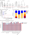
Figure 5. PLP1-IgG Titers in Serum and CSF, IgG Subclasses, and Complement-Dependent Cytotoxicity
(A) Serum and CSF PLP1-IgG titers tested in 41 serum and 24 CSF samples. The red dotted line indicates the cutoff for serum (1:40, left graph) and for CSF (1:5, right graph). (B) Serum:CSF PLP1-IgG titer ratio. The dotted arrow marks the 200:1 ratio expected in physiologic conditions. Values below the dotted line might associate with intrathecal synthesis. Blue dots represent patients positive in serum only, red dots patients positive in CSF only, and black dots patients positive in both serum and CSF. (C) PLP1-IgG subclasses in the whole cohort and in the 3 diagnostic groups of PLP1-IgG–positive patients. (D) PLP-IgG1 complement-dependent cytotoxicity. Data are normalized according to the amount of viable cells found in untreated cells (PLP1 cells, first 2 columns), which is considered 100%. The graph represents the finding after incubation with patient's sera. A reduction of viable cells is detected for 2 AQP4-IgG–positive serum samples (AQP4-01 and AQP4-02), which were used as positive controls, only when incubated with AQP4-transfected cells and in the presence of complement. Similarly, 3 of 8 PLP1-IgG–positive serum samples (other ADD#4, MS#5, and other ADD#18) showed reduced cell viability only when incubated with PLP1-transfected cells in the presence of complement. No effect is observed for the same samples in the absence of complement or after PLP1-IgG preadsorption performed on samples MS#5 and other ADD#18. The black dotted line represents the mean transfection rate for AQP4 and PLP1 (35%), which should theoretically set the limit for the maximum CDC effect in this experimental setting. CDC, which should involve only transfected cells, is unlikely below the cutoff. The numeric codes attributed to PLP1-IgG–positive patients correspond to those of Table 2, eTable 2, and eTable 3. ADD = autoimmune demyelinating disorder; AQP4 = aquaporin 4; Ig = immunoglobulin; MOGAD = myelin oligodendrocyte glycoprotein antibody–associated disease; MS = multiple sclerosis; PLP1 = proteolipid protein-1.

References

    1. GBD 2016 Multiple Sclerosis Collaborators. Global, regional, and national burden of multiple sclerosis 1990-2016: a systematic analysis for the Global Burden of Disease Study 2016. Lancet Neurol. 2019;18(3):269-285. doi:10.1016/S1474-4422(18)30443-5 - DOI - PMC - PubMed
    1. Wingerchuk DM, Banwell B, Bennett JL, et al. . International consensus diagnostic criteria for neuromyelitis optica spectrum disorders. Neurology. 2015;85(2):177-189. doi:10.1212/WNL.0000000000001729 - DOI - PMC - PubMed
    1. Banwell B, Bennett JL, Marignier R, et al. . Diagnosis of myelin oligodendrocyte glycoprotein antibody-associated disease: International MOGAD Panel proposed criteria. Lancet Neurol. 2023;22(3):268-282. doi:10.1016/S1474-4422(22)00431-8 - DOI - PubMed
    1. Ramanathan S, Dale RC, Brilot F. Anti-MOG antibody: the history, clinical phenotype, and pathogenicity of a serum biomarker for demyelination. Autoimmun Rev. 2016;15(4):307-324. doi:10.1016/j.autrev.2015.12.004 - DOI - PubMed
    1. Reindl M, Waters P. Myelin oligodendrocyte glycoprotein antibodies in neurological disease. Nat Rev Neurol. 2019;15(2):89-102. doi:10.1038/s41582-018-0112-x - DOI - PubMed

MeSH terms