

A large type IV-A choledochal cyst mimicking hydatid cyst of the liver: A case report
- PMID: 39823978
- PMCID: PMC11786652
- DOI: 10.1016/j.ijscr.2025.110898
A large type IV-A choledochal cyst mimicking hydatid cyst of the liver: A case report
Abstract
Introduction: Choledochal cysts are rare congenital anomalies of the bile ducts, with adult presentations being uncommon. This case is notable for its atypical presentation in a young adult, mimicking a hydatid cyst in a region where echinococcosis is endemic.
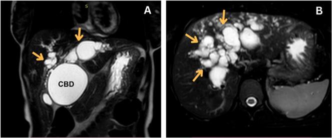
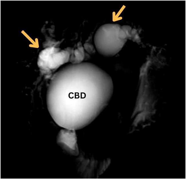
Case presentation: A 22-year-old female presented with a 3-month history of progressive jaundice, accompanied by 5 months of epigastric and right upper quadrant pain, dark urine, pale stools, pruritus, and significant weight loss. She reported a prior admission for cholangitis, treated with antibiotics. Examination revealed stable vital signs, icteric sclerae, right upper quadrant tenderness, and scratch marks on the skin. Laboratory investigations showed elevated liver enzymes and hyperbilirubinemia (total bilirubin = 26 mg/dL, direct bilirubin = 20.5 mg/dL). Initial imaging studies, including ultrasound and CT, suggested a hydatid cyst of the liver. However, MRCP revealed dilated intrahepatic and extrahepatic bile ducts, consistent with a Type IV-A choledochal cyst. The patient underwent cholecystectomy, extrahepatic bile duct excision, and Roux-en-Y cysto-jejunostomy. Histopathological analysis confirmed the diagnosis without evidence of malignancy. She recovered uneventfully, with no complications reported during a 6-month follow-up.
Discussion: This case highlights the diagnostic challenges in differentiating choledochal cysts from hydatid cysts, particularly in endemic regions. The use of MRCP was pivotal in achieving an accurate diagnosis and guiding definitive management. Early surgical intervention minimized the risks of complications and malignancy.
Conclusion: Type IV-A choledochal cysts can present atypically, mimicking hydatid cysts. Advanced imaging, especially MRCP, is critical for accurate diagnosis and management.
Keywords: Case report; Choledochal cyst; MRCP; Obstructive jaundice.
Copyright © 2025 The Authors. Published by Elsevier Ltd.. All rights reserved.
Conflict of interest statement
Declaration of competing interest The authors declare that they have no known competing financial interests or personal relationships that could have appeared to influence the work reported in this paper.
Figures
References
-
- Bandi Babita G, Prathamesh Vijay Kotawadekar, Pradeep S. Patil. Type IV-A choledochal cyst in newborn. Int J Sci Res. 2023;12(4):1–2. doi:10.36106/ijsr/1234567. - DOI
Publication types
LinkOut - more resources
Full Text Sources
Research Materials
