. 2025 Feb 6;112(2):414-427.
doi: 10.1016/j.ajhg.2024.12.020. Epub 2025 Jan 16.

Discovery of a DNA methylation profile in individuals with Sifrim-Hitz-Weiss syndrome

Affiliations

Discovery of a DNA methylation profile in individuals with Sifrim-Hitz-Weiss syndrome

Karim Karimi et al. Am J Hum Genet. .

Abstract

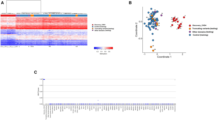
Pathogenic heterozygous variants in CHD4 cause Sifrim-Hitz-Weiss syndrome, a neurodevelopmental disorder associated with brain anomalies, heart defects, macrocephaly, hypogonadism, and additional features with variable expressivity. Most individuals have non-recurrent missense variants, complicating variant interpretation. A few were reported with truncating variants, and their role in disease is unclear. DNA methylation episignatures have emerged as highly accurate diagnostic biomarkers in a growing number of rare diseases. We aimed to study evidence for the existence of a CHD4-related DNA methylation episignature. We collected blood DNA samples and/or clinical information from 39 individuals with CHD4 variants, including missense and truncating variants. Genomic DNA methylation analysis was performed on 28 samples. We identified a sensitive and specific DNA methylation episignature in samples with pathogenic missense variants within the ATPase/helicase domain. The same episignature was observed in a family with variable expressivity, a de novo variant near the PHD domain, variants of uncertain significance within the ATPase/helicase domain, and a sample with compound heterozygous variants. DNA methylation data revealed higher percentages of shared probes with BAFopathies, CHD8, and the terminal ADNP variants encoding a protein known to form the ChAHP complex with CHD4. Truncating variants, as well as a sample with a recurrent pathogenic missense variant, exhibited DNA methylation profiles distinct from the ATPase/helicase domain episignature. These DNA methylation differences, together with the distinct clinical features observed in those individuals, provide preliminary evidence for clinical and molecular sub-types in the CHD4-related disorder.

Keywords: ADNP; CHD4; autism; chromatin remodeler; compound heterozygous; methylation; neurodevelopmental; truncating; variable expressivity.


Conflict of interest statement

Declaration of interests B.S. is a shareholder in EpiSign, Inc., a company involved in commercial uses of EpiSign technology. I.M.W. is an employee of GeneDx, LLC.

Figures

Figure 1
Figure 1
Genomic location of CHD4 variants and associated domains The top variants represent samples tested for DNA methylation and include variants with typical episignature (red), negative episignature similar to controls (black), and other inconclusive episignatures (turquoise, purple). The c. base position for each variant is detailed in Table S1. The bottom variants represent previously described pathogenic missense variants in different domains: c.606G>A (p.Met202Ile), c.1400G>A (p.Cys467Tyr), c.1409C>T (p.Ser470Phe), c.1933C>T (p.Arg645Trp), c.4018C>T (p.Arg1340Cys), and c.4033T>G (p.Tyr1345Asp).
Figure 2
Figure 2
Discovery of an episignature for variants within the ATPase/helicase domain (A) Euclidean hierarchical clustering (heatmap): each column presents a single SIHIWES sample or control, and each row represents one of the probes selected for episignature discovery. A clear separation was observed between ATPase/helicase domain samples (red) and controls (blue). A child and her father with the c.2903C>T (p.Ser968Phe) variant exhibiting a mild phenotype with variable expressivity (pink) were tested. The p.Ser968Phe variant associated with variable expressivity (pink) mapped with the other ATPase/helicase variants. The p.His448Tyr variant (sample 15, orange) clustered alongside the training samples. All truncating variants (purple) clustered with controls. (B) Multidimensional scaling (MDS) plot presents the differentiation between training samples (red) and controls (blue). The p.Ser968Phe variant (samples 11 and 12, pink) and p.His448Tyr variant (sample 15, orange) clustered alongside the training samples. (C) The methylation variant probability (MVP) scores created by the support vector machine (SVM): the ATPase/helicase samples provided MVP scores close to 1, indicating the high specificity of the classifier. Abbreviations are provided in Table S4.
Figure 3
Figure 3
Discovery of an episignature for truncating variants (A) Euclidean hierarchical clustering (heatmap): each column presents a single SIHIWES sample or control, and each row represents one of the probes selected for episignature discovery. Different methylation patterns were observed between truncating variants (red) and controls (blue). ATPase/helicase domain samples (orange), samples from other domains (purple), and samples with variable expressivity (pink) were used for testing. (B) Multidimensional scaling (MDS) plot presents the differentiation between case group (red) and controls (blue). ATPase/helicase domain samples (orange), samples from other domains (purple), and samples with variable expressivity (pink) were used as the testing set. A clear separation was observed between truncating variants (red), ATPase/helicase domains (orange), samples from other domains (purple), samples with variable expressivity (pink), and controls (blue). (C) The methylation variant probability (MVP) scores created by the support vector machine (SVM): the truncating variants provided MVP scores close to 1, indicating the high specificity of the classifier. Abbreviations are provided in Table S4.
Figure 4
Figure 4
Comprehensive model of SIHIWES episignature In total, 13 variants within ATPase/helicase domain and one sample with a variant near the PHD domain were used to train the model. (A) Euclidean hierarchical clustering (heatmap): each column presents a single SIHIWES sample or control, and each row represents one of 218 probes selected for episignature discovery. A clear separation was observed between discovery samples (red) and controls (blue). Truncating variants (orange) and other domains (purple) were clustered alongside the controls. (B) Multidimensional scaling (MDS) plot presents the differentiation between testing samples and controls. A clear separation was observed between discovery samples (red) and controls (blue). (C) The methylation variant probability (MVP) scores created by the support vector machine (SVM): the discovery samples provided MVP scores close to 1, indicating the high specificity of the classifier. Abbreviations are provided in Table S4.
Figure 5
Figure 5
Differentially methylated probes shared between the SIHIWES cohort and the 56 other neurodevelopmental disorders from the EKD Heatmap presenting the percentage of probes shared between each pair of cohorts. Colors show the percentage of y axis cohort’s probes shared with the x axis cohort’s probes. The heatmap uses a gradient from white (low) to red (high), indicating an increasing percentage.
Figure 6
Figure 6
Families with incomplete penetrance and variable expressivity (A) A child (III-1) with history of mild developmental delay and normal intelligence. The T allele was inherited from the father (II-1) with similar facial features, aortic root dilatation, and normal intelligence. (B) A child (III-1) with Chiari 1 malformation, aortic coarctation, developmental delay, learning disabilities, and normal intelligence. The A allele was inherited from the mother (II-2) with a history of developmental delay and learning disabilities with normal intelligence. Both probands demonstrate typical SIHIWES facial features, including a high and wide forehead, flared eyebrows, wide-spaced eyes, periorbital fullness, a short nose, and a squared face.

References

    1. Aref-Eshghi E., Kerkhof J., Pedro V.P., Barat-Houari M., Ruiz-Pallares N., Andrau J.C., Lacombe D., Van-Gils J., Fergelot P., et al. Groupe DI France Evaluation of DNA Methylation Episignatures for Diagnosis and Phenotype Correlations in 42 Mendelian Neurodevelopmental Disorders. Am. J. Hum. Genet. 2020;106:356–370. doi: 10.1016/j.ajhg.2020.01.019. - DOI - PMC - PubMed
    1. Aref-Eshghi E., Rodenhiser D.I., Schenkel L.C., Lin H., Skinner C., Ainsworth P., Paré G., Hood R.L., Bulman D.E., Kernohan K.D., et al. Genomic DNA Methylation Signatures Enable Concurrent Diagnosis and Clinical Genetic Variant Classification in Neurodevelopmental Syndromes. Am. J. Hum. Genet. 2018;102:156–174. doi: 10.1016/j.ajhg.2017.12.008. - DOI - PMC - PubMed
    1. van Jaarsveld R.H., Reilly J., Cornips M.C., Hadders M.A., Agolini E., Ahimaz P., Anyane-Yeboa K., Bellanger S.A., van Binsbergen E., van den Boogaard M.J., et al. Delineation of a KDM2B-related neurodevelopmental disorder and its associated DNA methylation signature. Genet. Med. 2023;25:49–62. doi: 10.1016/j.gim.2022.09.006. - DOI - PMC - PubMed
    1. Choufani S., Gibson W.T., Turinsky A.L., Chung B.H.Y., Wang T., Garg K., Vitriolo A., Cohen A.S.A., Cyrus S., Goodman S., et al. DNA Methylation Signature for EZH2 Functionally Classifies Sequence Variants in Three PRC2 Complex Genes. Am. J. Hum. Genet. 2020;106:596–610. doi: 10.1016/j.ajhg.2020.03.008. - DOI - PMC - PubMed
    1. Awamleh Z., Choufani S., Cytrynbaum C., Alkuraya F.S., Scherer S., Fernandes S., Rosas C., Louro P., Dias P., Neves M.T., et al. ANKRD11 pathogenic variants and 16q24.3 microdeletions share an altered DNA methylation signature in patients with KBG syndrome. Hum. Mol. Genet. 2023;32:1429–1438. doi: 10.1093/hmg/ddac289. - DOI - PMC - PubMed

LinkOut - more resources