. 2025 Jan 17;16(1):791.
doi: 10.1038/s41467-024-55579-y.

NKAPL facilitates transcription pause-release and bridges elongation to initiation during meiosis exit

Affiliations

NKAPL facilitates transcription pause-release and bridges elongation to initiation during meiosis exit

Zhenlong Kang et al. Nat Commun. .

Abstract

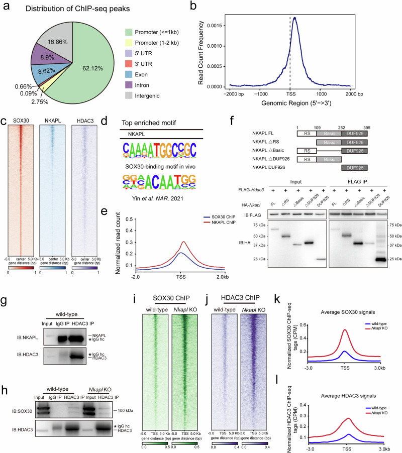
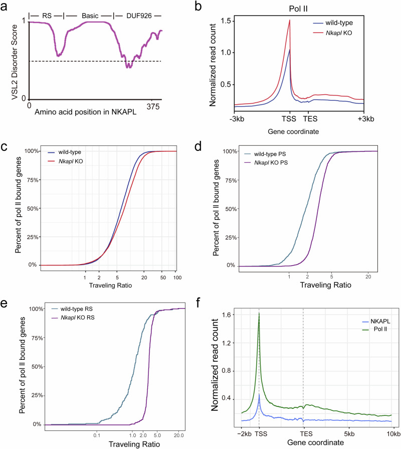
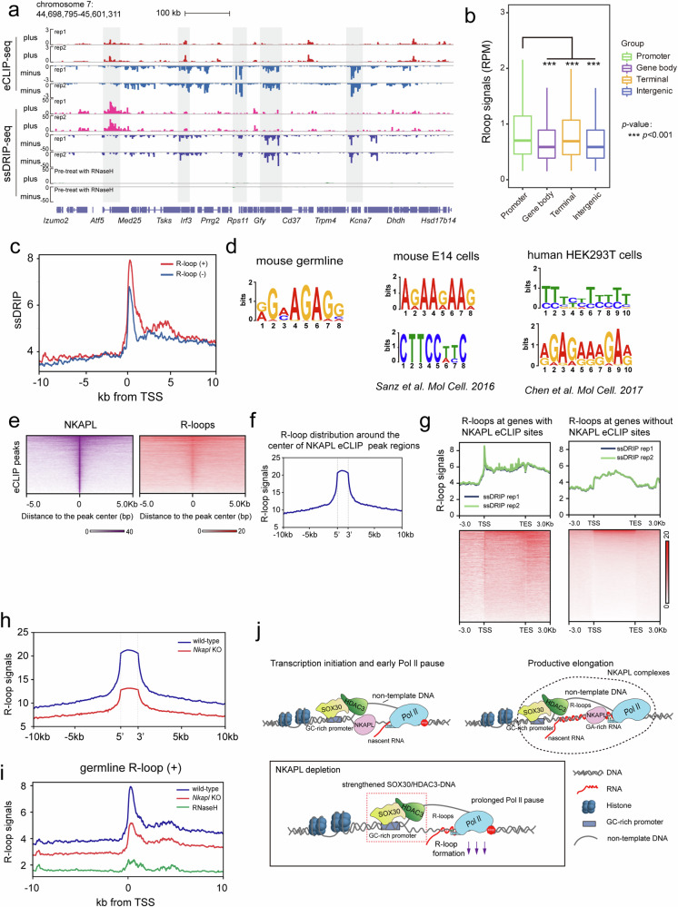
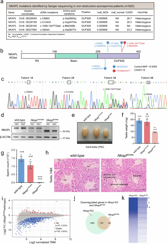
Transcription elongation, especially RNA polymerase II (Pol II) pause-release, is less studied than transcription initiation in regulating gene expression during meiosis. It is also unclear how transcription elongation interplays with transcription initiation. Here, we show that depletion of NKAPL, a testis-specific protein distantly related to RNA splicing factors, causes male infertility in mice by blocking the meiotic exit and downregulating haploid genes. NKAPL binds to promoter-associated nascent transcripts and co-localizes with DNA-RNA hybrid R-loop structures at GAA-rich loci to enhance R-loop formation and facilitate Pol II pause-release. NKAPL depletion prolongs Pol II pauses and stalls the SOX30/HDAC3 transcription initiation complex on the chromatin. Genetic variants in NKAPL are associated with azoospermia in humans, while mice carrying an NKAPL frameshift mutation (M349fs) show defective meiotic exit and transcriptomic changes similar to NKAPL depletion. These findings identify NKAPL as an R-loop-recognizing factor that regulates transcription elongation, which coordinates the meiotic-to-postmeiotic transcriptome switch in alliance with the SOX30/HDAC3-mediated transcription initiation.


Conflict of interest statement

Competing interests: The authors declare no competing interests.

Figures

Fig. 1
Fig. 1. Nkapl KO males show infertility and defects during meiotic exit.
a Schematic representation of mouse Nkap and Nkapl genes architecture. Exons were shown in black. b Evolution of the retrogene Nkapl by phylogenetic sequence analysis. c Western blot analysis of NKAPL protein in P21 wild-type and Nkapl KO testes. Experiments were performed in biological triplicates. d Significant size reduction in 8-week-old Nkapl KO males. e–g Testis weights (e), body weights (f) and epididymal sperm counts (g) of wild-type (n = 4) and Nkapl KO (n = 4) at 8-week-old. Data are presented as mean ± SD. ***p < 0.001, ****p < 0.0001, ns: not significant, two-tailed unpaired Student’s t test. h Histological analysis of testes from 8-week-old wild type and Nkapl KO mice. Scale bars, 20 μm. i Enlarged images of the Nkapl KO tubule. Apoptotic late stages of spermatocytes in stage XII tubules of Nkapl KO are marked by arrows, and black lines indicate round spermatids. Scale bar, 20 μm. j Histological analysis of juvenile Nkapl KO testes also revealed defects in meiotic exit in the first wave of spermatogenesis. Mouse testes from Nkapl KO mice at P18, P23 and P35 were collected. Biological duplicates were prepared for each time point during the first wave of spermatogenesis. Scale bars, 20 μm. k A diagram representing arrested stages of germ cell development in Nkapl KO mice. Blue and red crosses on lines indicate the earliest and ultimate time point of spermatogenic arrest, respectively. Lep: Leptotene; Zyg: Zygotene; e-Pac: early-Pachytene; m-Pac: middle-Pachytene; l-Pac: late-Pachytene; Dip: Diplotene; RS: round spermatids; ES: elongating spermatids.
Fig. 2
Fig. 2. Nkapl KO leads to defects in the meiotic-to-postmeiotic transition and predominant gene downregulation.
a, b MLH1 foci and the quantification of MLH1 foci in the wild-type and Nkapl KO spermatocytes at the pachytene stage. The number of MLH1 foci was comparable between wild type and Nkapl KO. Pachytene spermatocytes examined: wild-type, n = 54; Nkapl KO, n = 94. Data are presented as mean ± SD, two-tailed Student’s t test. Scale bars, 10 μm. c TUNEL assay of testicular sections from wild-type and Nkapl KO at 8-week-old. d Quantification of TUNEL-positive cells in adult wild-type and Nkapl KO. Many TUNEL-positive cells were observed in stage XII tubules. Tubules examined: wild-type, n = 296; Nkapl KO, n = 401. Data are presented as mean ± SD. ****P < 0.0001, two-tailed unpaired Student’s t test. Experiments were performed with biological triplicates. e Frozen sections from adult wild-type testes were immunolabelled with PNA (an acrosome marker, red) and γH2AX (green). The differentiation of round spermatids into mature sperm encompasses 16 steps, which are indicated by PNA. All the developmental steps were observed in the wild-type, and round spermatids at step 2-3, step 7-8 and step 11-12 were selectively shown. Biological duplicates were prepared. f Round spermatids of Nkapl KO mice were mainly arrested at step 2-3. Experiments were performed with biological duplicates. g Some spermatids in Nkapl KO developed beyond step 2-3 from biological duplicates. Scale bars, 20 μm. h Some spermatids in Nkapl KO displayed fragmented chromocenter and defective acrosome formation. Areas within the rectangles were enlarged in the right panel. i Scatter plot of RNA-seq data showing differentially expressed genes (fold change ≥ 1.5 up (red) or down (blue) and p < 0.05) in P21 wild-type and Nkapl KO testes using DESeq2, which employs a negative binomial distribution model along with the Wald test method. Experiments were performed in biological triplicates (n = 3 for each genotype). j Downregulated genes in Sox30 depleted testes (fold change ≥ 1.5, p < 0.01) were selected DESeq2 and used to produce a heatmap depicting their expression profile in Stra8-cre/Hdac3 KO and Nkapl KO testes.
Fig. 3
Fig. 3. NKAPL co-localizes with HDAC3/SOX30 on the genome and regulates the HDAC3/SOX30-DNA interaction.
a Annotations of NKAPL ChIP-seq peaks across different genomic regions in P21 wild-type testes. ChIP experiments were performed in biologically duplicates. b Caculated NKAPL ChIP-seq tags’ densities on UCSC mm10 RefSeq gene bodies (n = 20,460), and the genomic regions from -2 kb to +2 kb surrounding the TSS of genes were shown. c Heat map of NKAPL, SOX30 and HDAC3 ChIP-seq signals in P21 wild type testes depicting their co-localization at many binding sites. Regions from -5 kb to +5 kb surrounding the center of SOX30 sites were plotted in heatmap views. d The top-ranked motif in the binding sites of NKAPL with p values using Homer motif search. Sequences within ± 200 bp from the centers of all the binding sites were used for de novo motif analysis. p values = 1E-16. The top enriched motif at SOX30 binding sites in mouse testis was also shown. Hypergenomic Distribution Test, p values = 1E-132. e Position and distance between SOX30 and NKAPL average ChIP-seq signals. Average ChIP-seq signals of SOX30 and NKAPL, represented by counts per million, from -2 kb to +2 kb surrounding the TSS of genes were shown. f FLAG tagged HDAC3 was co-expressed with full-length HA-NKAPL or HA tagged NKAPL mutants with deleted fragments. Immunoprecipitation assay was performed with anti-FLAG antibody before western blot analysis with HA and FLAG antibodies. Representative images from biological duplicates were shown. g Testes protein lysates were immunoprecipitated either with HDAC3 or normal IgG antibodies followed by immunoblot analysis. h Co-immunoprecipitation of HDAC3 from wild-type and Nkapl KO testes at P21 and western blot with SOX30 antibodies. Protein lysates were prepared at P21. n = 3 for each genotype. Experiments were performed with biological triplicates. i, j Heat map of SOX30 (i) and HDAC3 (j) signals in Nkapl null testes at P21 from -5 kb to +5 kb surrounding the center of SOX30 sites. k Average SOX30 ChIP-seq signals across ±3 kb flanking TSS in Nkapl knockout versus their wild-types. CPM, counts per million. l Average HDAC3 ChIP-seq signals across ±3 kb flanking TSS in Nkapl knockout versus their wild-types.
Fig. 4
Fig. 4. NKAPL facilitates the release of paused Pol II at promoters.
a Predictions of intrinsic disorder of NKAPL as calculated by the VSL2 algorithm (http://www.pondr.com). b Pol II ChIP experiments with anti-total Pol II antibodies were performed in wild-type and Nkapl KO testes at P21. The average occupancy of total Pol II l along the length of genes occupied by SOX30. c Comparison of Pol II traveling ratios (the ratio between gene body and promoter-proximal polymerase) between wild-type and Nkapl KO testes. Pol II traveling ratio is defined as Pol II read density ratio between the promoter-proximal region (-80 bp to +250 bp around the transcription start site) and gene body (250 bp downstream of the TSS to the transcription end site). A higher traveling ratio value indicates a higher degree of pausing. The coverage of each region was calculated by featureCounts with the parameter ‘--fracOverlap 0.5’. The statistics by Mann-Whitney test was performed for the comparison of Pol II traveling ratios between wild-type and Nkapl KO testes. The statistics and plots were completed by customed scripts using R language. de Calculation of Pol II traveling ratios (the ratio between gene body and promoter-proximal polymerase) in PS (d) and RS populations (e) from wild-type and Nkapl KO. f Comparison of the binding signals of NKAPL and Pol II across genes.
Fig. 5
Fig. 5. NKAPL binds to promoter-associated RNAs.
a Schematic diagram of eCLIP-seq method. b RNA targets with NKAPL eCLIP peaks. c Genomic distribution of NKAPL-binding sites identified by eCLIP-seq. d Distribution of replicate NKAPL eCLIP peaks along the length of mRNA transcripts. e Averaged NKAPL eCLIP-seq signals across mRNA. Signals of ±200 bp flanking the TSS were shown. f NKAPL-binding motifs identified by MEME from all the NKAPL binding peaks, the top 50% of NKAPL eCLIP peaks, and NKAPL eCLIP peaks within promoter regions. g This GA-rich motif was enriched in R-loop peaks in the plant Arabidopsis in a previous study.
Fig. 6
Fig. 6. NKAPL co-localizes with R-loops at GA-rich sites and promotes R-loop formation.
a Representative genomic regions showing ssDRIP-seq signals in P21 wild-type testes. b Distribution of ssDRIP-seq signals for biological duplicates in different genomic regions. promoter, ±2 kb of TSS; terminal regions, ±2 kb of TTS. Bounds of box represent the interquartile range (IQR) from the 25th to the 75th percentile with the median as the horizontal line, while whiskers extend to the most extreme data points (outliers excluded) within 1.5×IQR.***p < 0.001. c ssDRIP-seq signals within ±10 kb of TSS. Red: forward strand signal; Blue: reverse strand signal. d The top enriched motif identified by MEME from germline R-loops in mouse testis as well as recently published R-loops in human and mouse cells. e NKAPL eCLIP binding signals at targeted loci generally co-localized with corresponding R-loop signals. f R-loop signal profiles within −10 kb/ +10 kb of NKAPL eCLIP binding sites. g R-loop signals by ssDRIP-seq at NKAPL target genes versus those genes without NKAPL eCLIP peaks. h Relative R-loop signals at NKAPL eCLIP binding sites in wild-type and Nkapl KO testes at P21. i Comparison of R-loop signal occupancy in wild-type and Nkapl KO on genes across ±10 kb genomic region flanking TSS. j Model for NKAPL function in the release of paused Pol II and R-loop formation. Transcription initiation complex containing SOX30/HDAC3 assembles at promoters and recruits Pol II. NKAPL interacts with the SOX30/HDAC3 initiation complex dynamically and transiently, and occupies at promoter regions indirectly. Initiation occurs with the opening of double-stranded DNA, and a short nascent RNA is synthesized before the transcription machinery pauses promoter proximally. Promoter-proximal stalled Pol II produces more levels of short RNAs, which re-anneal with its template DNA strand to form three-stranded RNA/DNA hybrid structures (R-loops). NKAPL binds promoter-associated nascent RNAs, and co-localizes with R-loops at GA-rich loci, where it interacts with RNA-DNA hybrid structures to enable R-loop formation and efficient Pol II elongation. Nkapl knockout causes prolonged Pol II pause and a pronounced reduction of R-loops, resulting in a stalled initiation complex containing SOX30/HDAC3.
Fig. 7
Fig. 7. Mutations in NKAPL contribute to human non-obstructive azoospermia.
a NKAPL mutations identified in four patients with azoospermia. Three heterozygous missense mutations and a frameshift mutation were identified: heterozygous missense mutations and a frameshift mutation (c.1046_1047TGdel [p.M349fs, referred to as NKAPLM349fs]). b NKAPL protein has an N-terminal arginine/serine-rich (RS) domain, repetitive basic sequences (the basic domain) and a C-terminal DUF926 domain of unknown functions. The deletion of TG base in NKAPL gene converts arginine 355 to a stop codon (R355*), thus causes a premature termination. c Sanger sequencing confirmed three heterozygous missense mutations and a frameshift mutation in azoospermia patients. d Western blot showing the presence of wild-type and truncated NKAPL proteins in testes from wild-type, Nkapl+/M349fs heterozygotes and NkaplM349fs homozygotes at P21. Experiments were performed with biological duplicates. e, f Testicular atrophy in NkaplM349fs mice at 8-week-old. Testis sizes (e), testis weights (f) of Nkapl+/+, Nkapl+/M349fs and NkaplM349fs mice. n = 4 for Nkapl+/+ and Nkapl+/M349fs, n = 3 for NkaplM349fs. Data are presented as mean ± SD. p < 0.001, two-tailed unpaired Student’s t test. g Absence of sperm in NkaplM349fs males. n = 3, 8-week-old males. Data are presented as mean ± SD. * p < 0.05, **** p < 0.0001, two-tailed unpaired Student’s t test. h Histological analysis of Nkapl+/+ and NkaplM349fs testes at 8 week-old. Apoptotic late stages of spermatocytes in stage XII tubules of NkaplM349fs are marked by black arrows. NkaplM349fs tubules were arrested either at late stages of spermatocytes (black arrows) or at the round spermatid stage (black lines). Scale bars, 20 μm. i RNA-seq analysis of differentially expressed genes in NkaplM349fs homozygotes compared with their wild-types at P21. Blue dots represent significantly downregulated transcripts, and red dots indicate upregulated transcripts (fold change ≥ 1.5, p < 0.05). DESeq2 with the Wald test method was used. j Venn diagram depicting the overlap between downregulated genes in Nkapl KO and NkaplM349fs homozygotes. k Downregulated genes in Nkapl KO (fold change ≥ 1.5, p < 0.05) with DESeq2 were used to produce a heatmap depicting their corresponding expression levels in NkaplM349fs homozygotes.

Similar articles

References

    1. Harlen, K. M. & Churchman, L. S. The code and beyond: transcription regulation by the RNA polymerase II carboxy-terminal domain. Nat. Rev. Mol. cell Biol.18, 263–273 (2017). - PubMed
    1. Noe Gonzalez, M., Blears, D. & Svejstrup, J. Q. Causes and consequences of RNA polymerase II stalling during transcript elongation. Nat. Rev. Mol. cell Biol.22, 3–21 (2021). - PubMed
    1. Rahl, P. B. et al. c-Myc regulates transcriptional pause release. Cell141, 432–445 (2010). - PMC - PubMed
    1. Mayer, A. et al. Native elongating transcript sequencing reveals human transcriptional activity at nucleotide resolution. Cell161, 541–554 (2015). - PMC - PubMed
    1. Jonkers, I. & Lis, J. T. Getting up to speed with transcription elongation by RNA polymerase II. Nat. Rev. Mol. cell Biol.16, 167–177 (2015). - PMC - PubMed

LinkOut - more resources