Review
. 2025 Jun;23(6):385-401.
doi: 10.1038/s41579-024-01144-9. Epub 2025 Jan 17.

Norovirus replication, host interactions and vaccine advances

Affiliations
Review

Norovirus replication, host interactions and vaccine advances

B V Venkataram Prasad et al. Nat Rev Microbiol. 2025 Jun.

Abstract

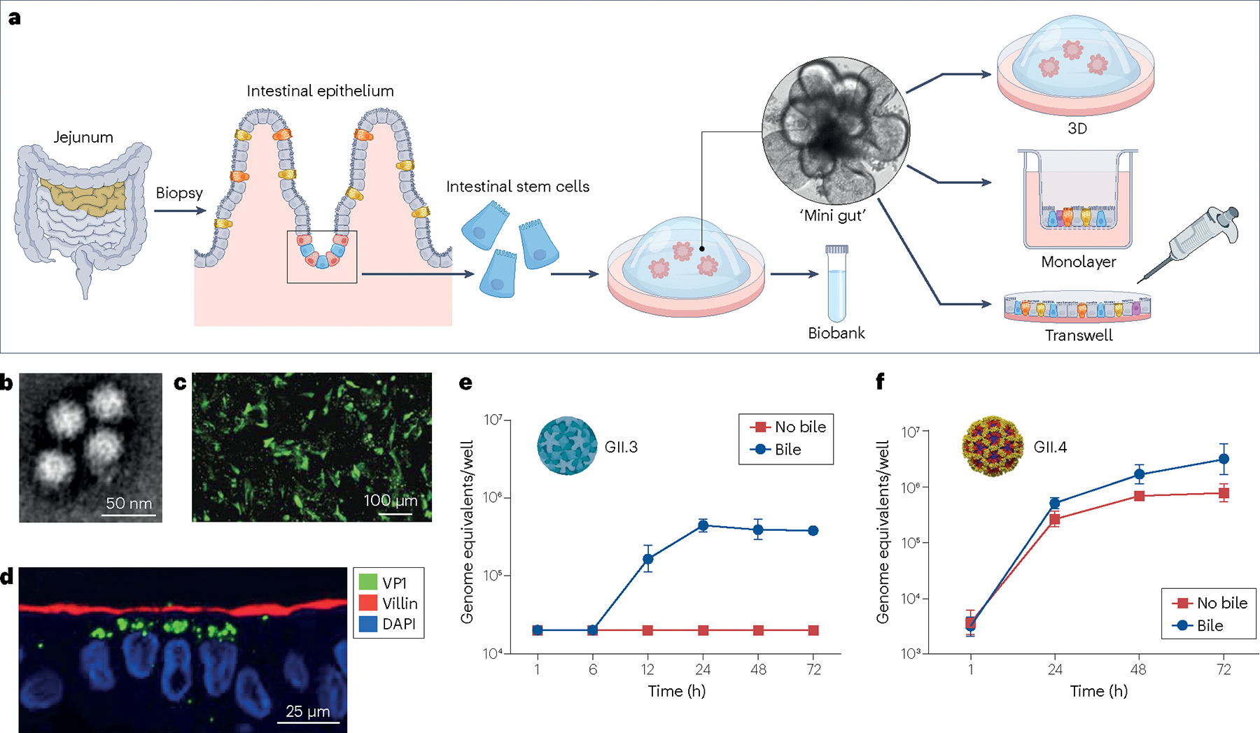
Human noroviruses (HuNoVs) are the leading cause of acute gastroenteritis worldwide in all age groups and cause significant disease and economic burden globally. To date, no approved vaccines or antiviral therapies are available to treat or prevent HuNoV illness. Several candidate vaccines are in clinical trials, although potential barriers to successful development must be overcome. Recently, significant advances have been made in understanding HuNoV biology owing to breakthroughs in virus cultivation using human intestinal tissue-derived organoid (or enteroid) cultures, advances in structural biology technology combined with epitope mapping and increased metagenomic sequencing. New and unexpected strain-specific differences in pandemic versus non-pandemic virus structures, replication properties and virus-host interactions, including host factors required for susceptibility to infection and pathogenesis, are discussed.


Conflict of interest statement

Competing interests: R.L.A., M.K.E. and B.V.V.P. have grant support from Hillevax, Inc., and R.L.A. and M.K.E. are consultants for that company. Baylor College of Medicine (R.L.A. and M.K.E. as inventors) has a patent for norovirus growth in human intestinal enteroids. M.K.E. has a patent on methods and reagents to detect and characterize Norwalk virus and related viruses. The other authors declare no competing interests.

Figures

Fig. 1 |
Fig. 1 |. Human norovirus genome, entry, replication and structure.
a, The human norovirus (HuNoV) genome (approximately 7.5 kb) is a viral protein (VPg)-linked positive-sense RNA with a poly(A) tail consisting of three open reading frames (ORFs). Replication of the HuNoV consists of several stages (1–8): (1) cell attachment involving interactions with histoblood group antigen (HBGA); (2) endocytic internalization; (3) disassembly; (4) ribosomal translation of the three ORFs; (5) cleavage of the polyprotein encoded by ORF1 by the viral protease (Pro) into six non-structural proteins (NSPs), synthesis of structural proteins VP1 and VP2 encoded by ORF2 and ORF3, respectively; (6) replication of the genomic and subgenomic RNA by the viral RNA-dependent RNA polymerase (RdRP); (7) encapsidation of the progeny VPg–RNA into capsids formed of VP1 and VP2 that are synthesized from subgenomic viral RNA; and the final stage of (8) viral capsid release. b, Among the NSPs, Pro and the RdRP are structurally the best characterized and have been the targets for developing a variety of classes of small molecule inhibitors. c, The T = 3 capsid of the HuNoV formed by 180 VP1 subunits in three quasi-equivalent positions (A, B and C) is shown along the icosahedral 3-fold axis with surrounding 5-fold axes denoted. The subunits A and B forming A/B dimers surrounding the 5-fold axes are shown in cyan and blue, whereas the C subunits, which form C–C dimers at the icosahedral 2-fold axes, are shown in green. Cartoon representations of the bent (A–B) and flat (C–C) dimers are shown on the right with protruding (P) and shell (S) domains and the N-terminal arm (NTA) denoted in the A/B dimer. d, HBGA (solid spheres in red) binding at the P2 subdomains of VP1 dimer (cyan and blue) in GI (left) and GII (right).
Fig. 2 |
Fig. 2 |. Phylogenetic trees of norovirus genogroups and genotypes.
a, VP1 amino acid sequences and b, RNA-dependent RNA polymerase (RdRP) nucleotide sequences using previously published sequences, reflect current classification systems (Supplementary methods). The natural hosts affected by each genotype are colour coded. c, GII.4 strains undergo epochal evolution and emergent variants are shown by their time of circulation,,,,. The Sydney variant has two major P types (P31 for the 2012-like strains and P16 for the 2015-like strains).
Fig. 3 |
Fig. 3 |. The human intestinal enteroid culture system for human norovirus.
a, Intestinal enteroids are produced from intestinal tissue samples that contain intestinal stem cells that will grow and self-assemble into 3D cultures within several days of being cultivated in media containing Matrigel and growth factors that support proliferation of the stem cells. After expansion of the ‘mini-gut’ cultures, some are frozen down to create a biobank. Removal of growth factors from the media of proliferating cultures induces the stem cells to differentiate into all the multiple cell types present in the mature intestinal epithelium including absorptive (enterocytes) and secretory cells (enteroendocrine, goblet and Paneth cells) and these cultures can be plated in different formats (3D, on transwells or 2D on plastic wells for infection studies. Inside each 3D mini-gut culture, enterocytes form the brush border surrounding a single luminal compartment, which receives secretions from the other cells. b, Virus produced from infected human intestinal enteroids (HIEs) visualized by negative stain electron microscopy. c, Immunofluorescence detects the VP1 capsid protein in infected HIEs plated on 96 wells. d, Immunofluorescence detects the VP1 capsid protein (green), villin (red) and nuclei (4′,6-diamidino-2-phenylindole (DAPI), blue) in infected HIEs plated on transwells. e, A time course illustrates the kinetics of RNA replication and shows bile is required for GII.3 replication. f, A time course illustrates the kinetics of RNA replication and shows bile enhances GII.4 replication. Parts bf adapted with permission from ref. , AAAS.
Fig. 4 |
Fig. 4 |. Distinct entry pathways and replication of bile acid-dependent GII.3 and bile acid-independent GII.4 into human intestinal enteroids.
a, Each virus uses a different entry pathway. GII.3 requires sphingosine-1-phosphate receptor 2 (S1PR2) and bile acid to induce endocytosis (left panel), whereas GII.4 causes initial membrane injury and wound repair mechanisms to induce clathrin-independent carrier (CLIC)-mediated endocytosis (right panel). Common elements include initial binding to histoblood group antigens (HBGAs), putative receptor clustering in lipid rafts, endosomal acidification and lysosomal exocytosis that results in ceramide (Cer) appearance on the cell surface. b, The table highlights strain-specific differences in the biology of GII.3 and GII.4 viruses related to the requirement for FUT2 gene expression,, cell entry,, bile acid dependence,, acid sphingomyelinase involvement,, effects of JAK1/JAK2 inhibition, effects of knockout of innate immune factors and host transcriptional responses,,,. ASM, acid sphingomyelinase; Gal-3, Galectin-3; IFN, interferon; SM, sphingomyelinase; V-ATPase, vacuolar-type ATPase.
Fig. 5 |
Fig. 5 |. Mechanisms of neutralization of human norovirus by antibodies.
a, Neutralization of GI.1 by the human antibody 5I2 (yellow) by directly competing with the histoblood group antigen (HBGA) binding site in the protruding (P) domain dimer (grey). b, Neutralization of GI.1 by the human antibody 10E9 (purple) by directly competing with the HBGA binding site in the P domain dimer (grey). c, Human antibody A1431 (blue) binds to an epitope slightly away from the HBGA binding site in the GII.4 P domain dimer (grey) and neutralizes GII.4 by clumping or crosslinking particles. d, Resting and raised conformations of the P domain (shell (S), P1, and P2 subdomains are shown in blue, red, and yellow, respectively). e, Fragment antigen-binding region (Fab) of the human antibody NORO-320 (yellow) targets capsid plasticity by binding an epitope in the GII.4 P domain (grey) that is exposed only in the raised conformation and probably neutralizes the virus by blocking co-receptor interactions or VP2 externalization. f, The llama nanobody M4 (green) also binds to an epitope exposed only when the P domain (grey) transits to the raised confirmation and neutralizes by compromising capsid integrity.

References

    1. Pires SM et al. Aetiology-specific estimates of the global and regional incidence and mortality of diarrhoeal diseases commonly transmitted through food. PLoS ONE 10, e0142927 (2015). - PMC - PubMed
    1. Bartsch SM, Lopman BA, Ozawa S, Hall AJ & Lee BY Global economic burden of norovirus gastroenteritis. PLoS ONE 11, e0151219 (2016). - PMC - PubMed
    1. Bok K & Green KY Norovirus gastroenteritis in immunocompromised patients. N. Engl. J. Med. 367, 2126–2132 (2012). - PMC - PubMed
    1. Thorne LG & Goodfellow IG Norovirus gene expression and replication. J. Gen. Virol. 95, 278–291 (2014). - PubMed
    1. Kroneman A et al. Proposal for a unified norovirus nomenclature and genotyping. Arch. Virol. 158, 2059–2068 (2013). - PMC - PubMed

MeSH terms

LinkOut - more resources