. 2025 Jan 17;9(1):20.
doi: 10.1038/s41698-025-00800-4.

Defective homologous recombination and genomic instability predict increased responsiveness to carbon ion radiotherapy in pancreatic cancer

Affiliations

Defective homologous recombination and genomic instability predict increased responsiveness to carbon ion radiotherapy in pancreatic cancer

Brock J Sishc et al. NPJ Precis Oncol. .

Abstract

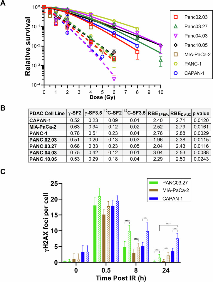
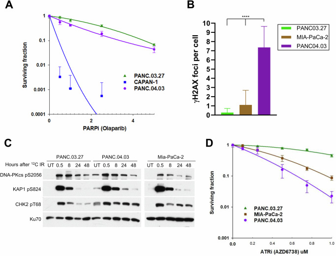
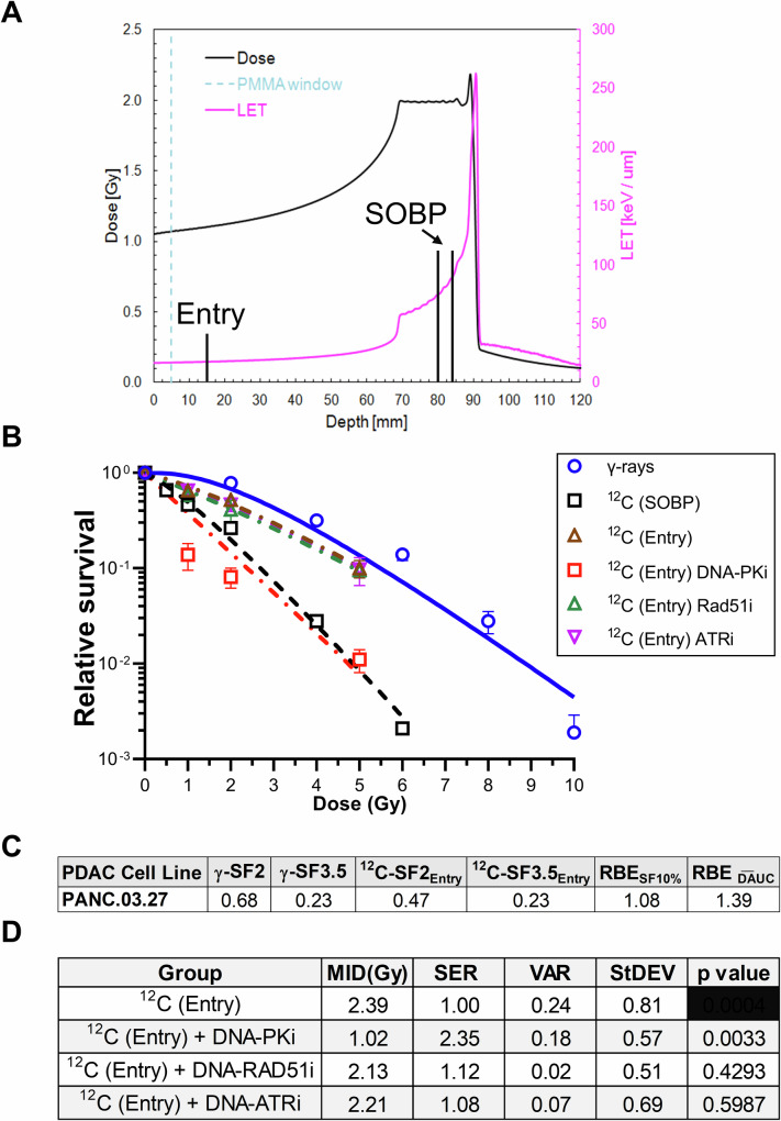
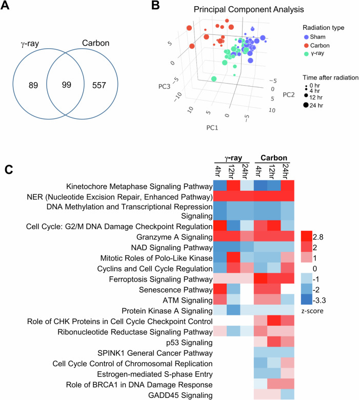
Pancreatic ductal adenocarcinoma (PDAC) is notably resistant to conventional chemotherapy and radiation treatment. However, clinical trials indicate that carbon ion radiotherapy (CIRT) with concurrent gemcitabine is effective for unresectable locally advanced PDAC. This study aimed to identify patient characteristics predictive of CIRT response. We utilized a panel of human PDAC cell lines with diverse genetic profiles to determine their sensitivity to CIRT compared to γ-rays, assessing relative biological effectiveness (RBE) at 10% survival, which ranged from 1.96 to 3.04. Increased radiosensitivity was linked to impaired DNA double-strand break (DSB) repair, particularly in cell lines with deficiencies in the homologous recombination (HR) repair pathway and/or elevated genomic instability from replication stress. Furthermore, pretreatment with the HR inhibitor B02 significantly enhanced CIRT sensitivity in a radioresistant PDAC cell line when irradiated in the spread-out Bragg peak but not at the entry position of the beam. These findings suggest that PDAC tumors with HR pathway mutations or high replication stress are more likely to benefit from CIRT while minimizing normal tissue toxicity.


Conflict of interest statement

Competing interests: The authors declare no competing interests.

Figures

Fig. 1
Fig. 1. Response of PDAC cell lines to γ-ray and 12C ion irradiation.
A Clonogenic survival assays were performed to compare the radiation sensitivities of seven PDAC cell lines. Cells were irradiated at the indicated doses of γ-rays (solid) or carbon ions (dashed) and plated for analysis of survival and colony-forming ability. B Survival fraction (SF) at 2 Gy and 3.5 Gy in response to γ-rays and carbon ions. Relative biological effectiveness (RBE) is calculated using multiple methods. RBESF10%, RBE calculated using 10% survival and RBEDAUC, RBE calculated using mean inactivation dose derived from Reimann sum. Statistical comparisons (p values) of clonogenic survival curves of cells treated with γ-rays vs γ-rays were conducted using a student’s t-test comparing the MID for each irradiation treatment. C Immunostaining of γH2AX foci in PANC03.27, MIA PaCA-2, and CAPAN-1 cells after exposure to 1 Gy of γ-rays (solid) or carbon ions (crosshatch). Cells were fixed at 0.5 h, 8 h, and 24 h after IR and immunostained for γH2AX foci. γH2AX foci were counted for each cell and averaged. Student’s t-test (two-sided) was performed to assess statistical significance (****p < 0.0001).
Fig. 2
Fig. 2. PANC.04.03 cell line exhibits increased DNA replication stress.
A Clonogenic survival assays were performed to compare the sensitivities of PANC.03.27, PANC.04.03, and CAPAN-1 to increase doses of the PARP inhibitor olaparib. B Immunostaining of γH2AX foci in PANC03.27, MIA-PaCA-2, and PANC.04.03 in G1 cells in the absence of exogenous DNA damage. γH2AX foci were counted for each cell and averaged. Student’s t-test (two-sided) was performed to assess statistical significance (****p < 0.0001). C PANC03.27, MIA-PaCA-2, and PANC.04.03 were irradiated using a dose of 0 (Untreated, UT) or 1 Gy carbon ions and then allowed to recover at times indicated in the figure. Phosphorylation of DNA-PKcs at S2056, KAP1 at S824, and CHK2 at T68 were assessed via immunoblotting. Immunoblotting for Ku70 was used as a loading control. D Clonogenic survival assays were performed to compare the sensitivities of PANC03.27, MIA-PaCA-2, and PANC.04.03 to increasing doses of the ATR inhibitor AZD6738.
Fig. 3
Fig. 3. NHEJ and HR pathway inhibition selectively sensitizes PDAC cells to CIRT.
Clonogenic survival assays were performed to compare the radiation sensitivities of PANC03.27 (A) and MIA-PaCa-2 (D) cells in the presence of DNA damage response inhibitors. Cells were pretreated with DMSO (Control), 3 μM NU7441 (DNA-PKi), 20 μM B02 (RAD51i), or 100 nM AZD6738 (ATRi) and then irradiated at the indicated doses of γ-rays (solid) or carbon ions (dashed) and plated for analysis of survival and colony-forming ability. B, E Sensitization enhancement ratio (SER) was calculated using the mean inactivation dose (MID) for each clonogenic survival (γ-rays or carbon ions) and calculating the ratio of the control (DMSO) vs each inhibitor individually. Variance (VAR), standard deviation (StDEV), and p values as generated by a student’s t-test are also shown. Immunostaining of γH2AX foci in PANC03.27 (C) and MIA PaCA-2 (F) cells after pretreatment with DMSO (control), 3 μM NU7441 (DNA-PKi), 20 μM B02 (RAD51i), or 100 nM AZD6738 (ATRi), and then exposed to 1 Gy of γ-rays (solid) or carbon ions (crosshatch). Cells were fixed at 0.5 h, 8 h, and 24 h after IR and immunostained for γH2AX foci. γH2AX foci were counted for each cell and averaged. Student’s t-test (two-sided) was performed to assess statistical significance (****p < 0.0001).
Fig. 4
Fig. 4. Comparing responses to irradiation in the SOBP to the entry of the CIRT beam.
A Schematic showing the placement of the cells at the entry and spread-out Bragg peak (SOBP). B Clonogenic survival assays were performed to compare the radiation sensitivities of PANC03.27 and MIA-PaCa-2 cells in the presence of DNA damage response inhibitors when exposed to carbon ions at the beam entry. Cells were pretreated with DMSO (Control), 3 μM NU7441 (DNA-PKi), 20 μM B02 (RAD51i), or 100 nM AZD6738 (ATRi) and then irradiated at the indicated doses of γ-rays (solid) or carbon ions (dashed) and plated for analysis of survival and colony-forming ability. C Survival fraction (SF) at 2 and 3.5 Gy in response to γ-rays and 12C ions. Relative biological effectiveness (RBE) is calculated using multiple methods. RBESF10%, RBE calculated using 10% survival and RBEDAUC, RBE calculated using mean inactivation dose derived from Reimann sum. D Sensitization enhancement ratio (SER) was calculated using the Mean Inactivation Dose (MID) for each clonogenic survival (carbon ions at the entry) and calculating the ratio of the control (DMSO) vs each inhibitor individually. Variance (VAR), standard deviation (StDEV), and p values as generated by a student’s t-test are also shown.
Fig. 5
Fig. 5. Gene expression analysis of pancreatic cancer cell lines irradiated with γ-rays or carbon particles.
A Venn-diagram showing numbers of differentially expressed genes after γ-ray or carbon ion irradiations in comparison to sham-irradiated cell lines (FDR < 0.1). B Plot of cell line samples with the top 3 principal components after supervised PCA analysis using 745 differentially expressed genes. C Pathway analysis of differentially expressed genes in response to radiations showed significantly changed signaling pathways (FDR < 0.1 in at least one condition) with activation z-scores. A positive z-score indicated up-regulation and a negative z-score indicated down-regulation of the pathway.

References

    1. Siegel, R. L., Miller, K. D., Wagle, N. S. & Jemal, A. Cancer statistics, 2023. CA Cancer J. Clin.73, 17–48 (2023). - PubMed
    1. Kolbeinsson, H. M., Chandana, S., Wright, G. P. & Chung, M. Pancreatic cancer: a review of current treatment and novel therapies. J. Invest Surg.36, 2129884 (2023). - PubMed
    1. Conroy, T. et al. FOLFIRINOX versus gemcitabine for metastatic pancreatic cancer. N. Engl. J. Med.364, 1817–1825 (2011). - PubMed
    1. Von Hoff, D. D. et al. Increased survival in pancreatic cancer with nab-paclitaxel plus gemcitabine. N. Engl. J. Med.369, 1691–1703 (2013). - PMC - PubMed
    1. Conroy, T. et al. FOLFIRINOX or gemcitabine as adjuvant therapy for pancreatic cancer. N. Engl. J. Med.379, 2395–2406 (2018). - PubMed

LinkOut - more resources