. 2025 Sep;203(9):4789-4806.
doi: 10.1007/s12011-024-04503-y. Epub 2025 Jan 17.

The Role of SIRT1-BDNF Signaling Pathway in Fluoride-Induced Toxicity for Glial BV-2 Cells

Affiliations

The Role of SIRT1-BDNF Signaling Pathway in Fluoride-Induced Toxicity for Glial BV-2 Cells

Bo Yang et al. Biol Trace Elem Res. 2025 Sep.

Abstract

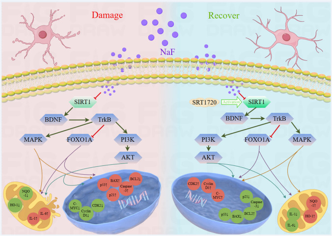
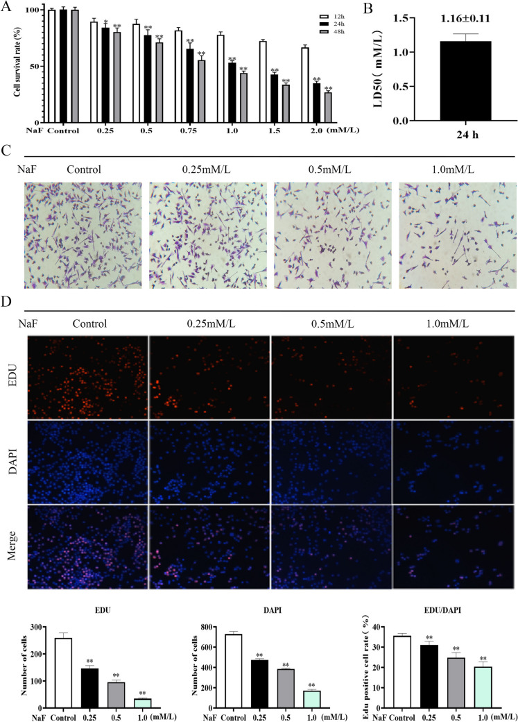
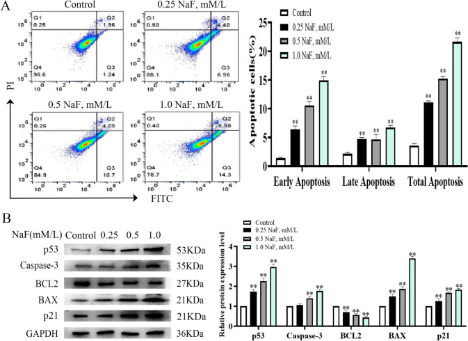
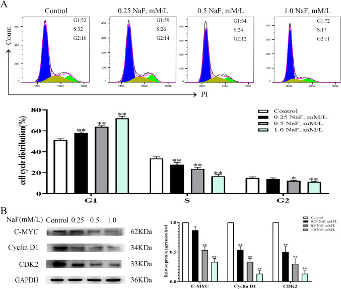
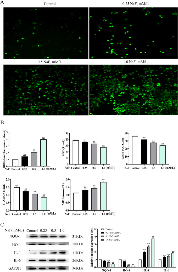
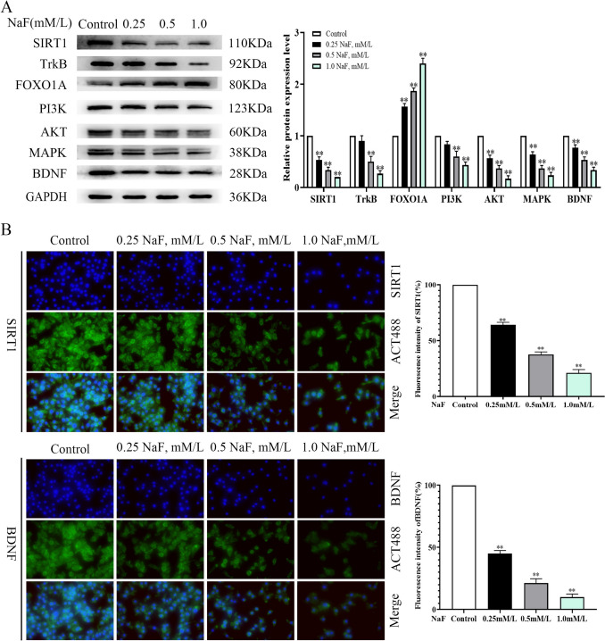
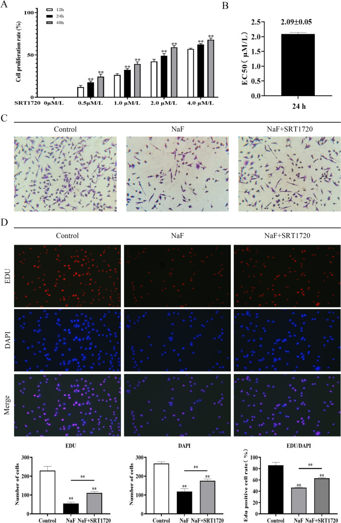
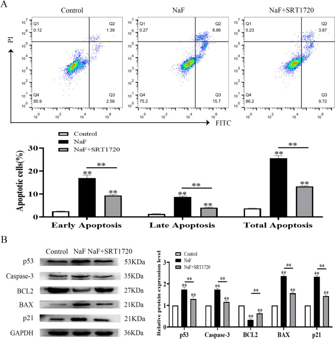
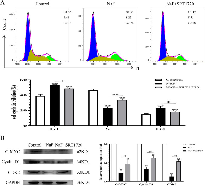
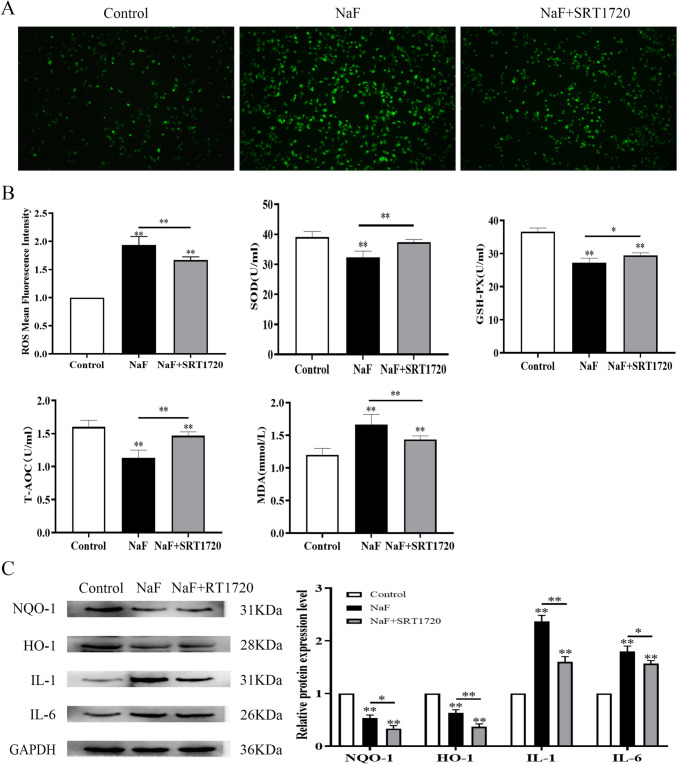
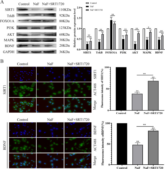
Chronic fluorosis is often accompanied by neurological symptoms, leading to attention, memory and learning ability decline and causing tension, anxiety, depression, and other mental symptoms. In the present study, we analyzed the molecular mechanisms of SIRT1-BDNF regulation of PI3K-AKT, MAPK, and FOXO1A in F-treated BV2 cells. The cytotoxic effect of sodium fluoride (NaF) on BV2 cells was assessed using Cell Counting Kit-8 (CCK-8), crystal violet, and 5-ethynyl-2'-deoxyuridine (EdU) staining. Cell cycle progression and apoptosis were evaluated through flow cytometry and western blotting. Reactive oxygen species (ROS) levels, oxidative stress, and inflammatory markers were measured by ROS staining, microplate reader assays, and western blotting. The role of SIRT1 in fluoride-induced toxicity for glial cells was determined using the SIRT1 activator SRT1720. The experiments demonstrated that NaF was toxic to BV2 cells, inhibited their proliferative ability, halted their cell cycle progression, triggered cellular apoptosis, promoted cellular oxidative stress (detected by ROS, SOD, MDA, GSH-Px, T-AOC) and associated protein NQO-1 and HO-1, and elevated inflammatory mediator associated protein IL-1and IL-6 expression). The fluoride-exposed groups had reduced SIRT1, BDNF, TrkB, PI3K, AKT, and MAPK protein expression levels, and increased FOXO1A protein expression. SRT1720 mitigated the harmful effects of NaF, stimulated cell proliferation and cell cycle progression, decreased apoptosis, reduced oxidative stress and inflammatory factors, elevated SIRT1, BDNF, TrkB, PI3K, AKT, and MAPK protein levels, and suppressed FOXO1A protein expression. The results indicate that NaF potentially harms glial cells by suppressing SIRT1 activation, and SIRT1 significantly mitigated the damage. Furthermore, the SIRT1 signaling pathway might regulate the nerve damage caused by fluoride poisoning and may be a protective factor in treating fluoride-induced brain injury.

Keywords: Fluoride; Inflammation Factors; Nerve Damage; Oxidative Stress; SIRT1.


Conflict of interest statement

Declarations. Consent to Participate: The consent of all authors to participate has been fully confirmed. Consent for Publication: All authors have agreed to submit this paper for publication. Conflict of Interest: The authors declare that the research was conducted in the absence of any commercial or financial relationships that could be construed as a potential conflict of interest. Clinical Trial Number: Not applicable.

Figures

Fig. 1
Fig. 1
This research mechanism diagram
Fig. 2
Fig. 2
Effect of NaF (0, 0.25, 0.5 and 1.0 mM/mL) on BV2 cell proliferation. A CCK-8 analysis of BV2 cells after 12-, 24-, and 48-h treatment with NaF. * p < 0.05, **p < 0.01 vs. control (B) LD50 of 24-h NaF exposure on BV2 cells. C Morphological changes of cells observed by light microscopy after 24-h NaF application. *p < 0.05, **p < 0.01 vs. control. (D) EdU assay measurement of the proliferation capacity of BV2 cells treated for 24-h with NaF (× 100). **p < 0.01 vs. Control
Fig. 3
Fig. 3
NaF significantly promotes apoptosis in BV2 cells. A Flow cytometry analysis demonstrating that NaF promoted early and late apoptosis in BV2 cells (n = 3 per group). B BCL2 expression decreased and p53, caspase-3, BAX, and p21 expression increased as NaF concentrations increased (n = 3 per group). Values are the mean ± SD. ** p < 0.01 vs. control
Fig. 4
Fig. 4
NaF significantly inhibits the cell cycle of BV2 cells. A Flow cytometric analysis demonstrating that NaF significantly inhibited the cell cycle progression of BV2 cells, stopping them in the G1-phase (n = 3 per group). B C-MYC, cyclin D1, and CDK2 expression decreased as NaF concentrations increased (n = 3 per group). Values are the mean ± SD. * p < 0.05, ** p < 0.01 vs. control
Fig. 5
Fig. 5
NaF suppresses oxidative stress and promotes inflammatory factors in BV2 cells. A ROS assay measurement of the oxidative stress of BV2 cells treated for 24 h with NaF (× 100). B ROS, SOD, GSH-PX, T-AOC, and SOD activity in the NaF groups (n = 3 per group). Values are the mean ± SD. * p < 0.05, ** p < 0.01 vs. control. (C) NQO-1 and HO-1 expression decreased and IL-1 and IL-6 expression increased as NaF concentrations increased (n = 3 per group). Values are the mean ± SD. ** p < 0.01 vs. control
Fig. 6
Fig. 6
The NaF pathway influences BV2 cells by modulating SIRT1, which regulates the FOXO, PI3K–AKT, and BDNF pathways. A FOXO1A expression increased and SIRT1, TrkB, PI3K, AKT, MAPK, and BDNF expression decreased as NaF concentrations increased (n = 3 per group). Values are the mean ± SD. ** p < 0.01 vs. control. B Immunofluorescence demonstrated that SIRT1 and BDNF expression decreased as NaF concentrations increased. Fluorescence intensity values are the mean ± SD. ** p < 0.01 vs. control
Fig. 7
Fig. 7
SRT1720 alleviates the toxic effect of NaF on BV2 cells. A CCK-8 analysis of BV2 cells after 12-, 24-, and 48-h treatment with 0.5 mM/L NaF and 0, 0.5, 1.0, 2.0, or 4.0 μM/L SRT1720. B EC50 of 24-h SRT1720 to recover BV2 cells. C Light micromorphological changes of cells in the control, NaF, and NaF + SRT1720 groups. ** p < 0.01 vs. control. D EdU proliferation was measured in the control, NaF, and NaF + SRT1720 groups (× 100). ** p < 0.01 vs. control
Fig. 8
Fig. 8
SRT1720 significantly alleviates apoptosis in NaF-exposed BV2 cells. A Flow cytometry analysis demonstrating that SRT1720 alleviated early and late apoptosis (n = 3 per group). B BCL2 expression increased and p53, caspase-3, BAX, and p21 expression were decreased in the NaF + SRT1720 group compared with the NaF group (n = 3 per group). Values are the mean ± SD, ** p < 0.01 vs. NaF group
Fig. 9
Fig. 9
SRT1720 significantly recovers the cell cycle of BV2 cells. A Flow cytometric analysis demonstrating that SRT1720 recovers the cell cycle progression of BV2 cells: the S-phase cells increase while the G1/G0-phase cells decrease (n = 3 per group). B SRT1720 increased C-MYC, cyclin D1, and CDK2 expression (n = 3 per group). Values are the mean ± SD, * p < 0.05, ** p < 0.01 vs. NaF group
Fig. 10
Fig. 10
SRT1720 alleviates the inhibition of oxidative stress and activation of inflammatory factors in BV2 cells by NaF. A ROS assay measurement of the oxidative stress of BV2 cells treated for 24 h with control, NaF, or NaF + SRT1720 (× 100). B ROS, SOD, GSH-PX, T-AOC, and MDA activity (n = 3 per group). Values are the mean ± SD. * p < 0.05, ** p < 0.01 vs. NaF group. C SRT1720 decreased IL-1 and IL-6 expression, increased NQO-1 and HO-1 expression (n = 3 per group). * p < 0.05, ** p < 0.01 vs. control
Fig. 11
Fig. 11
SRT1720 mediation of SIRT1 relieves NaF damage to BV2 cells. A SRT1720 decreased FOXO1A expression and increased SIRT1, TrkB, PI3K, AKT, MAPK, and BDNF expression (n = 3 per group). Values are the mean ± SD. * p < 0.05, ** p < 0.01 vs. NaF group. B Immunofluorescence staining demonstrating that SRT1720 increased SIRT1 and BDNF expression. Fluorescence intensity values are the mean ± SD. ** p < 0.01 vs. NaF group

Similar articles

References

    1. Velez-León E, Pacheco-Quito EM, Díaz-Dosque M et al (2023) Worldwide Variations in Fluoride Content in Beverages for Infants. Children (Basel) 10:1896 - PMC - PubMed
    1. D’Hollander W, Voogt P, Coen W et al (2010) Perfluorinated substances in human food and other sources of human exposure. Rev Environ Contam Toxicol 208:179–215 - PubMed
    1. Zhou J, Sun D, Wei W (2023) Necessity to Pay Attention to the Effects of Low Fluoride on Human Health: an Overview of Skeletal and Non-skeletal Damages in Epidemiologic Investigations and Laboratory Studies. Biol Trace Elem Res 201:1627–1638 - PubMed
    1. Dec K, Łukomska A, Maciejewska D et al (2017) The Influence of Fluorine on the Disturbances of Homeostasis in the Central Nervous System. Biol Trace Elem Res 177:224–234 - PMC - PubMed
    1. Guth S, Hüser S, Roth A et al (2020) Toxicity of fluoride: critical evaluation of evidence for human developmental neurotoxicity in epidemiological studies, animal experiments and in vitro analyses. Arch Toxicol 94:1375–1415 - PMC - PubMed

LinkOut - more resources