. 2025 Feb;57(1):221-234.
doi: 10.1038/s12276-024-01376-y. Epub 2025 Jan 17.

mtSTAT3 suppresses rheumatoid arthritis by regulating Th17 and synovial fibroblast inflammatory cell death with IL-17-mediated autophagy dysfunction

Affiliations

mtSTAT3 suppresses rheumatoid arthritis by regulating Th17 and synovial fibroblast inflammatory cell death with IL-17-mediated autophagy dysfunction

Seon-Yeong Lee et al. Exp Mol Med. 2025 Feb.

Abstract

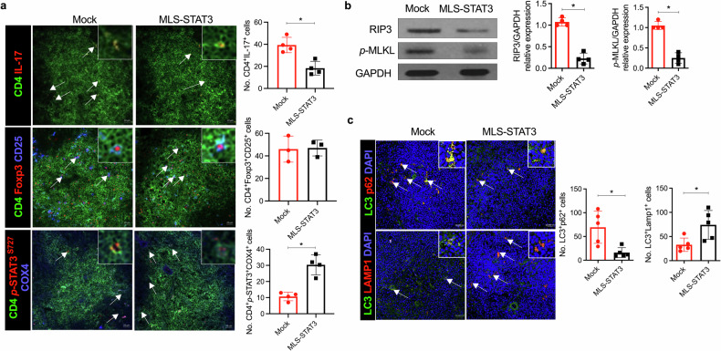
Th17 cells are activated by STAT3 factors in the nucleus, and these factors are correlated with the pathologic progression of rheumatoid arthritis (RA). Recent studies have demonstrated the presence of STAT3 in mitochondria, but its function is unclear. We investigated the novel role of mitochondrial STAT3 (mitoSTAT3) in Th17 cells and fibroblast-like synoviocytes (FLSs) and analyzed the correlation of mitoSTAT3 with RA. We used a collagen-induced arthritis (CIA) mouse model to determine the effect of mitochondrial STAT3. We observed changes in the RA mouse model via the use of a mitochondrial STAT3-inducing vector and inhibitor. We observed the accumulation of abnormal autophagosomes, increased inflammatory cell death signaling, and decreased mitoSTAT3 activity in FLSs from both patients with RA and patients with IL-17-treated FLSs. We first discovered that IL-17 increased the accumulation of abnormal autophagosomes and the expression of inflammatory cell death factors in synovial fibroblasts and decreased mitoSTAT3 activation. In a mouse model of CIA, arthritis and joint inflammation were decreased by injection vectors that induced mitoSTAT3 overexpression. The abnormal accumulation of autophagosomes and the expression of inflammatory cell death factors were also decreased in these mice. In mouse and human immune cells, ZnSO4, an inducer of mitochondrial STAT3, decreases the production of reactive oxygen species, the IL-17 concentration, and differentiation into Th17 cells. However, mitoSTAT3 blockade accelerated the development of arthritis, inflammatory cell death, and abnormal autophagosome/autophagolysosome formation. Therefore, this study suggests a novel inhibitory mechanism of RA using mitoSTAT3 via the regulation of autophagy, Th17 differentiation, and inflammatory cell death.


Conflict of interest statement

Competing interests: The authors have no financial conflicts of interest. All the authors agree with the submission. This work has not been published or submitted for publication elsewhere, either completely or in part or in another form or language. If material has been reproduced from another source, the authors have authorization from the copyright holder (usually the Publisher) to use it and have included this authorization with their submission. Experiments using animal or human materials were approved by national or local authorities.

Figures

Fig. 1
Fig. 1. IL-17 suppressed mitoSTAT3 in RA FLSs and induced mitochondrial dysfunction, inflammatory cell death, and autophagosome accumulation.
a Autophagosomes and autophagolysosomes were analyzed in OA and RA FLSs via transmission electron microscopy (scale bars = 0.5 μm, AP autophagosome, AL autophagolysosome). b RA FLSs were cultured with 10 μM rapamycin and 20 μM chloroquine for 24 h. Autophagosomes and autophagolysosomes were analyzed with anti-LC3-APC antibodies, anti-p62-FITC antibodies, anti-LAMP1-PE antibodies, and DAPI via confocal microscopy. c Mitochondria were isolated from IL-17-stimulated RA FLSs. The mitochondrial lysates were analyzed via western blotting with anti-p-STAT3Y705, anti-p-STAT3S727, anti-STAT3, anti-tubulin, and anti-COX4 antibodies. d RA FLSs were cultured with IL-17 for 12 h and then lysed to obtain proteins. The expression levels of MFN2, DRP1, and p-DRP1, which are dynamic mitochondrial molecules, were measured. e RA FLSs were transfected with mock or MLS-STAT3 vectors. P-STAT3S727 expression in cytoplasmic lysates and mitochondrial lysates was analyzed via western blotting. f RA FLSs were transfected with mock or MLS-STAT3 DNA vectors. The cells were cultured with IL-17 for 24 h posttransfection. Autophagosomes and autophagolysosomes were analyzed via confocal microscopy using anti-LC3-APC, anti-p62-FITC, and anti-LAMP1-PE antibodies and DAPI. g RA FLSs were cultured with IL-17 (10 ng/mL) for 24 h posttransfection. Inflammatory cell death molecules in the cell lysates were analyzed by western blotting. All the experiments were performed in triplicate. Bars represent the mean ± standard deviation (*p < 0.05, **p < 0.01, ***p < 0.005, ****p < 0.001).
Fig. 2
Fig. 2. mitoSTAT3 suppressed the pathological changes associated with arthritis in CIA-affected mice.
a Schematic map of the mouse MLS-STAT3-FLAG vector. b Western blot of mitochondria from mock- and MLS-STAT3-transfected NIH3T3 cells. Lanes were derived from the same gel and rearranged. c Schematic map of the schedule for arthritis induction. A total of 100 μg of mock or MLS-STAT3 DNA vector was injected every 10 days for 9 weeks after the first immunization. The severity scores and incidence of arthritis were analyzed every week. d, e Ankle joints were obtained 9 weeks after the first immunization, and the tissues were stained with H&E or antibodies against IL-17, IL-6, TNF-α, IL-1β, RIP1, RIP3, or p-MLKL. Representative images (scale bar = 100 μm) are displayed as the number of positive cells (dark brown) in five mice from each group. All the experiments were performed in triplicate. Arthritis scores are displayed as the mean ± standard error of the mean, and the bars represent the mean ± standard deviation (*p < 0.05, **p < 0.01).
Fig. 3
Fig. 3. mitoSTAT3 regulated the Th17 cell population, inflammatory cell death, and autophagosome accumulation.
ac Mock- or MLS-STAT3-injected mice were sacrificed at 9 weeks after the first immunization. a Splenic tissues were stained with specific antibodies for the analysis of Th17 cells (anti-IL-17-PE and anti-CD4-FITC), Tregs (anti-Foxp3-PE, anti-CD25-APC, and anti-CD4-FITC), and mitochondrial STAT3 (anti-p-STAT3S727-PE, anti-CD4-FITC, and anti-Cox4-APC). Representative numbers of positive cells from five different tissues are shown (left). b The expression levels of RIP3, p-MLKL, and GAPDH were measured in splenocyte lysates from the mice. c Splenic tissues were stained with anti-LC3-FITC, anti-p62-PE, and anti-LAMP1-PE antibodies and DAPI for autophagosome and autophagolysosome analysis. Bars represent the mean ± standard deviation (*p < 0.05).
Fig. 4
Fig. 4. Activation of cytoplasmic STAT3 induced the development of arthritis.
ae 100 μg of the 705 mutant (STAT3Y705F) or the 705 mutant or the MLS-STAT3 DNA vector was injected every 10 days for 9 weeks after the first immunization. a Severity scores and incidence of arthritis were analyzed every week. b Ankle joints were obtained at 9 weeks after the first immunization, and the tissues were stained with H&E and safranin O for analysis of inflammation and bone damage. c Populations of IFN-γ+, IL-4+, IL-17+, and CD25+Foxp3+ cells among splenic CD4+ T cells were analyzed by flow cytometry. d The expression levels of RIP1, RIP3, p-MLKL, and GAPDH were measured in splenocyte lysates from each group. Lanes were derived from the same gel and rearranged. e Ankle joint tissues were stained with antibodies against IL-17, IL-6, TNF-α, and IL-1β. Representative images (scale bar = 100 μm) are displayed as the number of positive cells (dark brown) in three mice from each group. All the experiments were performed in triplicate. Arthritis scores are displayed as the mean ± standard error of the mean, and the bars represent the mean ± standard deviation (*p < 0.05, **p < 0.01).
Fig. 5
Fig. 5. mitoSTAT3 altered Th17 differentiation by regulating mitochondrial function in mouse and human cells.
a, b Healthy peripheral blood mononuclear cells were cultured under T-cell activation conditions in the presence or absence of a mitoSTAT3 inhibitor (0.1 μM). Three days later, the cells were stained with antibodies against CD4, IL-17, and IL-10 and analyzed via flow cytometry. The concentrations of IL-17 and IL-10 were measured via ELISA. c Human RA synovial fluid mononuclear cells were cultured with an anti-CD3 antibody in the presence or absence of a mitoSTAT3 inhibitor. The concentrations of IL-17 and IL-10 were measured via ELISA. d Th1, Th2, Th17 and Treg cells were analyzed by flow cytometry in splenocytes from mitoSTAT3 TG mice. e, f Splenic CD4+ T cells from C57BL/6 mice were cultured under Th17-inducing conditions in the presence or absence of ZnSO4 (10 μM). e At 3 days poststimulation, the cells were stained with antibodies against CD4, IL-17, CD25, and Foxp3 and analyzed via flow cytometry. f Concentrations of IL-17 and IL-10 in culture supernatants were measured via ELISA. All the experiments were performed in triplicate. The bars represent the mean ± standard deviation (*p < 0.05, **p < 0.01, ***p < 0.005, ****p < 0.001).
Fig. 6
Fig. 6. ZnSO4, a mitoSTAT3 inducer, alleviated the pathogenesis of arthritis through T-cell homeostasis.
af Seven days after CIA induction, ZnSO4 (5 mg/kg) was orally administered for 9 weeks. a Severity scores and incidence of arthritis were analyzed every week. b Ankle joints were obtained at 9 weeks, and the tissues were stained with H&E and safranin O for analysis of inflammation and bone damage. c Splenic tissues were stained with specific antibodies for the analysis of Th17 cells (anti-IL-17-PE and anti-CD4-FITC) and mitochondrial STAT3 (anti-p-STAT3S727-PE, anti-CD4-FITC, and anti-Cox4-APC). d Splenocytes were stained with anti-CD4 and anti-IL-17 antibodies at 9 weeks after the first immunization and analyzed by flow cytometry. e The sectioned ankle joint tissues were immunohistochemically stained to identify cells positive for IL-6, IL-1β, IL-17, TNF-α, RIP1, and RIP3. Representative images (scale bar = 100 μm) are displayed as the number of positive cells (dark brown) in five mice per group. f Splenic tissues were stained with anti-LC3-FITC, anti-p62-PE, and anti-LAMP1-PE antibodies and DAPI for autophagosome and autophagolysosome analysis. Arthritis scores are displayed as the mean ± standard error of the mean, and the bars represent the mean ± standard deviation (*p < 0.05, **p < 0.01, ****p < 0.001).
Fig. 7
Fig. 7. mitoSTAT3 blockade exacerbated arthritis pathology in CIA-affected mice through increased IL-17 expression.
ae, Seven days after CIA induction, a mitoSTAT3 inhibitor (10 mg/kg) was intraperitoneally injected into CIA-affected mice 3 times weekly. Joint and spleen tissues were obtained 7 weeks after the first immunization. a Severity scores and incidence of arthritis were analyzed every week. b Western blot data demonstrating the expression of mitoSTAT3 in splenocytes from mitoSTAT3 inhibitor-injected or vehicle-injected mice with CIA. Lanes were derived from the same gel and rearranged. c Splenocytes were stained with anti-CD4, anti-IL-17, anti-IFN-γ, anti-IL-4, anti-CD25, and anti-Foxp3 antibodies. d, e Sectioned ankle joint tissues were stained with H&E and safranin O and immunohistochemically stained with antibodies against IL-17, IL-1β, IL-6, TNF-α, RIP1, RIP3, and p-MLKL. All the experiments were performed in triplicate. Arthritis scores are displayed as the mean ± standard error of the mean, and the bars represent the mean ± standard deviation (*p < 0.05, **p < 0.01).

References

    1. Guo, Q. et al. Rheumatoid arthritis: pathological mechanisms and modern pharmacologic therapies. Bone Res.6, 15, 10.1038/s41413-018-0016-9 (2018). - PMC - PubMed
    1. Golebski, K. et al. IL-1beta, IL-23, and TGF-beta drive plasticity of human ILC2s towards IL-17-producing ILCs in nasal inflammation. Nat. Commun.10, 2162, 10.1038/s41467-019-09883-7 (2019). - PMC - PubMed
    1. Yang, C. et al. Altered CD4+ T cell and cytokine levels in peripheral blood and skin samples from systemic sclerosis patients and IL-35 in CD4+ T cell growth. Rheumatology61, 794–805, 10.1093/rheumatology/keab359 (2022). - PubMed
    1. Dong, C. Genetic controls of Th17 cell differentiation and plasticity. Exp. Mol. Med.43, 1–6, 10.3858/emm.2011.43.1.007 (2011). - PMC - PubMed
    1. Hata, H. et al. Distinct contribution of IL-6, TNF-alpha, IL-1, and IL-10 to T cell-mediated spontaneous autoimmune arthritis in mice. J. Clin. Invest.114, 582–588, 10.1172/JCI21795 (2004). - PMC - PubMed

MeSH terms